SlideShare a Scribd company logo
1 of 165
Basic Immunology 2018’
Lecture 1st
Introduction
Requirements of the Department.
Historical overview. Composition of the
immune system. General characteristics of
the immune machinery.
While immunology is
important the for
pharmacists?
Basic terms
Basic terms
• Immunis,- e (Julius Caesar) = exempt, free of
burden (E.g. tax, law, or diseases)
• IMMUNE: individuals who do not capitulate to a
disease when infected;
• IMMUNITY: status of specific resistance to a
disease;
• IMMUNOLOGY: branch of theoretical biology
focuses on mechanisms responsible for self and
non-self recognition, elimination of the foreign
invaders or altered self structures with
protection of the basic structural elements.
History
• Athen (B.C. 5th century Thukidites - plaque survivors),
ancient Chinese papers about the pox immunity
• Infections, epidemies, vaccination
Edward Jenner Louis Pasteur
(1749 - 1823) (1822 - 1895)
Edward Jenner (1749 - 1823)
• He was a doctor in Berkeley, Gloucestershire. In 1796 he
carried out his now famous experiment on eight-year-old
James Phipps. Jenner inserted pus taken from a cowpox
pustule on the hand of milkmaid Sarah Nelmes and inserted
it into an incision on the boy's arm. He was testing his
theory, drawn from the folklore of the countryside, that
milkmaids who suffered the mild disease of cowpox never
contracted smallpox.
• Jenner subsequently proved that having been inoculated
with cowpox Phipps was now immune to smallpox. He
submitted a paper to the Royal Society in 1797 describing
his experiment but was told that his ideas were too
revolutionary and that he needed more proof. Undaunted,
Jenner experimented on several other children, including his
own 11-month-old son. In 1798 the results were finally
published and Jenner coined the word vaccine from the
Latin vacca for cow, and called the process vaccination.
Smallpox vaccination (1796 – 1979)
THE NOBEL PRIZE LAUREATES IN IMMUNOLOGY
1901 E.A. Von Behring (Germany) for the work on serum therapy especially its application against diphtheria.
1905 R. Koch (Germany) for the investigations concerning tuberculosis.
1908 E. Metchnikoff (Russia) and P. Ehrlich (Germany) for their work on immunity (respectively,
phagocytosis/cellular theory and humoral theory).
1913 C.R. Richet (France) for the work on anaphylaxis.
1919 J. Bordet (Belgium) for the discoveries relating to immunity (complement).
1930 K. Landsteiner (Austria/USA) for the discovery of human blood groups.
1951 M. Theiler (South Africa) for the discoveries and developments concerning yellow fever.
1957 D. Bovet (Italy/Switzerland) for the discoveries related to histamine and compounds, which inhibit action of
histamine and other substances on the vascular system and the skeleton muscles.
1960 Sir F.McFarlane Burnet (Australia) and Sir P.B. Medawar (Great Britain) for the discovery of acquired
immunological tolerance.
1972 G.M. Edelman (USA) and R.R. Porter (Great Britain) for their discovery concerning the chemical structure of
antibodies.
1977 R. Yalow (USA) for the development of radioimmunoassays of peptide hormones.
1980 B. Benacerraf (USA), J. Dausset (France) and G.D. Snell (USA) for their discoveries concerning genetically
determined structures on the cell surface (major histocompatibility complex) that regulate immunological
reactions.
1982 S. K. Bergstrom (Sweden), B. I. Samuelsson (Sweden) and J. R. Vane (UK) for their discoveries concerning
prostaglandins and related biologically active substances.
1984 N.K. Jerne (Denmark/Switzerland) for theories concerning the specificity in development (lymphocyte
clonality) and control of the immune system; G.J.F. Köhler (Germany/Switzerland) and C. Milstein
(Argentina/Great Britain) for the discovery of the principle for production of monoclonal antibodies.
1987 S. Tonegawa (Japan/USA) for the discovery of the genetic principle for generation of antibody diversity.
1990 J.E. Murray and E.D. Thomas (USA) for their discovery concerning organ and cell transplantation in the
treatment of human diseases.
1996 P.C. Doherty (Australia/USA) and R.M. Zinkernagel (Switzerland) for their discoveries concerning the
specificity of the cell mediated immune defense ("dual recognition").
1997 S.B. Prusiner (USA) for the discovery of prions as a new biological principle of infection.
1999 G. Blobel (USA) for discoveries concerning signal transduction.
Main fields of applied immunology
• Infectious immunity
Basic empirical observations on survivors during the big epidemics (plague, pox,
cholera, etc) in the Middle Age. New aspects occurred in the end of the 21st
century: sever viral infections (HIV, influenza), fungal infections, antibiotic
resistance in different bacteria.
• Tumor immunology
Animal experiments with tumor transplantation clarified the genetic mechanisms
of graft rejection, and the correlation between the blood groups and the
transplantation immunity (Gorer, 1927). New immunological concept developed in
biology and medicine in the first decades of 20th century: immune system is
responsible for the self integrity of individuals. The defense against tumors is not
known in details yet, however, the role and heredity of major histocompatibility
complex (MHC) was discovered during the tumor-transplantation experiments
establishing the immunogenetics.
• Transplantation immunology
Immunonological aspects of organ transplantaions
• Cellular and molecular immunology (Basic and applied
immunological research, related innovations and R&D) diagnostics ant drug
designe.
• Immunological biotechnology (increased need for individual
diagnostics ant therapy)
• Biological therapies (Therapeutic monoclonal antibodies, recombinant
cytokines)
What is the main function
of the immune system?
Saving the individual integrity against foreign
invaders (pathogens) and the modification of
self structures by mutations, tumorous
transformations, physical or chemical
effects, or virus infections.
Immune system
 Individuals and species
 Organs
 Cells
 Molecules
 Functions
Immune system is a general structural
and functional network composed by
molecular and cellular elements of the
body.
Organs of the
immune system
Primary Secondary
(central) (peripheral)
• Bone marrow
• Thymus
• (Embryonic
liver)
• Lymph nodes
• Spleen
• MALT
• SALT
Red and yellow bone marrow
Normal bone marrow (HE)
Strómasejt
B és T sejt előalakok
Neutrophil granulocytes
Eosinophil granulocyte and a small
lymphocyte
Bazophil granulocyte, neutrophil
granulocite and a large lymphocyte
Monocyte
White blood cells in
peripheral blood smears
T cell
T cell migration
Thymus involution during aging
Thymus
Lymph node
B cells
Spleen
Macrophage
Haemopoiesis in embryonic life
Blue: stem cells
Dark blue: immature cells
Brown: matured cells
Hematopoietic
differentiation
Cells of the immune system
Antigen-presenting cells: “professional” or “accidental”
Antigen-binding cells: T- and B lymphocytes
Effektor cells: T, NK, granulocytes, mast cells,
monocytes/macrophages
Organ distribution of T and B lymphocytes
------------------------------------------------------------------------------------------------
Organ % lymphocyte
T B
------------------------------------------------------------------------------------------------
Tymus >99 <0.5
Lymph node 75 25
Spleen 50 50
Peripheral blood 55-75 15-30
Bone marrow 7 >75
------------------------------------------------------------------------------------------------
Theoretical scheme of the innate immunity
ANTIGEN
RECOGNITION RESPONSE
Theoretical scheme of the adaptive immunity
ANTIGEN
RECOGNITION DIFFERENTIATION EFFECTOR FUNCTIONS
MEMORY
Composition of the immune system
Natural
Innate-like immunity with adaptive features
Innate
•None antigen specific
•No immunological memory
•Rapid reactivity
•Linear amplification of the
reaction
Adaptive
•Antigen specific
•Immunological memory
•Activated after a latency
•Exponential amplification of
the reaction
Basic Immunology
Lecture 3rd-4th
Molecular components of
immunological recognition.
Definition of the antigen. Antibodies, T- and
B-cell receptors: molecular structure, functions,
subcalsses
Definition of the antigen
László Detre (Detsch) : antibody generator
- Old definition: foreign agent induces
immune reaction
- Modern definition: substance recognized by
T- or B-cell receptor and induces tolerating
or active type immune response according
to the MHC haplotype of the individual.
Factors determining the immunogenity
• immunodominant regions
• chemical structure (inorganic molecules are not antigens at
general, but e.g. heavy metals in protein complex are able to
induce specific metal allergies). The best antigens are
proteins>polypeptides>polysaccharides>lipides>nucleic acids
• physico-chemical nature (D and L configuration; ortho-,
para,- meta position; hydrophilic and hydrophobic amino acid
sequence)
• molecular weight (not an absolute category)
• conformation sensitivity (folding and refolding)
• Origin auto-, allo-, xenoantigen
• mode and anatomic region of the administration (e.g.
peripheral immune reaction and oral tolerance for the same
antigen depending from the place of the antigen presentation)
• dose dependence (large and low dose)
• Valency: monovalent, bivalent, and multivalent antigens
Basic terms
immunogen (fine chemical structure can induce
specific immune response)
epitope (antigen determinant) well circumscribed
region of the antigen molecule targeted by Ig/BcR or
TcR
paratop (ligand pair of the epitope)
hapten (small molecular weight antigen can not
induce immune reaction itself, but specifically
recognized by immunoglobulins)
carrier (indifferent, large molecular weight molecule,
hold on the surface hapten molecules; carrier
molecules did not participate in the anti-hapten
immune reaction only hapten)
Antigen recognition
Innate immunity Natural immunity Acquired immunity
• Pattern recognition • Pattern recognition • Antigen recognition
• Mainly sugar
recognition
• Mainly peptide patter
recognition
• Mainly peptide
pattern recognition
• PRR • MHC • MHC
• PAMP • iTcR, γδTcR, BcR,
IgM
• αβTcR, γδTcR, BcR,
• IgM/G/A/E/D
• Low number of
molecularly distinct
receptors and high
number of
recognized patterns
• Limited number of
molecularly distinct
receptors and high
number of
recognized patterns
• High number of
distinct antigen
receptors and high
number of
specifically
recognized antigens
Recognition molecules
Immunoglobulins
B cell receptors (BcR)
T cell receptors (TcR)
MHC class I and class II
Specialized molecules manage antigen recognition.
The common structural features of these molecules
are the well-conserved (constant) basic elements
(designed by 110 amino acids domain units)
containing variable, antigen specific parts (binding
sites) for the recognition and ligand formation.
Antigen specific recognition molecules
Domain structure
Well conserved amino acid sequence designed by 110 amino acids
closed to a “ring shape” with disulphide bound.
Immune recognition molecules
Antigen
specific
recognition
molceules
Accessory
molecules
Immunoglobulin molecule
CDR
Variable region
Idiotype
Fab fragment
Constant region
Isotype
Fc fragment
Ig domains: intra-chain disulphide
bonds form loops in the peptide
chain, the loops are globular,
constructed from beta-plated
sheets and beta-turn loops.
Immunoglobulins
Monofunctional character (specific antigen
recognition and binding) before the antigen
administration. Fab dependent function.
Polyfunctional character after the
antigen administration (signal transduction,
complement fixation, opsonization,
immunocomplex formation, FcR binding, etc).
Fc dependent functions.
Immunoglobulin isotypes
• Based upon the constant structures of heavy
(H) and light (L) chains
• CH isotypes: called Ig classes and
subclasses as IgG, IgM, IgA, IgD and IgE.
All classes are represented in a normal
serum (except the membrane bound IgD) as
isotype variants.
• CL chain exists in two isotypic forms:
kappa (κ) and lambda (λ), which can
associate with all heavy chain isotypes.
IgA and IgM
Immunoglobulin idiotype
Individual determinants in V regions, specific for each
antibody.
The N terminal Ig domain contains V region forming
the antigen binding site: clustering the 3 hyper variable
sequences close to each other on both chains - the
variation of 3 x 3 results tremendous diversity.
Structure of IgG
IgG – blood, lymph, make up 80% of Ig
• only Ig of maternal origin to pass the placenta wall give newborns
(Mw 150 kD)
• neutralize toxins and viruses
IgM – Blood, lymph (cell surface) pentamer structure (Mw 900 kD)
• First antibodies formed in response to initial infection.
IgA – Mucosal surfaces, blood (active in dimeric or tetrameric form)
(Mw 150-600 kD)
IgD – only membrane-bounded form in B-cell surfaces (Mw 150 kD)
• may function in initiation of antibody-antigen response
IgE – blood (bound to basophiles, mast cells)
(Mw 190 kD) initiation of allergic reactions
Kinetics of antibody production
Antigen – antibody reactions
• Neutralization (e.g. toxins)
• Precipitation (soluble molecules)
• Agglutination (particles, cells)
• Opsonization (large particles)
• Complement fixation
B cell receptor complex
T cell receptor complex expressed in mature T cells
ab TcR – SP(CD4+ or CD8+)
gd TcR – DN (CD4-CD8-)
T Cell Receptor
complex
ITAMs
Immunoreceptor
Tyrosine-based
Activation
Motifs
Basic Immunology
Lecture 5-6th
MHC
Structure, genetics, role in the
immunological recognition. Antigen
presenatation.
The Immune System
• Innate immunity (complement, cytokines,
antibacterial peptides, macrophages,
neutrophil granulocytes, NK cells, etc.)
• Adaptive immunity (antibodies, T and B
lymphocytes, lymphoid cytokines, etc.)
• Natural immunity (natural autoantibodies,
ILCs, iNKT cells, γδT cells, MAIT cells, etc.)
Recognition of the antigen in
the adaptive immunity
Native antigens are recognized by
immunoglobulins or B cell receptors.
T cells can recognize in denatured
(presented by MHC) forms of the
antigens exclusively.
Major Histocompatibility Complex
Self and foreign antigens are presented on
the cell surface by specialized host-cell
glycoproteins encoded in a large cluster of
genes that were first identified by their
effects on the immune response to
transplanted tissues. For that reason, the
gene complex was termed the Major
Histocompatibility Complex (MHC). The
antigen binding glycoproteins are called
MHC molecules/antigens. (MHC vs. HLA,
H2, BoLA, ChLA etc.)
Tolerated skin grafts on MHC (H2) identical mice
MHC Class I
Present in all
nucleated cells
and platelets
(in different
density).
MHC class II
molecule
a and b chains
Expressed on
professional or
facultative antigen
presenting cells
(APCs):
•Constitutive
•Induced
Class I and class II MHC Molecules
MHC class I and class II
molecules with bound peptides
MHC Class I presents antigen
for T cell
Structure of MHC genes
HLA map
Polygenic: (there are several different class I and
class II genes encoding proteins with different
specificities:– HLA-A, B, C, HLA-DR, DP, DQ)
Polymorphic: there are multiple alleles of each gene
(6-50)
Co-dominant: haplotypes (allel variants) of BOTH
parents are expressed
Characteristics of MHC
What type of cells express MHC
class I and MHC class II?
MHC I Any cell type with nucleus
MHC II Mainly professional antigen presenting
cells
Dendritic cell, Follicular dendritic cells
B cells
Macrophages, monocytes
(Thymic epithelial cells)
Facultative antigen presenting cells
Inflammatory epithel
Antigen presentation though
MHC class I
Antigen Processing and
Presentation on MHC I
Transporter Associated with
Antigen Processing
Molecular Immunol. 2002
Chaperons in the MHC-I Antigen
Presentation
Calnexin, calreticulin, Erp57, tapasin
Antigen Presentation on
MHC class I
-Cytosolic, mainly normal or viral/modified proteins
-Proteasomal degradation
Peptide transfer to the ER (TAP1&2)
MHC I chains produced into ER by ribosomes
Chaperons: calnexin, calreticulin, Erp57
Tapasin and TAP1&2
MHCI & peptide binding within the ER
Antigen presentation through
MHC class II
Generation of Antigenic Peptides in
the Endocytic Pathway
Peptide Loading of Class II
Molecules
HLA-DM: MHCII chaperon
CLIP=class II associated invariant chain peptide
Antigen Presentation on MHC II
-HLA-DM: MHC II specific chaperon
-invariant chain
-Endocytosed proteins: bacteria, bacterial
product, internalised receptor bound
peptide, parts of another cell
-Endosomal degradation
-MHCII chains produced into the ER by ribosomes
-CLIP=class II associated invariant chain peptide
-MHC II & peptide binding in endosomes outside
the ER
a2
a3
a1
a2a1
b2
b1
b2 mgl
MHC Restriction
Herpes simplex – produces a protein which inhibits TAP
Adenovirus – produces a protein, which binds to and
retains MHC-I in the ER
Cytomegalovirus – accelerates MHC-I translocation to the
cytosol for degradation
HIV – accumulate mutations faster than the adaptive
immune system can cope with
MHC-I
MHC-II
Helicobacter pylori – encodes a 95kD protein toxin, which
increases the pH of the lysosomes, inhibiting protease activity
How do pathogens avoid detection?
Septic schock - superantigens
Activated T cells produce cytokines randomly - systemic
collaps of several biological functions („Cytokine tsunami”)
Compared to a normal
antigen-induced T-cell
response where low number
of the body’s T-cells are
activated, SAgs (endotoxins)
of viral or bacterial origin are
capable of activating large set
(up to 20%) of the body’s T-
cells randomly. This causes a
massive and irregular immune
response (toxic shock
syndrome) that is not specific
to any particular epitope on
the SAg.
Basic Immunology
Lecture 7th - 8th
Communication between cellular
components of the immune
system.
Co-receptors and adhesion molecules.
Cytokines, chemokines and their receptors.
Mediators of cell-cell interactions:
„cross-talk”
Cell-cell interactions play basic biological role in
development and function of multicellular organisms.
These interactions allow cells to communicate with
each other. This ability to send and receive signals is
essential for the further functions of the cells.
- Direct interactions: adhesion molecules
- Microparticles: microvesicules, microtubes
- Soluble mediators perform indirect
interactions: cytokines, chemokines,
interleukins, interferons, growth factors, tissue
hormons, complement factors, etc.
Immunological „cross-talk”
-Haematopoiesis: adhesion between stromal cells of the
bone marrow and the differentiating leukocytes
-Lymphocyte recirculation and recruitment: adhesion
between endothelial cells and the circulating leukocytes,
recruiting immune active cells into the inflammatory tissues
-Immune response: T cell and APC/B cell interactions
during antigen presentation, activation and differentiation of
immune cells, cytotoxic effector reactions
Adhesion molecules
Cell surface molecules whose function is to
promote adhesive interactions with other
cells or the extracellular matrix and initiate
signal transduction.
Leukocytes express various types of
adhesion molecules, such as selectins,
integrins, and members of the Ig superfamyli,
and these molecules play crucial role in cell
migration and cellular activation both in
innate and adaptive immune response.
Cell adhesion molecules
Accessory molecules on T cells
Family of accessory molecules, adhesion
molecules, co-receptors
Common characteristics:
1. Molecules, responsible for the direct
interaction of the immune cells
2. Their interaction is not antigen-
specific
2. Low-affinity, reversible association
4. Increase the antigen-specific
interaction
5. Co-receptors: - signaling function
6. Co-stimulatory molecules: help cell
activation
7. Non-polymorphic
T cell
Activation of adheseion molecules
• Lymphocyte Function-associated Antigen
• IntraCellular Adhesion Mloecule
Families of adhesion molecules
CD2
CD4
CD8
B7
CD28
CTLA 4
ICAM
L selectin
E selectin
P selectin
VLA
LFA
Mac1
„vascular
addressins”
„other”
accessory
molecules
CD45
CD44
CD40, CD40L
CD19/CD21/CD81
CD22
Ig-superfamily
members
Selectins Integrins
Mucin-like
molecules
Lectin
domain
SCR
domains
cysteins
90% of
T cells
„T cell rosette”
CD2
„sheep red-blood cell receptor”
Adhesion
Cell activation
Ig-superfamily members
Binds CD58
(LFA3)
T cell activation, CTL- and NK-mediated lysis
Differentiation markers:
At different stages of
T cell maturation
CD4 and CD8 together
„double positive”
in thymus
At the periphery:
„single positive”
T helper: CD4
T cytotoxic: CD8
CD4 and CD8:
extracellular domain: binding to MHC constant domain
intracellular domain: signal transduction, binding kinases
CD4 - MHCII CD8 - MHCI
CD4 - HIV-receptor as well
55 kDa
Th, Mo,
Mf, DC
35 kDa
Tc
CD4+ T cell CD8+ T cell
B7 (CD80, CD86), CD28 and CTLA-4 molecules
CD28: - co-stimulatory molecule in T
cell activation
- Increases IL-2 and IL-2R expression,
- Induces T cell proliferation
CTLA-4 (CD152): - expressed in
a later phase of the T cell activation
- inhibitory function
CTLA: Cytolytic T lymphocyte associated Antigen
CD28 and CTLA-4 of T cells
bind to the B7-1 (CD80), B7-2
(CD86) molecules of the APC
T cell
APC
„OTHER” accessory molecules
Plays important role in cell activation and in
regulation of signal transduction
- tyrosine–phosphatase domain:
dephosphorylation
CD45
Expressed on every leukocyte
“pan-leukocyte marker”
- Highly glycosylated,
- More isoforms (180, 190, 200, 205, 220 kDa)
- alternate splicing
CD45
CD45 isoforms
CD45
CD44
Expressed on activated and memory T- and
B-cells, phagocytes, fibroblasts, neuronal
cells
Important in „homing” of leukocytes
More isoforms - alternate splicing
CD44
„OTHER” accessory molecules
POSSIBLE MECHANISMS
BY WHICH NK CELLS
DISTINGUISH INFECTED
FROM UNINFECTED CELLS
NK cells can use several different receptors that
signal them to kill, including lectinlike
activating receptors, or ‘killer receptors,’ that
recognize carbohydrate on self cells. However,
another set of receptors, called Ly49 in the
mouse and killer inhibitory receptors (KIRs) in
the human, recognize MHC class I molecules
and inhibit killing by NK cells by overruling the
actions of the killer receptors. This inhibitory
signal is lost when cells do not express MHC
class I and perhaps also in cells infected with
virus, which might inhibit MHC class I
expression or alter its conformation. Another
possibility is that normal uninfected cells
respond to IFN-α and IFN-β by increasing
expression of MHC class I molecules, making
them resistant to killing by activated NK cells.
In contrast, infected cells can fail to increase
MHC class I expression, making them targets
for activated NK cells. Ly49 and KIR belong to
different protein families—the C-type lectins in
the case of Ly49 and the immuno-globulin
superfamily for KIRs. The KIRs are made in two
forms, p58 and p70, which differ by the
presence of one immunoglobulin domain.
PHAGOCYTE ADHESION
TO VASCULAR
ENDOTHELIUM IS
MEDIATED BY
INTEGRINS
Vascular endothelium, when it is
activated by inflammatory
mediators, expresses two
adhesion molecules—ICAM-1 and
ICAM-2. These are ligands for
integrins expressed by
phagocytes—αL:β2 (also called
LFA-1 or CD11a: CD18) and αM:β2
(also called Mac-1, CR3, or
CD11b:CD18).
Lymphocyte recirculation:continuos migration of cells from
the blood flow and lymph to the lymhatic organs and to the inflammation = HOMING
Role:
- Promots the antigen capturing
- Promots the development of
inflammatory reactions
Mechanism:
-Extravasation: leucocyte adhesion
to the endothel, and migration
across the wall of the blodd vessels
to the tissue
1-2 total circle managed by all white blood cells pro day
Neutrophils leave the blood and migrate to sites of infection in a multistep
process mediated through adhesive interactions that are regulated by
macrophage-derived cytokines and chemokines.
Naive lymphocytes migrating to the
peripheral lymphatis tissues:
The role of the hugh endothelial venules (HEV), and the
adhesion molecules:
1. Selectin-mediated 2. chemoattractant 3. Integrin-mediated
mediated
Migration of neutrophil granulocytes to the
inflammed tissues throgh the endothel
Different adhesion molecules determine
the migration of naive and memory
(effector) cells
Peripheral lymphatic tissue Inflammatory tissue
Some important accessory
molecules
• CD3
• CD4 and CD8
• CD28
• CD80/86 (B7.1 and B7.2)
• CD152 (CTLA4)
• CD25 (IL-2 Receptor)
• CD45RA/RO
• CD154 (CD40 Ligand)
Direct cell-cell
communication
Cell-cell communication through
cytokines and their receptors
• Cytokines
• Chemokines
• Interferons
• growth factors
Mechanism of cytokine action I.:
Cytokine producing cell Target cell:
Cytokine producing cell
Nearby cell
Distant cell
Autocrine action
Paracrine actiont
Endocrine action
Cytokines act in each phase of the immune response
Recognition:
Activation:
Effector phase:
Mechanism of cytokine action II.:
A cytokine induces different
effects on different target
cells
The action of more
cytokine on the target cell
is similar
The effect of two
cytokines is stronger than
their additive effects
One cytokine inhibits the
effects of another
cytokine
Pleiotropy
Redundancy
Synergy
Antagonism
Starting a cascade
Basic characteristics of cytokies
• Low molecular weight (10-40 kDa), and genetically
well conserved glycoproteins
• Isolated cells secrete them, due to gene activation
• They mediate cell-cell interaction:
• - sending information
• - regulation of immune response
• Mechanism of action:
- produced after transient gene activation
- act through receptors triggering signal-transduction
- high affinity
- picomolar concentration
- they act mostly locally
Functional groups of cytokines
I. Regulators of natural
immunity and inflammation
IFNa, IFNb, TNFa, TNFb (LT),
IL-1a, IL-1b, IL-6, IL-12,
MIF, chemokines
II. Regulators of lymphocyte
activation and differentiation
IL-2, IL-4, IL-5, IL-6, IL-13, IL-15,
INFg,
IL-10 and TGFb
III. Regulators of
haematopoiesis
IL-3, IL-7, GM-CSF, SCF
MHC
IL-1
bakteriális
endotoxin
makrofág
T-sejtTCR
citotoxicitás
monokinek
adhéziós molekulák



aktiváció
IL-2R
limfokinek



prosztaglandinok
láz
aluszékonyság
fájdalomküszöb
fogyás


autokrin parakrin
endokrin
Autocrine, paracrine and endocrine action of IL-1
macrophage
T cell
Activation,
IL-2R expression
Cytokine secretion.
Bacterial
endotoxin
Enhanced cytotoxicity
Cytokine production
Adhesion mol. Expr.
Prostaglandin
Production, fever
Acute phase protein
Secretion by hepatocytes
Cytokine receptors
Characteristics of multichain cytokine
receptors
Chemokines
- 90-130 aa. Polypeptides
- Receptorial action
- Produced by lymhatic and none-lymphatic tissues
Functions:
- chemotaxis for different
leukocytes
- regulation of normal leukocyte
traffic
- recruitment of cells to
inflammatory sites
- enhancement of cell adhesion
- activation of effectors leukocytes
- development of the inflammatory
reaction
- development of normal lymphoid
tissues
Basic Immunology
Lectures 9-10
Lymphocyte Development and
Antigen Receptor Gene
Rearrangement
OVERVIEW OF LYMPHOCYTE DEVELOPMENT
The maturation of B and T lymphocytes involves a series of
events that occur in the generative lymphoid organs. These
events include the following:
1. The commitment of progenitor cells to the B cell or T cell
lineage.
2. Proliferation of progenitors and immature committed cells at
specific early stages of development, providing a large pool of
cells that can generate useful lymphocytes.
3. The sequential and ordered rearrangement of antigen
receptor genes and the expression of antigen receptor proteins.
OVERVIEW OF LYMPHOCYTE DEVELOPMENT (cont.)
4. Selection events that preserve cells that have produced
correct antigen receptor proteins and eliminate potentially
dangerous cells that strongly recognize self antigens. These
checkpoints during development ensure that lymphocytes that
express functional receptors with useful specificities will mature
and enter the peripheral immune system.
5. Differentiation of B and T cells into functionally and
phenotypically distinct subpopulations. B cells develop into
follicular, marginal zone, and B-1 B cells, and T cells develop into
CD4+ and CD8+ T lymphocytes and γδ T cells. This
differentiation into distinct classes provides the specialization that
is an important characteristic of the adaptive immune system.
Development of B cells
B cells develop in the bone marrow and migrate to peripheral lymphoid organs,
where they can be activated by antigens.
Development of T cells
T cells undergo development in the thymus and migrate to the peripheral lymphoid
organs, where they are activated by foreign antigens.
Copyright © 2011 by Saunders, an imprint of Elsevier Inc.Abbas, Lichtman, and Pillai. Cellular and Molecular Immunology, 7th edition. Copyright © 2012 by Saunders, an imprint o
Stages of Lymphocyte Maturation
Fig. 8-1
Development of both B and T lymphocytes involves the sequence of maturational
stages shown. B cell maturation is illustrated, but the basic stages of T cell maturation
are similar.
Copyright © 2011 by Saunders, an imprint of Elsevier Inc.Abbas, Lichtman, and Pillai. Cellular and Molecular Immunology, 7th edition. Copyright © 2012 by Saunders, an imprint o
Distinct Lineages of Lymphocyte Maturation
Fig. 8-2
Copyright © 2011 by Saunders, an imprint of Elsevier Inc.Abbas, Lichtman, and Pillai. Cellular and Molecular Immunology, 7th edition. Copyright © 2012 by Saunders, an imprint o
Checkpoints in Lymphocyte Maturation
Fig. 8-3
During development, the lymphocytes that express receptors required for continued proliferation and
maturation are selected to survive, and cells that do not express functional receptors die by apoptosis.
Positive selection and negative selection further preserve cells with useful specificities. The presence of
multiple checkpoints ensures that only cells with useful receptors complete their maturation.
Copyright © 2011 by Saunders, an imprint of Elsevier Inc.Abbas, Lichtman, and Pillai. Cellular and Molecular Immunology, 7th edition. Copyright © 2012 by Saunders, an imprint o
Pos. and Neg. Selection of Lymphocytes
Fig. 8-4
Copyright © 2011 by Saunders, an imprint of Elsevier Inc.Abbas, Lichtman, and Pillai. Cellular and Molecular Immunology, 7th edition. Copyright © 2012 by Saunders, an imprint o
Germline Organization of Human Ig Loci
Fig. 8-5
The human heavy chain, κ light chain, and λ light chain loci are shown. Only functional genes are shown;
pseudogenes have been omitted for simplicity. Exons and introns are not drawn to scale. Each CH gene is
shown as a single box but is composed of several exons, as illustrated for Cμ. Gene segments are indicated as
follows: L, leader (often called signal sequence); V, variable; D, diversity; J, joining; C, constant; enh, enhancer.
Copyright © 2011 by Saunders, an imprint of Elsevier Inc.Abbas, Lichtman, and Pillai. Cellular and Molecular Immunology, 7th edition. Copyright © 2012 by Saunders, an imprint o
Domains of Ig and TCR proteins
Fig. 8-6
The domains of Ig heavy and light chains are shown in A, and the domains of TCR α and β chains are shown in B.
The relationships between the Ig and TCR gene segments and the domain structure of the antigen receptor
polypeptide chains are indicated. The V and C regions of each polypeptide are encoded by different gene
segments. The locations of intrachain and interchain disulfide bonds (S-S) are approximate. Areas in the dashed
boxes are the hypervariable (complementarity-determining) regions. In the Ig μ chain and the TCR α and β chains,
transmembrane (TM) and cytoplasmic (CYT) domains are encoded by separate exons.
Copyright © 2011 by Saunders, an imprint of Elsevier Inc.Abbas, Lichtman, and Pillai. Cellular and Molecular Immunology, 7th edition. Copyright © 2012 by Saunders, an imprint o
Germline Organization of Human TCR Loci (1)
Fig. 8-7
The human TCR β, α, γ, and δ chain loci are shown, as indicated. Exons and introns are not
drawn to scale, and nonfunctional pseudogenes are not shown. Each C gene is shown as a single
box but is composed of several exons, as illustrated for Cβ1. Gene segments are indicated as
follows: L, leader (usually called signal sequence); V, variable; D, diversity; J, joining; C, constant;
enh, enhancer; sil, silencer (sequences that regulate TCR gene transcription).
Copyright © 2011 by Saunders, an imprint of Elsevier Inc.Abbas, Lichtman, and Pillai. Cellular and Molecular Immunology, 7th edition. Copyright © 2012 by Saunders, an imprint o
Germline Organization of Human TCR Loci (2)
Fig. 8-7
Copyright © 2011 by Saunders, an imprint of Elsevier Inc.Abbas, Lichtman, and Pillai. Cellular and Molecular Immunology, 7th edition. Copyright © 2012 by Saunders, an imprint o
Antigen receptor Gene Rearrangements
Fig. 8-8
Southern blot analysis of DNA from nonlymphoid (liver) cells and from a monoclonal population of B
lymphocyte lineage origin (e.g., a B cellt umor) is shown in schematic fashion. The DNA is digested
with a restriction enzyme (EcoRI as depicted), different-sized fragments are separated by
electrophoresis, and the fragments are transferred onto a filter. The sites at which the EcoRI
restriction enzyme cleaves the DNA are indicated by arrows. The size of the fragments containing
the Jκ3 segment of the Ig κ light chain gene or the Vκ29 V region gene was determined by use of a
radioactive probe that specifically binds to Jκ3 segment DNA or to Vκ29 DNA. In the hypothetical
example shown, Vκ29 is part of a 5-kb EcoRI fragment in liver cells but is on a 3-kb fragment in the
B cell clone studied. Similarly, the Jκ3 fragment is 8 kb in liver cells but 3 kb in the B cell clone.
www.nobelprize.org
Copyright © 2011 by Saunders, an imprint of Elsevier Inc.Abbas, Lichtman, and Pillai. Cellular and Molecular Immunology, 7th edition. Copyright © 2012 by Saunders, an imprint o
Diversity of Antigen Receptor Genes
Fig. 8-9
From the same germline DNA, it is possible to generate recombined DNA sequences
and mRNAs that differ in their V-D-J junctions. In the example shown, three distinct
antigen receptor mRNAs are produced from the same germline DNA by the use of
different gene segments and the addition of nucleotides to the junctions.
Copyright © 2011 by Saunders, an imprint of Elsevier Inc.Abbas, Lichtman, and Pillai. Cellular and Molecular Immunology, 7th edition. Copyright © 2012 by Saunders, an imprint o
V(D)J Recombination (1)
Fig. 8-10A
The DNA sequences and mechanisms involved in recombination in the Ig gene loci are depicted.
The same sequences and mechanisms apply to recombinations in the TCR loci. A, Conserved
heptamer (7 bp) and nonamer (9 bp) sequences, separated by 12- or 23-bp spacers, are located
adjacent to V and J exons (for κ and λ loci) or to V, D, and J exons (in the H chain locus). The
V(D)J recombinase recognizes these recombination signal sequences and brings the exons
together.
Copyright © 2011 by Saunders, an imprint of Elsevier Inc.Abbas, Lichtman, and Pillai. Cellular and Molecular Immunology, 7th edition. Copyright © 2012 by Saunders, an imprint o
V(D)J Recombination (2)
Fig. 8-10B
B, C, Recombination of V and J
exons may occur by deletion of
intervening DNA and ligation of
the V and J segments (B) or, if
the V gene is in the opposite
orientation, by inversion of the
DNA followed by ligation
of adjacent gene segments (C).
Red arrows indicate the sites
where germline sequences are
cleaved before their ligation to
other Ig or TCR gene segments.
Copyright © 2011 by Saunders, an imprint of Elsevier Inc.Abbas, Lichtman, and Pillai. Cellular and Molecular Immunology, 7th edition. Copyright © 2012 by Saunders, an imprint o
V(D)J Recombination (3)
Fig. 8-10C
B, C, Recombination of V and J
exons may occur by deletion of
intervening DNA and ligation of the
V and J segments (B) or, if the V
gene is in the opposite orientation,
by inversion of the DNA followed by
ligation of adjacent gene segments
(C). Red arrows indicate the sites
where germline sequences are
cleaved before their ligation to
other Ig or TCR gene segments.
Contributions of different mechanisms to the
generation of diversity in Ig and TCR genes
The rearrangement of antigen receptor genes is the key event in lymphocyte
development that is responsible for the generation of a diverse repertoire.
Copyright © 2011 by Saunders, an imprint of Elsevier Inc.Abbas, Lichtman, and Pillai. Cellular and Molecular Immunology, 7th edition. Copyright © 2012 by Saunders, an imprint o
Transcriptional Regulation of Ig Genes
Fig. 8-11
V-D-J recombination brings promoter sequences (shown as P) close to the enhancer
(enh). The enhancer promotes transcription of the rearranged V gene (V2, whose active
promoter is indicated by a bold green arrow). Many receptor genes have an enhancer in
the J-C intron and another 3′ of the C region. Only the 3′ enhancer is depicted
here.
Copyright © 2011 by Saunders, an imprint of Elsevier Inc.Abbas, Lichtman, and Pillai. Cellular and Molecular Immunology, 7th edition. Copyright © 2012 by Saunders, an imprint o
Events During V(D)J Recombination (1)
Fig. 8-12
Sequential events during V(D)J recombination. Synapsis and cleavage of DNA at the
heptamer/coding segment boundary are mediated by Rag-1 and Rag-2. The coding
end hairpin is opened by the Artemis endonuclease, and broken ends are repaired by
the NHEJ machinery.
Copyright © 2011 by Saunders, an imprint of Elsevier Inc.Abbas, Lichtman, and Pillai. Cellular and Molecular Immunology, 7th edition. Copyright © 2012 by Saunders, an imprint o
Events During V(D)J Recombination (2)
Fig. 8-12
Sequential events during V(D)J
recombination. Synapsis and
cleavage of DNA at the
heptamer/coding segment boundary
are mediated by Rag-1 and Rag-2.
The coding end hairpin is opened by
the Artemis endonuclease, and
broken ends are repaired by the
NHEJ machinery.
Copyright © 2011 by Saunders, an imprint of Elsevier Inc.Abbas, Lichtman, and Pillai. Cellular and Molecular Immunology, 7th edition. Copyright © 2012 by Saunders, an imprint o
Events During V(D)J Recombination (3)
Fig. 8-12
Sequential events during V(D)J
recombination. Synapsis and
cleavage of DNA at the
heptamer/coding segment
boundary are mediated by Rag-
1 and Rag-2. The coding end
hairpin is opened by the
Artemis endonuclease, and
broken ends are repaired by the
NHEJ machinery.
Copyright © 2011 by Saunders, an imprint of Elsevier Inc.Abbas, Lichtman, and Pillai. Cellular and Molecular Immunology, 7th edition. Copyright © 2012 by Saunders, an imprint o
Junctional Diversity
Fig. 8-13
During the joining of different
gene segments, addition or
removal of nucleotides may lead
to the generation of novel
nucleotide and amino acid
sequences at the junction.
Nucleotides (P sequences) may
be added to asymmetrically
cleaved hairpins in a templated
manner. Other nucleotides (N
regions) may be added to the
sites of VD, VJ, or DJ junctions in
a nontemplated manner by the
action of the enzyme TdT. These
additions generate new
sequences that are not present in
the germline.
Copyright © 2011 by Saunders, an imprint of Elsevier Inc.Abbas, Lichtman, and Pillai. Cellular and Molecular Immunology, 7th edition. Copyright © 2012 by Saunders, an imprint o
Stages of B Cell Maturation
Fig. 8-14
Events corresponding to each stage of B cell maturation from a bone marrow stem cell to a mature
B lymphocyte are illustrated. Several surface markers in addition to those shown have been used to
define distinct stages of B cell maturation.
Copyright © 2011 by Saunders, an imprint of Elsevier Inc.Abbas, Lichtman, and Pillai. Cellular and Molecular Immunology, 7th edition. Copyright © 2012 by Saunders, an imprint o
Ig Gene Recombination
Fig. 8-15A
Ig heavy and light chain gene recombination and expression. The sequence of DNA
recombination and gene expression events is shown for the Ig μ heavy chain (A) and the Ig
κ light chain (B). In the example shown in A, the V region of the μ heavy chain is encoded by
the exons V1, D2, and J1. In the example shown in B, the V region of the κ chain is encoded
by the exons V2 and J1.
Copyright © 2011 by Saunders, an imprint of Elsevier Inc.Abbas, Lichtman, and Pillai. Cellular and Molecular Immunology, 7th edition. Copyright © 2012 by Saunders, an imprint o
Ig Gene Expression
Fig. 8-15B
Ig heavy and light chain
gene recombination and
expression. The
sequence of DNA
recombination and gene
expression events is
shown for the Ig μ
heavy chain (A) and the
Ig κ light chain (B). In
the example shown in
A, the V region of the μ
heavy chain
is encoded by the exons
V1, D2, and J1. In the
example shown in B,
the V region of the κ
chain is encoded by the
exons V2 and J1.
Copyright © 2011 by Saunders, an imprint of Elsevier Inc.Abbas, Lichtman, and Pillai. Cellular and Molecular Immunology, 7th edition. Copyright © 2012 by Saunders, an imprint o
Pre-B cell and Pre-T cell Receptors
Fig. 8-16
The pre-B cell receptor (A) and the
pre-T cell receptor (B) are expressed
during the pre-B and pre-T cell stages
of maturation, respectively, and both
receptors share similar structures and
functions. The pre-B cell receptor is
composed of the μ heavy chain and
an invariant surrogate light chain. The
surrogate light chain is composed of
two proteins, the V pre-B protein,
which is homologous to a light chain V
domain, and a λ5 protein that is
covalently attached to the μ heavy
chain by a disulfide bond. The pre-T
cell receptor is composed of the TCR
β chain and the invariant pre-T α
(pTα)chain. The pre-B cell receptor is
associated with the Igα and Igβ
signaling molecules that are part of
the BCR complex in mature B cells
(see Chapter 9), and the pre-T cell
receptor associates with the CD3 and
ζ proteins that are part of the TCR
complex in mature T cells
Copyright © 2011 by Saunders, an imprint of Elsevier Inc.Abbas, Lichtman, and Pillai. Cellular and Molecular Immunology, 7th edition. Copyright © 2012 by Saunders, an imprint o
B Lymphocyte Subsets
Fig. 8-17
A, Most B cells that develop from fetal liver–derived stem cells differentiate into the B-1
lineage. B, B lymphocytes that arise from bone marrow precursors after birth give rise
to the B-2 lineage. Two major subsets of B lymphocytes are derived from B-2 B cell
precursors. Follicular B cells are recirculating lymphocytes; marginal zone B cells are
abundant in the spleen in rodents but can also be found in lymph nodes in humans.
Copyright © 2011 by Saunders, an imprint of Elsevier Inc.Abbas, Lichtman, and Pillai. Cellular and Molecular Immunology, 7th edition. Copyright © 2012 by Saunders, an imprint o
Coexpression of IgM and IgD
Fig. 8-18
Alternative processing of a primary RNA transcript results in the formation of a μ or δ
mRNA. Dashed lines indicate the H chain segments that are joined by RNA splicing.
Copyright © 2011 by Saunders, an imprint of Elsevier Inc.Abbas, Lichtman, and Pillai. Cellular and Molecular Immunology, 7th edition. Copyright © 2012 by Saunders, an imprint o
Stages of T Cell Maturation
Fig. 8-19
Events corresponding to each stage of T cell maturation from a bone marrow stem cell to a mature
T lymphocyte are illustrated. Several surface markers in addition to those shown have been used
to define distinct stages of T cell maturation.
Copyright © 2011 by Saunders, an imprint of Elsevier Inc.Abbas, Lichtman, and Pillai. Cellular and Molecular Immunology, 7th edition. Copyright © 2012 by Saunders, an imprint o
Maturation of T cells in the Thymus
Fig. 8-20
Precursors of T cells travel from the bone marrow through the blood to the thymus. In the thymic cortex,
progenitors of αβ T cells express TCRs and CD4 and CD8 coreceptors. Selection processes eliminate self-
reactive T cells in the cortex at the double-positive (DP) stage and also single-positive (SP) medullary thymocytes.
They promote survival of thymocytes whose TCRs bind self MHC molecules with low affinity. Functional and
phenotypic differentiation into CD4+CD8− or CD8+CD4− T cells occurs in the medulla, and mature T cells are
released into the circulation.
Copyright © 2011 by Saunders, an imprint of Elsevier Inc.Abbas, Lichtman, and Pillai. Cellular and Molecular Immunology, 7th edition. Copyright © 2012 by Saunders, an imprint o
TCR α and β Chain Gene Recombination
Fig. 8-21A
The sequence of recombination and gene expression events is shown for the TCR β chain (A) and the TCR α
chain (B). In the example shown in A, the variable (V) region of the rearranged TCR β chain includes the Vβ1
and Dβ1 gene segments and the third J segment in the Jβ1 cluster. The constant (C) region is encoded by the
Cβ1 exon. Note that at the TCR β chain locus, rearrangement begins with D-to-J joining followed by V-to-DJ
joining. In humans, 14 Jβ segments have been identified, and not all are shown in the figure. In the example
shown in B, the V region of the TCR α chain includes the Vα1 gene and the second J segment in the Jα cluster
(this cluster is made up of at least 61 Jα segments in humans; not all are shown here).
Copyright © 2011 by Saunders, an imprint of Elsevier Inc.Abbas, Lichtman, and Pillai. Cellular and Molecular Immunology, 7th edition. Copyright © 2012 by Saunders, an imprint o
TCR α and β Chain Gene Expression
Fig. 8-21B
The sequence of recombination and gene expression events is shown for the TCR β chain (A) and the TCR α
chain (B). In the example shown in A, the variable (V) region of the rearranged TCR β chain includes the Vβ1
and Dβ1 gene segments and the third J segment in the Jβ1 cluster. The constant (C) region is encoded by the
Cβ1 exon. Note that at the TCR β chain locus, rearrangement begins with D-to-J joining followed by V-to-DJ
joining. In humans, 14 Jβ segments have been identified, and not all are shown in the figure. In the example
shown in B, the V region of the TCR α chain includes the Vα1 gene and the second J segment in the Jα cluster
(this cluster is made up of at least 61 Jα segments in humans; not all are shown here).
Copyright © 2011 by Saunders, an imprint of Elsevier Inc.Abbas, Lichtman, and Pillai. Cellular and Molecular Immunology, 7th edition. Copyright © 2012 by Saunders, an imprint o
T Cell CD4 and CD8 Expression in the Thymus
Fig. 8-22
A, The maturation of thymocytes can be followed by changes in expression of the CD4 and CD8 coreceptors. A
two-color flow cytometric analysis of thymocytes using anti-CD4 and anti-CD8 antibodies, each tagged with a
different fluorochrome, is illustrated. The percentages of all thymocytes contributed by each major population are
shown in the four quadrants. The least mature subset is the CD4−CD8− (double-negative) cells. Arrows indicate
the sequence of maturation. B, Positive selection of T cells. Double-positive T cells differentiate into a
CD4+CD8low stage and are instructed to become CD4+ cells if the TCR on a double-positive T cell recognizes
self class II MHC with moderate avidity and therefore receives adequate coreceptor signals. A CD4+CD8low T cell
whose TCR recognizes MHC class I molecules fails to receive strong coreceptor signals and differentiates into a
CD8+ T cell, silencing CD4 expression.
Summary of the development of human conventional B-lineage cells
Summary of the development of human conventional B-lineage cells (cont.)
Summary of the development of human α:β T cells
Summary of the development of human α:β T cells (cont.)

More Related Content

What's hot

What's hot (20)

Effector functions of immune system
Effector functions of immune systemEffector functions of immune system
Effector functions of immune system
 
Immunological tolerance
Immunological toleranceImmunological tolerance
Immunological tolerance
 
Ch 6 diseases of the immune system part 1
Ch 6 diseases of the immune system part 1Ch 6 diseases of the immune system part 1
Ch 6 diseases of the immune system part 1
 
Natural killer cells
Natural killer cellsNatural killer cells
Natural killer cells
 
Humoral Immunity Lecture (2)
Humoral Immunity Lecture (2)Humoral Immunity Lecture (2)
Humoral Immunity Lecture (2)
 
Autoimmune disorders
Autoimmune disordersAutoimmune disorders
Autoimmune disorders
 
Cell mediated & humoral immunity
Cell mediated & humoral immunityCell mediated & humoral immunity
Cell mediated & humoral immunity
 
Immune response
Immune responseImmune response
Immune response
 
Immunopathology
ImmunopathologyImmunopathology
Immunopathology
 
Secondary Immunodeficiency
Secondary ImmunodeficiencySecondary Immunodeficiency
Secondary Immunodeficiency
 
Autoimmunity and Tolerance
Autoimmunity and ToleranceAutoimmunity and Tolerance
Autoimmunity and Tolerance
 
Immunology lec
Immunology lecImmunology lec
Immunology lec
 
02.13.09(b): T-Cell Development
02.13.09(b): T-Cell Development02.13.09(b): T-Cell Development
02.13.09(b): T-Cell Development
 
Antibodies
AntibodiesAntibodies
Antibodies
 
Cd4 and cd8 cells by RK
Cd4 and cd8 cells by RKCd4 and cd8 cells by RK
Cd4 and cd8 cells by RK
 
Immunology of parasitic diseases
Immunology of parasitic diseasesImmunology of parasitic diseases
Immunology of parasitic diseases
 
Macrophages
MacrophagesMacrophages
Macrophages
 
Autoimmunity
AutoimmunityAutoimmunity
Autoimmunity
 
B cell(Immunology)
B cell(Immunology)B cell(Immunology)
B cell(Immunology)
 
Innate immunity lecture
Innate immunity lectureInnate immunity lecture
Innate immunity lecture
 

Similar to Basic immunology 1 10

Concepts of immunology
Concepts of immunologyConcepts of immunology
Concepts of immunologyNdiranguNancy1
 
Final history lecture
Final history lectureFinal history lecture
Final history lecturemehr92
 
Overview of the immune system
Overview of the immune systemOverview of the immune system
Overview of the immune systemAmi Desai
 
historyandoverviewofimmunology-110703093250-phpapp02.pptx
historyandoverviewofimmunology-110703093250-phpapp02.pptxhistoryandoverviewofimmunology-110703093250-phpapp02.pptx
historyandoverviewofimmunology-110703093250-phpapp02.pptxShinee13
 
CAR-T Cell Therapy (Aliakbar Esfahani,Bsc of clinical laboratory sciences)
CAR-T Cell Therapy (Aliakbar Esfahani,Bsc of clinical laboratory sciences)CAR-T Cell Therapy (Aliakbar Esfahani,Bsc of clinical laboratory sciences)
CAR-T Cell Therapy (Aliakbar Esfahani,Bsc of clinical laboratory sciences)Mashhad University of Medical Sciences
 
History and overview of immunology
History and overview of immunologyHistory and overview of immunology
History and overview of immunologyBruno Mmassy
 
History and development of immunology by Akash.M.P
History and development of immunology by Akash.M.PHistory and development of immunology by Akash.M.P
History and development of immunology by Akash.M.PSri Paramakalyani College
 
EVIDENCE FOR EFFECTS ON THE IMMUNE SYSTEM - EMF Sensitivity
EVIDENCE FOR EFFECTS ON THE IMMUNE SYSTEM - EMF Sensitivity EVIDENCE FOR EFFECTS ON THE IMMUNE SYSTEM - EMF Sensitivity
EVIDENCE FOR EFFECTS ON THE IMMUNE SYSTEM - EMF Sensitivity Not Relevant
 
Introduction to Immunity Antibody Function & Diversity 2006 L1&2-overview & Ab
Introduction to Immunity Antibody Function & Diversity 2006 L1&2-overview & AbIntroduction to Immunity Antibody Function & Diversity 2006 L1&2-overview & Ab
Introduction to Immunity Antibody Function & Diversity 2006 L1&2-overview & AbLionel Wolberger
 
History and development of Immunology by Akash M.P
History and development of Immunology by Akash M.PHistory and development of Immunology by Akash M.P
History and development of Immunology by Akash M.PSri Paramakalyani College
 
Introduction to immune system and inflammatory response
Introduction to immune system and inflammatory responseIntroduction to immune system and inflammatory response
Introduction to immune system and inflammatory responseSRQSRQ
 
IMMUNOLOGY - HISTORY - BASIC DEFENCE MECHANISM - CELLS AND ORGANS INVOLVED IN...
IMMUNOLOGY - HISTORY - BASIC DEFENCE MECHANISM - CELLS AND ORGANS INVOLVED IN...IMMUNOLOGY - HISTORY - BASIC DEFENCE MECHANISM - CELLS AND ORGANS INVOLVED IN...
IMMUNOLOGY - HISTORY - BASIC DEFENCE MECHANISM - CELLS AND ORGANS INVOLVED IN...Meenatchisundaram Subramani
 
1 Phylogenetic Persp
1 Phylogenetic Persp1 Phylogenetic Persp
1 Phylogenetic PerspAlfonso Islas
 
Nobel Discoveries in Immunology (1901-Till date)
Nobel Discoveries in Immunology (1901-Till date)Nobel Discoveries in Immunology (1901-Till date)
Nobel Discoveries in Immunology (1901-Till date)Madhukar Vedantham
 
Chapter 1 Basic Immunology ppts DZ 2010.ppt
Chapter 1  Basic Immunology ppts DZ 2010.pptChapter 1  Basic Immunology ppts DZ 2010.ppt
Chapter 1 Basic Immunology ppts DZ 2010.pptKhotchawanBangpanwim
 
Chapter 1 Basic Immunology ppts DZ 2010.ppt
Chapter 1  Basic Immunology ppts DZ 2010.pptChapter 1  Basic Immunology ppts DZ 2010.ppt
Chapter 1 Basic Immunology ppts DZ 2010.pptDRSUDEEPSOLANKI
 
Chapter 1 Basic Immunology ppts DZ 2010.ppt
Chapter 1  Basic Immunology ppts DZ 2010.pptChapter 1  Basic Immunology ppts DZ 2010.ppt
Chapter 1 Basic Immunology ppts DZ 2010.pptDhandapanikuppusamy
 

Similar to Basic immunology 1 10 (20)

Concepts of immunology
Concepts of immunologyConcepts of immunology
Concepts of immunology
 
Final history lecture
Final history lectureFinal history lecture
Final history lecture
 
Overview of the immune system
Overview of the immune systemOverview of the immune system
Overview of the immune system
 
Chapter 1 The Basic Immunology
Chapter 1  The Basic ImmunologyChapter 1  The Basic Immunology
Chapter 1 The Basic Immunology
 
historyandoverviewofimmunology-110703093250-phpapp02.pptx
historyandoverviewofimmunology-110703093250-phpapp02.pptxhistoryandoverviewofimmunology-110703093250-phpapp02.pptx
historyandoverviewofimmunology-110703093250-phpapp02.pptx
 
CAR-T Cell Therapy (Aliakbar Esfahani,Bsc of clinical laboratory sciences)
CAR-T Cell Therapy (Aliakbar Esfahani,Bsc of clinical laboratory sciences)CAR-T Cell Therapy (Aliakbar Esfahani,Bsc of clinical laboratory sciences)
CAR-T Cell Therapy (Aliakbar Esfahani,Bsc of clinical laboratory sciences)
 
1. History of immunology.pptx
1. History of immunology.pptx1. History of immunology.pptx
1. History of immunology.pptx
 
History and overview of immunology
History and overview of immunologyHistory and overview of immunology
History and overview of immunology
 
History and development of immunology by Akash.M.P
History and development of immunology by Akash.M.PHistory and development of immunology by Akash.M.P
History and development of immunology by Akash.M.P
 
EVIDENCE FOR EFFECTS ON THE IMMUNE SYSTEM - EMF Sensitivity
EVIDENCE FOR EFFECTS ON THE IMMUNE SYSTEM - EMF Sensitivity EVIDENCE FOR EFFECTS ON THE IMMUNE SYSTEM - EMF Sensitivity
EVIDENCE FOR EFFECTS ON THE IMMUNE SYSTEM - EMF Sensitivity
 
Introduction to Immunity Antibody Function & Diversity 2006 L1&2-overview & Ab
Introduction to Immunity Antibody Function & Diversity 2006 L1&2-overview & AbIntroduction to Immunity Antibody Function & Diversity 2006 L1&2-overview & Ab
Introduction to Immunity Antibody Function & Diversity 2006 L1&2-overview & Ab
 
History and development of Immunology by Akash M.P
History and development of Immunology by Akash M.PHistory and development of Immunology by Akash M.P
History and development of Immunology by Akash M.P
 
Introduction to immune system and inflammatory response
Introduction to immune system and inflammatory responseIntroduction to immune system and inflammatory response
Introduction to immune system and inflammatory response
 
Immunology_overview.ppt
Immunology_overview.pptImmunology_overview.ppt
Immunology_overview.ppt
 
IMMUNOLOGY - HISTORY - BASIC DEFENCE MECHANISM - CELLS AND ORGANS INVOLVED IN...
IMMUNOLOGY - HISTORY - BASIC DEFENCE MECHANISM - CELLS AND ORGANS INVOLVED IN...IMMUNOLOGY - HISTORY - BASIC DEFENCE MECHANISM - CELLS AND ORGANS INVOLVED IN...
IMMUNOLOGY - HISTORY - BASIC DEFENCE MECHANISM - CELLS AND ORGANS INVOLVED IN...
 
1 Phylogenetic Persp
1 Phylogenetic Persp1 Phylogenetic Persp
1 Phylogenetic Persp
 
Nobel Discoveries in Immunology (1901-Till date)
Nobel Discoveries in Immunology (1901-Till date)Nobel Discoveries in Immunology (1901-Till date)
Nobel Discoveries in Immunology (1901-Till date)
 
Chapter 1 Basic Immunology ppts DZ 2010.ppt
Chapter 1  Basic Immunology ppts DZ 2010.pptChapter 1  Basic Immunology ppts DZ 2010.ppt
Chapter 1 Basic Immunology ppts DZ 2010.ppt
 
Chapter 1 Basic Immunology ppts DZ 2010.ppt
Chapter 1  Basic Immunology ppts DZ 2010.pptChapter 1  Basic Immunology ppts DZ 2010.ppt
Chapter 1 Basic Immunology ppts DZ 2010.ppt
 
Chapter 1 Basic Immunology ppts DZ 2010.ppt
Chapter 1  Basic Immunology ppts DZ 2010.pptChapter 1  Basic Immunology ppts DZ 2010.ppt
Chapter 1 Basic Immunology ppts DZ 2010.ppt
 

More from improvemed

2019 2020 predavanje letenje, ronjenje drenjancevic
2019 2020 predavanje letenje, ronjenje drenjancevic2019 2020 predavanje letenje, ronjenje drenjancevic
2019 2020 predavanje letenje, ronjenje drenjancevicimprovemed
 
In vitro models of hepatotoxicity
In vitro models of hepatotoxicityIn vitro models of hepatotoxicity
In vitro models of hepatotoxicityimprovemed
 
Etiology of liver diseases
Etiology of liver diseasesEtiology of liver diseases
Etiology of liver diseasesimprovemed
 
An introduction to experimental epidemiology
An introduction to experimental epidemiology An introduction to experimental epidemiology
An introduction to experimental epidemiology improvemed
 
Genotyping methods of nosocomial infections pathogen
Genotyping methods of nosocomial infections pathogenGenotyping methods of nosocomial infections pathogen
Genotyping methods of nosocomial infections pathogenimprovemed
 
Use of MALDI-TOF in the diagnosis of infectious diseases
Use of MALDI-TOF in the diagnosis of infectious diseasesUse of MALDI-TOF in the diagnosis of infectious diseases
Use of MALDI-TOF in the diagnosis of infectious diseasesimprovemed
 
Molecular microbiology methods
Molecular microbiology methodsMolecular microbiology methods
Molecular microbiology methodsimprovemed
 
Isolated vascular rings
Isolated vascular ringsIsolated vascular rings
Isolated vascular ringsimprovemed
 
Isolated blood vessels
Isolated blood vesselsIsolated blood vessels
Isolated blood vesselsimprovemed
 
Notes for Measuring blood flow and reactivity of the blood vessels in the ski...
Notes for Measuring blood flow and reactivity of the blood vessels in the ski...Notes for Measuring blood flow and reactivity of the blood vessels in the ski...
Notes for Measuring blood flow and reactivity of the blood vessels in the ski...improvemed
 
Notes for STAINING AND ANALYSIS of HISTOLOGICAL PREPARATIONS
Notes for STAINING AND ANALYSIS of HISTOLOGICAL PREPARATIONSNotes for STAINING AND ANALYSIS of HISTOLOGICAL PREPARATIONS
Notes for STAINING AND ANALYSIS of HISTOLOGICAL PREPARATIONSimprovemed
 
Notes for Fixation of tissues and organs for educational and scientific purposes
Notes for Fixation of tissues and organs for educational and scientific purposesNotes for Fixation of tissues and organs for educational and scientific purposes
Notes for Fixation of tissues and organs for educational and scientific purposesimprovemed
 
Notes for The principle and performance of capillary electrophoresis
Notes for The principle and performance of capillary electrophoresisNotes for The principle and performance of capillary electrophoresis
Notes for The principle and performance of capillary electrophoresisimprovemed
 
Notes for The principle and performance of liquid chromatography–mass spectro...
Notes for The principle and performance of liquid chromatography–mass spectro...Notes for The principle and performance of liquid chromatography–mass spectro...
Notes for The principle and performance of liquid chromatography–mass spectro...improvemed
 
Notes for Cell Culture Basic Techniques
Notes for Cell Culture Basic TechniquesNotes for Cell Culture Basic Techniques
Notes for Cell Culture Basic Techniquesimprovemed
 
Systems biology for Medicine' is 'Experimental methods and the big datasets
Systems biology for Medicine' is 'Experimental methods and the big datasetsSystems biology for Medicine' is 'Experimental methods and the big datasets
Systems biology for Medicine' is 'Experimental methods and the big datasetsimprovemed
 
Systems biology for medical students/Systems medicine
Systems biology for medical students/Systems medicineSystems biology for medical students/Systems medicine
Systems biology for medical students/Systems medicineimprovemed
 

More from improvemed (20)

2019 2020 predavanje letenje, ronjenje drenjancevic
2019 2020 predavanje letenje, ronjenje drenjancevic2019 2020 predavanje letenje, ronjenje drenjancevic
2019 2020 predavanje letenje, ronjenje drenjancevic
 
In vitro models of hepatotoxicity
In vitro models of hepatotoxicityIn vitro models of hepatotoxicity
In vitro models of hepatotoxicity
 
Etiology of liver diseases
Etiology of liver diseasesEtiology of liver diseases
Etiology of liver diseases
 
An introduction to experimental epidemiology
An introduction to experimental epidemiology An introduction to experimental epidemiology
An introduction to experimental epidemiology
 
Genotyping methods of nosocomial infections pathogen
Genotyping methods of nosocomial infections pathogenGenotyping methods of nosocomial infections pathogen
Genotyping methods of nosocomial infections pathogen
 
Use of MALDI-TOF in the diagnosis of infectious diseases
Use of MALDI-TOF in the diagnosis of infectious diseasesUse of MALDI-TOF in the diagnosis of infectious diseases
Use of MALDI-TOF in the diagnosis of infectious diseases
 
Molecular microbiology methods
Molecular microbiology methodsMolecular microbiology methods
Molecular microbiology methods
 
Isolated vascular rings
Isolated vascular ringsIsolated vascular rings
Isolated vascular rings
 
Isolated blood vessels
Isolated blood vesselsIsolated blood vessels
Isolated blood vessels
 
Notes for Measuring blood flow and reactivity of the blood vessels in the ski...
Notes for Measuring blood flow and reactivity of the blood vessels in the ski...Notes for Measuring blood flow and reactivity of the blood vessels in the ski...
Notes for Measuring blood flow and reactivity of the blood vessels in the ski...
 
Notes for STAINING AND ANALYSIS of HISTOLOGICAL PREPARATIONS
Notes for STAINING AND ANALYSIS of HISTOLOGICAL PREPARATIONSNotes for STAINING AND ANALYSIS of HISTOLOGICAL PREPARATIONS
Notes for STAINING AND ANALYSIS of HISTOLOGICAL PREPARATIONS
 
Notes for Fixation of tissues and organs for educational and scientific purposes
Notes for Fixation of tissues and organs for educational and scientific purposesNotes for Fixation of tissues and organs for educational and scientific purposes
Notes for Fixation of tissues and organs for educational and scientific purposes
 
Notes for
Notes for Notes for
Notes for
 
Notes for The principle and performance of capillary electrophoresis
Notes for The principle and performance of capillary electrophoresisNotes for The principle and performance of capillary electrophoresis
Notes for The principle and performance of capillary electrophoresis
 
Notes for The principle and performance of liquid chromatography–mass spectro...
Notes for The principle and performance of liquid chromatography–mass spectro...Notes for The principle and performance of liquid chromatography–mass spectro...
Notes for The principle and performance of liquid chromatography–mass spectro...
 
Notes for Cell Culture Basic Techniques
Notes for Cell Culture Basic TechniquesNotes for Cell Culture Basic Techniques
Notes for Cell Culture Basic Techniques
 
Big datasets
Big datasetsBig datasets
Big datasets
 
Systems biology for Medicine' is 'Experimental methods and the big datasets
Systems biology for Medicine' is 'Experimental methods and the big datasetsSystems biology for Medicine' is 'Experimental methods and the big datasets
Systems biology for Medicine' is 'Experimental methods and the big datasets
 
Systems biology for medical students/Systems medicine
Systems biology for medical students/Systems medicineSystems biology for medical students/Systems medicine
Systems biology for medical students/Systems medicine
 
Use cases
Use casesUse cases
Use cases
 

Recently uploaded

ROOT CAUSE ANALYSIS PowerPoint Presentation
ROOT CAUSE ANALYSIS PowerPoint PresentationROOT CAUSE ANALYSIS PowerPoint Presentation
ROOT CAUSE ANALYSIS PowerPoint PresentationAadityaSharma884161
 
Quarter 4 Peace-education.pptx Catch Up Friday
Quarter 4 Peace-education.pptx Catch Up FridayQuarter 4 Peace-education.pptx Catch Up Friday
Quarter 4 Peace-education.pptx Catch Up FridayMakMakNepo
 
EPANDING THE CONTENT OF AN OUTLINE using notes.pptx
EPANDING THE CONTENT OF AN OUTLINE using notes.pptxEPANDING THE CONTENT OF AN OUTLINE using notes.pptx
EPANDING THE CONTENT OF AN OUTLINE using notes.pptxRaymartEstabillo3
 
MULTIDISCIPLINRY NATURE OF THE ENVIRONMENTAL STUDIES.pptx
MULTIDISCIPLINRY NATURE OF THE ENVIRONMENTAL STUDIES.pptxMULTIDISCIPLINRY NATURE OF THE ENVIRONMENTAL STUDIES.pptx
MULTIDISCIPLINRY NATURE OF THE ENVIRONMENTAL STUDIES.pptxAnupkumar Sharma
 
ACC 2024 Chronicles. Cardiology. Exam.pdf
ACC 2024 Chronicles. Cardiology. Exam.pdfACC 2024 Chronicles. Cardiology. Exam.pdf
ACC 2024 Chronicles. Cardiology. Exam.pdfSpandanaRallapalli
 
AMERICAN LANGUAGE HUB_Level2_Student'sBook_Answerkey.pdf
AMERICAN LANGUAGE HUB_Level2_Student'sBook_Answerkey.pdfAMERICAN LANGUAGE HUB_Level2_Student'sBook_Answerkey.pdf
AMERICAN LANGUAGE HUB_Level2_Student'sBook_Answerkey.pdfphamnguyenenglishnb
 
How to Configure Email Server in Odoo 17
How to Configure Email Server in Odoo 17How to Configure Email Server in Odoo 17
How to Configure Email Server in Odoo 17Celine George
 
Atmosphere science 7 quarter 4 .........
Atmosphere science 7 quarter 4 .........Atmosphere science 7 quarter 4 .........
Atmosphere science 7 quarter 4 .........LeaCamillePacle
 
HỌC TỐT TIẾNG ANH 11 THEO CHƯƠNG TRÌNH GLOBAL SUCCESS ĐÁP ÁN CHI TIẾT - CẢ NĂ...
HỌC TỐT TIẾNG ANH 11 THEO CHƯƠNG TRÌNH GLOBAL SUCCESS ĐÁP ÁN CHI TIẾT - CẢ NĂ...HỌC TỐT TIẾNG ANH 11 THEO CHƯƠNG TRÌNH GLOBAL SUCCESS ĐÁP ÁN CHI TIẾT - CẢ NĂ...
HỌC TỐT TIẾNG ANH 11 THEO CHƯƠNG TRÌNH GLOBAL SUCCESS ĐÁP ÁN CHI TIẾT - CẢ NĂ...Nguyen Thanh Tu Collection
 
Keynote by Prof. Wurzer at Nordex about IP-design
Keynote by Prof. Wurzer at Nordex about IP-designKeynote by Prof. Wurzer at Nordex about IP-design
Keynote by Prof. Wurzer at Nordex about IP-designMIPLM
 
Alper Gobel In Media Res Media Component
Alper Gobel In Media Res Media ComponentAlper Gobel In Media Res Media Component
Alper Gobel In Media Res Media ComponentInMediaRes1
 
Proudly South Africa powerpoint Thorisha.pptx
Proudly South Africa powerpoint Thorisha.pptxProudly South Africa powerpoint Thorisha.pptx
Proudly South Africa powerpoint Thorisha.pptxthorishapillay1
 
Framing an Appropriate Research Question 6b9b26d93da94caf993c038d9efcdedb.pdf
Framing an Appropriate Research Question 6b9b26d93da94caf993c038d9efcdedb.pdfFraming an Appropriate Research Question 6b9b26d93da94caf993c038d9efcdedb.pdf
Framing an Appropriate Research Question 6b9b26d93da94caf993c038d9efcdedb.pdfUjwalaBharambe
 
What is Model Inheritance in Odoo 17 ERP
What is Model Inheritance in Odoo 17 ERPWhat is Model Inheritance in Odoo 17 ERP
What is Model Inheritance in Odoo 17 ERPCeline George
 
Judging the Relevance and worth of ideas part 2.pptx
Judging the Relevance  and worth of ideas part 2.pptxJudging the Relevance  and worth of ideas part 2.pptx
Judging the Relevance and worth of ideas part 2.pptxSherlyMaeNeri
 
Gas measurement O2,Co2,& ph) 04/2024.pptx
Gas measurement O2,Co2,& ph) 04/2024.pptxGas measurement O2,Co2,& ph) 04/2024.pptx
Gas measurement O2,Co2,& ph) 04/2024.pptxDr.Ibrahim Hassaan
 
ECONOMIC CONTEXT - LONG FORM TV DRAMA - PPT
ECONOMIC CONTEXT - LONG FORM TV DRAMA - PPTECONOMIC CONTEXT - LONG FORM TV DRAMA - PPT
ECONOMIC CONTEXT - LONG FORM TV DRAMA - PPTiammrhaywood
 
Solving Puzzles Benefits Everyone (English).pptx
Solving Puzzles Benefits Everyone (English).pptxSolving Puzzles Benefits Everyone (English).pptx
Solving Puzzles Benefits Everyone (English).pptxOH TEIK BIN
 
Influencing policy (training slides from Fast Track Impact)
Influencing policy (training slides from Fast Track Impact)Influencing policy (training slides from Fast Track Impact)
Influencing policy (training slides from Fast Track Impact)Mark Reed
 
AmericanHighSchoolsprezentacijaoskolama.
AmericanHighSchoolsprezentacijaoskolama.AmericanHighSchoolsprezentacijaoskolama.
AmericanHighSchoolsprezentacijaoskolama.arsicmarija21
 

Recently uploaded (20)

ROOT CAUSE ANALYSIS PowerPoint Presentation
ROOT CAUSE ANALYSIS PowerPoint PresentationROOT CAUSE ANALYSIS PowerPoint Presentation
ROOT CAUSE ANALYSIS PowerPoint Presentation
 
Quarter 4 Peace-education.pptx Catch Up Friday
Quarter 4 Peace-education.pptx Catch Up FridayQuarter 4 Peace-education.pptx Catch Up Friday
Quarter 4 Peace-education.pptx Catch Up Friday
 
EPANDING THE CONTENT OF AN OUTLINE using notes.pptx
EPANDING THE CONTENT OF AN OUTLINE using notes.pptxEPANDING THE CONTENT OF AN OUTLINE using notes.pptx
EPANDING THE CONTENT OF AN OUTLINE using notes.pptx
 
MULTIDISCIPLINRY NATURE OF THE ENVIRONMENTAL STUDIES.pptx
MULTIDISCIPLINRY NATURE OF THE ENVIRONMENTAL STUDIES.pptxMULTIDISCIPLINRY NATURE OF THE ENVIRONMENTAL STUDIES.pptx
MULTIDISCIPLINRY NATURE OF THE ENVIRONMENTAL STUDIES.pptx
 
ACC 2024 Chronicles. Cardiology. Exam.pdf
ACC 2024 Chronicles. Cardiology. Exam.pdfACC 2024 Chronicles. Cardiology. Exam.pdf
ACC 2024 Chronicles. Cardiology. Exam.pdf
 
AMERICAN LANGUAGE HUB_Level2_Student'sBook_Answerkey.pdf
AMERICAN LANGUAGE HUB_Level2_Student'sBook_Answerkey.pdfAMERICAN LANGUAGE HUB_Level2_Student'sBook_Answerkey.pdf
AMERICAN LANGUAGE HUB_Level2_Student'sBook_Answerkey.pdf
 
How to Configure Email Server in Odoo 17
How to Configure Email Server in Odoo 17How to Configure Email Server in Odoo 17
How to Configure Email Server in Odoo 17
 
Atmosphere science 7 quarter 4 .........
Atmosphere science 7 quarter 4 .........Atmosphere science 7 quarter 4 .........
Atmosphere science 7 quarter 4 .........
 
HỌC TỐT TIẾNG ANH 11 THEO CHƯƠNG TRÌNH GLOBAL SUCCESS ĐÁP ÁN CHI TIẾT - CẢ NĂ...
HỌC TỐT TIẾNG ANH 11 THEO CHƯƠNG TRÌNH GLOBAL SUCCESS ĐÁP ÁN CHI TIẾT - CẢ NĂ...HỌC TỐT TIẾNG ANH 11 THEO CHƯƠNG TRÌNH GLOBAL SUCCESS ĐÁP ÁN CHI TIẾT - CẢ NĂ...
HỌC TỐT TIẾNG ANH 11 THEO CHƯƠNG TRÌNH GLOBAL SUCCESS ĐÁP ÁN CHI TIẾT - CẢ NĂ...
 
Keynote by Prof. Wurzer at Nordex about IP-design
Keynote by Prof. Wurzer at Nordex about IP-designKeynote by Prof. Wurzer at Nordex about IP-design
Keynote by Prof. Wurzer at Nordex about IP-design
 
Alper Gobel In Media Res Media Component
Alper Gobel In Media Res Media ComponentAlper Gobel In Media Res Media Component
Alper Gobel In Media Res Media Component
 
Proudly South Africa powerpoint Thorisha.pptx
Proudly South Africa powerpoint Thorisha.pptxProudly South Africa powerpoint Thorisha.pptx
Proudly South Africa powerpoint Thorisha.pptx
 
Framing an Appropriate Research Question 6b9b26d93da94caf993c038d9efcdedb.pdf
Framing an Appropriate Research Question 6b9b26d93da94caf993c038d9efcdedb.pdfFraming an Appropriate Research Question 6b9b26d93da94caf993c038d9efcdedb.pdf
Framing an Appropriate Research Question 6b9b26d93da94caf993c038d9efcdedb.pdf
 
What is Model Inheritance in Odoo 17 ERP
What is Model Inheritance in Odoo 17 ERPWhat is Model Inheritance in Odoo 17 ERP
What is Model Inheritance in Odoo 17 ERP
 
Judging the Relevance and worth of ideas part 2.pptx
Judging the Relevance  and worth of ideas part 2.pptxJudging the Relevance  and worth of ideas part 2.pptx
Judging the Relevance and worth of ideas part 2.pptx
 
Gas measurement O2,Co2,& ph) 04/2024.pptx
Gas measurement O2,Co2,& ph) 04/2024.pptxGas measurement O2,Co2,& ph) 04/2024.pptx
Gas measurement O2,Co2,& ph) 04/2024.pptx
 
ECONOMIC CONTEXT - LONG FORM TV DRAMA - PPT
ECONOMIC CONTEXT - LONG FORM TV DRAMA - PPTECONOMIC CONTEXT - LONG FORM TV DRAMA - PPT
ECONOMIC CONTEXT - LONG FORM TV DRAMA - PPT
 
Solving Puzzles Benefits Everyone (English).pptx
Solving Puzzles Benefits Everyone (English).pptxSolving Puzzles Benefits Everyone (English).pptx
Solving Puzzles Benefits Everyone (English).pptx
 
Influencing policy (training slides from Fast Track Impact)
Influencing policy (training slides from Fast Track Impact)Influencing policy (training slides from Fast Track Impact)
Influencing policy (training slides from Fast Track Impact)
 
AmericanHighSchoolsprezentacijaoskolama.
AmericanHighSchoolsprezentacijaoskolama.AmericanHighSchoolsprezentacijaoskolama.
AmericanHighSchoolsprezentacijaoskolama.
 

Basic immunology 1 10

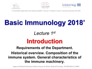
  • 1. Basic Immunology 2018’ Lecture 1st Introduction Requirements of the Department. Historical overview. Composition of the immune system. General characteristics of the immune machinery.
  • 2. While immunology is important the for pharmacists?
  • 4. Basic terms • Immunis,- e (Julius Caesar) = exempt, free of burden (E.g. tax, law, or diseases) • IMMUNE: individuals who do not capitulate to a disease when infected; • IMMUNITY: status of specific resistance to a disease; • IMMUNOLOGY: branch of theoretical biology focuses on mechanisms responsible for self and non-self recognition, elimination of the foreign invaders or altered self structures with protection of the basic structural elements.
  • 5. History • Athen (B.C. 5th century Thukidites - plaque survivors), ancient Chinese papers about the pox immunity • Infections, epidemies, vaccination Edward Jenner Louis Pasteur (1749 - 1823) (1822 - 1895)
  • 6. Edward Jenner (1749 - 1823) • He was a doctor in Berkeley, Gloucestershire. In 1796 he carried out his now famous experiment on eight-year-old James Phipps. Jenner inserted pus taken from a cowpox pustule on the hand of milkmaid Sarah Nelmes and inserted it into an incision on the boy's arm. He was testing his theory, drawn from the folklore of the countryside, that milkmaids who suffered the mild disease of cowpox never contracted smallpox. • Jenner subsequently proved that having been inoculated with cowpox Phipps was now immune to smallpox. He submitted a paper to the Royal Society in 1797 describing his experiment but was told that his ideas were too revolutionary and that he needed more proof. Undaunted, Jenner experimented on several other children, including his own 11-month-old son. In 1798 the results were finally published and Jenner coined the word vaccine from the Latin vacca for cow, and called the process vaccination.
  • 8. THE NOBEL PRIZE LAUREATES IN IMMUNOLOGY 1901 E.A. Von Behring (Germany) for the work on serum therapy especially its application against diphtheria. 1905 R. Koch (Germany) for the investigations concerning tuberculosis. 1908 E. Metchnikoff (Russia) and P. Ehrlich (Germany) for their work on immunity (respectively, phagocytosis/cellular theory and humoral theory). 1913 C.R. Richet (France) for the work on anaphylaxis. 1919 J. Bordet (Belgium) for the discoveries relating to immunity (complement). 1930 K. Landsteiner (Austria/USA) for the discovery of human blood groups. 1951 M. Theiler (South Africa) for the discoveries and developments concerning yellow fever. 1957 D. Bovet (Italy/Switzerland) for the discoveries related to histamine and compounds, which inhibit action of histamine and other substances on the vascular system and the skeleton muscles. 1960 Sir F.McFarlane Burnet (Australia) and Sir P.B. Medawar (Great Britain) for the discovery of acquired immunological tolerance. 1972 G.M. Edelman (USA) and R.R. Porter (Great Britain) for their discovery concerning the chemical structure of antibodies. 1977 R. Yalow (USA) for the development of radioimmunoassays of peptide hormones. 1980 B. Benacerraf (USA), J. Dausset (France) and G.D. Snell (USA) for their discoveries concerning genetically determined structures on the cell surface (major histocompatibility complex) that regulate immunological reactions. 1982 S. K. Bergstrom (Sweden), B. I. Samuelsson (Sweden) and J. R. Vane (UK) for their discoveries concerning prostaglandins and related biologically active substances. 1984 N.K. Jerne (Denmark/Switzerland) for theories concerning the specificity in development (lymphocyte clonality) and control of the immune system; G.J.F. Köhler (Germany/Switzerland) and C. Milstein (Argentina/Great Britain) for the discovery of the principle for production of monoclonal antibodies. 1987 S. Tonegawa (Japan/USA) for the discovery of the genetic principle for generation of antibody diversity. 1990 J.E. Murray and E.D. Thomas (USA) for their discovery concerning organ and cell transplantation in the treatment of human diseases. 1996 P.C. Doherty (Australia/USA) and R.M. Zinkernagel (Switzerland) for their discoveries concerning the specificity of the cell mediated immune defense ("dual recognition"). 1997 S.B. Prusiner (USA) for the discovery of prions as a new biological principle of infection. 1999 G. Blobel (USA) for discoveries concerning signal transduction.
  • 9. Main fields of applied immunology • Infectious immunity Basic empirical observations on survivors during the big epidemics (plague, pox, cholera, etc) in the Middle Age. New aspects occurred in the end of the 21st century: sever viral infections (HIV, influenza), fungal infections, antibiotic resistance in different bacteria. • Tumor immunology Animal experiments with tumor transplantation clarified the genetic mechanisms of graft rejection, and the correlation between the blood groups and the transplantation immunity (Gorer, 1927). New immunological concept developed in biology and medicine in the first decades of 20th century: immune system is responsible for the self integrity of individuals. The defense against tumors is not known in details yet, however, the role and heredity of major histocompatibility complex (MHC) was discovered during the tumor-transplantation experiments establishing the immunogenetics. • Transplantation immunology Immunonological aspects of organ transplantaions • Cellular and molecular immunology (Basic and applied immunological research, related innovations and R&D) diagnostics ant drug designe. • Immunological biotechnology (increased need for individual diagnostics ant therapy) • Biological therapies (Therapeutic monoclonal antibodies, recombinant cytokines)
  • 10. What is the main function of the immune system? Saving the individual integrity against foreign invaders (pathogens) and the modification of self structures by mutations, tumorous transformations, physical or chemical effects, or virus infections.
  • 11. Immune system  Individuals and species  Organs  Cells  Molecules  Functions Immune system is a general structural and functional network composed by molecular and cellular elements of the body.
  • 12. Organs of the immune system Primary Secondary (central) (peripheral) • Bone marrow • Thymus • (Embryonic liver) • Lymph nodes • Spleen • MALT • SALT
  • 13. Red and yellow bone marrow
  • 14. Normal bone marrow (HE) Strómasejt B és T sejt előalakok
  • 16. Eosinophil granulocyte and a small lymphocyte
  • 19. White blood cells in peripheral blood smears
  • 29. Blue: stem cells Dark blue: immature cells Brown: matured cells Hematopoietic differentiation
  • 30. Cells of the immune system Antigen-presenting cells: “professional” or “accidental” Antigen-binding cells: T- and B lymphocytes Effektor cells: T, NK, granulocytes, mast cells, monocytes/macrophages Organ distribution of T and B lymphocytes ------------------------------------------------------------------------------------------------ Organ % lymphocyte T B ------------------------------------------------------------------------------------------------ Tymus >99 <0.5 Lymph node 75 25 Spleen 50 50 Peripheral blood 55-75 15-30 Bone marrow 7 >75 ------------------------------------------------------------------------------------------------
  • 31. Theoretical scheme of the innate immunity ANTIGEN RECOGNITION RESPONSE Theoretical scheme of the adaptive immunity ANTIGEN RECOGNITION DIFFERENTIATION EFFECTOR FUNCTIONS MEMORY
  • 32. Composition of the immune system Natural Innate-like immunity with adaptive features Innate •None antigen specific •No immunological memory •Rapid reactivity •Linear amplification of the reaction Adaptive •Antigen specific •Immunological memory •Activated after a latency •Exponential amplification of the reaction
  • 33. Basic Immunology Lecture 3rd-4th Molecular components of immunological recognition. Definition of the antigen. Antibodies, T- and B-cell receptors: molecular structure, functions, subcalsses
  • 34. Definition of the antigen László Detre (Detsch) : antibody generator - Old definition: foreign agent induces immune reaction - Modern definition: substance recognized by T- or B-cell receptor and induces tolerating or active type immune response according to the MHC haplotype of the individual.
  • 35. Factors determining the immunogenity • immunodominant regions • chemical structure (inorganic molecules are not antigens at general, but e.g. heavy metals in protein complex are able to induce specific metal allergies). The best antigens are proteins>polypeptides>polysaccharides>lipides>nucleic acids • physico-chemical nature (D and L configuration; ortho-, para,- meta position; hydrophilic and hydrophobic amino acid sequence) • molecular weight (not an absolute category) • conformation sensitivity (folding and refolding) • Origin auto-, allo-, xenoantigen • mode and anatomic region of the administration (e.g. peripheral immune reaction and oral tolerance for the same antigen depending from the place of the antigen presentation) • dose dependence (large and low dose) • Valency: monovalent, bivalent, and multivalent antigens
  • 36. Basic terms immunogen (fine chemical structure can induce specific immune response) epitope (antigen determinant) well circumscribed region of the antigen molecule targeted by Ig/BcR or TcR paratop (ligand pair of the epitope) hapten (small molecular weight antigen can not induce immune reaction itself, but specifically recognized by immunoglobulins) carrier (indifferent, large molecular weight molecule, hold on the surface hapten molecules; carrier molecules did not participate in the anti-hapten immune reaction only hapten)
  • 37. Antigen recognition Innate immunity Natural immunity Acquired immunity • Pattern recognition • Pattern recognition • Antigen recognition • Mainly sugar recognition • Mainly peptide patter recognition • Mainly peptide pattern recognition • PRR • MHC • MHC • PAMP • iTcR, γδTcR, BcR, IgM • αβTcR, γδTcR, BcR, • IgM/G/A/E/D • Low number of molecularly distinct receptors and high number of recognized patterns • Limited number of molecularly distinct receptors and high number of recognized patterns • High number of distinct antigen receptors and high number of specifically recognized antigens
  • 38. Recognition molecules Immunoglobulins B cell receptors (BcR) T cell receptors (TcR) MHC class I and class II Specialized molecules manage antigen recognition. The common structural features of these molecules are the well-conserved (constant) basic elements (designed by 110 amino acids domain units) containing variable, antigen specific parts (binding sites) for the recognition and ligand formation.
  • 40. Domain structure Well conserved amino acid sequence designed by 110 amino acids closed to a “ring shape” with disulphide bound.
  • 42. Immunoglobulin molecule CDR Variable region Idiotype Fab fragment Constant region Isotype Fc fragment
  • 43. Ig domains: intra-chain disulphide bonds form loops in the peptide chain, the loops are globular, constructed from beta-plated sheets and beta-turn loops.
  • 44. Immunoglobulins Monofunctional character (specific antigen recognition and binding) before the antigen administration. Fab dependent function. Polyfunctional character after the antigen administration (signal transduction, complement fixation, opsonization, immunocomplex formation, FcR binding, etc). Fc dependent functions.
  • 45. Immunoglobulin isotypes • Based upon the constant structures of heavy (H) and light (L) chains • CH isotypes: called Ig classes and subclasses as IgG, IgM, IgA, IgD and IgE. All classes are represented in a normal serum (except the membrane bound IgD) as isotype variants. • CL chain exists in two isotypic forms: kappa (κ) and lambda (λ), which can associate with all heavy chain isotypes.
  • 46.
  • 47.
  • 49.
  • 50.
  • 51. Immunoglobulin idiotype Individual determinants in V regions, specific for each antibody. The N terminal Ig domain contains V region forming the antigen binding site: clustering the 3 hyper variable sequences close to each other on both chains - the variation of 3 x 3 results tremendous diversity.
  • 53.
  • 54.
  • 55.
  • 56. IgG – blood, lymph, make up 80% of Ig • only Ig of maternal origin to pass the placenta wall give newborns (Mw 150 kD) • neutralize toxins and viruses IgM – Blood, lymph (cell surface) pentamer structure (Mw 900 kD) • First antibodies formed in response to initial infection. IgA – Mucosal surfaces, blood (active in dimeric or tetrameric form) (Mw 150-600 kD) IgD – only membrane-bounded form in B-cell surfaces (Mw 150 kD) • may function in initiation of antibody-antigen response IgE – blood (bound to basophiles, mast cells) (Mw 190 kD) initiation of allergic reactions
  • 57. Kinetics of antibody production
  • 58. Antigen – antibody reactions • Neutralization (e.g. toxins) • Precipitation (soluble molecules) • Agglutination (particles, cells) • Opsonization (large particles) • Complement fixation
  • 59. B cell receptor complex
  • 60. T cell receptor complex expressed in mature T cells ab TcR – SP(CD4+ or CD8+) gd TcR – DN (CD4-CD8-)
  • 62. Basic Immunology Lecture 5-6th MHC Structure, genetics, role in the immunological recognition. Antigen presenatation.
  • 63. The Immune System • Innate immunity (complement, cytokines, antibacterial peptides, macrophages, neutrophil granulocytes, NK cells, etc.) • Adaptive immunity (antibodies, T and B lymphocytes, lymphoid cytokines, etc.) • Natural immunity (natural autoantibodies, ILCs, iNKT cells, γδT cells, MAIT cells, etc.)
  • 64. Recognition of the antigen in the adaptive immunity Native antigens are recognized by immunoglobulins or B cell receptors. T cells can recognize in denatured (presented by MHC) forms of the antigens exclusively.
  • 65. Major Histocompatibility Complex Self and foreign antigens are presented on the cell surface by specialized host-cell glycoproteins encoded in a large cluster of genes that were first identified by their effects on the immune response to transplanted tissues. For that reason, the gene complex was termed the Major Histocompatibility Complex (MHC). The antigen binding glycoproteins are called MHC molecules/antigens. (MHC vs. HLA, H2, BoLA, ChLA etc.)
  • 66.
  • 67. Tolerated skin grafts on MHC (H2) identical mice
  • 68. MHC Class I Present in all nucleated cells and platelets (in different density).
  • 69. MHC class II molecule a and b chains Expressed on professional or facultative antigen presenting cells (APCs): •Constitutive •Induced
  • 70. Class I and class II MHC Molecules
  • 71. MHC class I and class II molecules with bound peptides
  • 72. MHC Class I presents antigen for T cell
  • 75. Polygenic: (there are several different class I and class II genes encoding proteins with different specificities:– HLA-A, B, C, HLA-DR, DP, DQ) Polymorphic: there are multiple alleles of each gene (6-50) Co-dominant: haplotypes (allel variants) of BOTH parents are expressed Characteristics of MHC
  • 76. What type of cells express MHC class I and MHC class II? MHC I Any cell type with nucleus MHC II Mainly professional antigen presenting cells Dendritic cell, Follicular dendritic cells B cells Macrophages, monocytes (Thymic epithelial cells) Facultative antigen presenting cells Inflammatory epithel
  • 79. Transporter Associated with Antigen Processing Molecular Immunol. 2002
  • 80. Chaperons in the MHC-I Antigen Presentation Calnexin, calreticulin, Erp57, tapasin
  • 81. Antigen Presentation on MHC class I -Cytosolic, mainly normal or viral/modified proteins -Proteasomal degradation Peptide transfer to the ER (TAP1&2) MHC I chains produced into ER by ribosomes Chaperons: calnexin, calreticulin, Erp57 Tapasin and TAP1&2 MHCI & peptide binding within the ER
  • 83. Generation of Antigenic Peptides in the Endocytic Pathway
  • 84. Peptide Loading of Class II Molecules HLA-DM: MHCII chaperon CLIP=class II associated invariant chain peptide
  • 85. Antigen Presentation on MHC II -HLA-DM: MHC II specific chaperon -invariant chain -Endocytosed proteins: bacteria, bacterial product, internalised receptor bound peptide, parts of another cell -Endosomal degradation -MHCII chains produced into the ER by ribosomes -CLIP=class II associated invariant chain peptide -MHC II & peptide binding in endosomes outside the ER
  • 86.
  • 88. Herpes simplex – produces a protein which inhibits TAP Adenovirus – produces a protein, which binds to and retains MHC-I in the ER Cytomegalovirus – accelerates MHC-I translocation to the cytosol for degradation HIV – accumulate mutations faster than the adaptive immune system can cope with MHC-I MHC-II Helicobacter pylori – encodes a 95kD protein toxin, which increases the pH of the lysosomes, inhibiting protease activity How do pathogens avoid detection?
  • 89. Septic schock - superantigens Activated T cells produce cytokines randomly - systemic collaps of several biological functions („Cytokine tsunami”) Compared to a normal antigen-induced T-cell response where low number of the body’s T-cells are activated, SAgs (endotoxins) of viral or bacterial origin are capable of activating large set (up to 20%) of the body’s T- cells randomly. This causes a massive and irregular immune response (toxic shock syndrome) that is not specific to any particular epitope on the SAg.
  • 90. Basic Immunology Lecture 7th - 8th Communication between cellular components of the immune system. Co-receptors and adhesion molecules. Cytokines, chemokines and their receptors.
  • 91. Mediators of cell-cell interactions: „cross-talk” Cell-cell interactions play basic biological role in development and function of multicellular organisms. These interactions allow cells to communicate with each other. This ability to send and receive signals is essential for the further functions of the cells. - Direct interactions: adhesion molecules - Microparticles: microvesicules, microtubes - Soluble mediators perform indirect interactions: cytokines, chemokines, interleukins, interferons, growth factors, tissue hormons, complement factors, etc.
  • 92. Immunological „cross-talk” -Haematopoiesis: adhesion between stromal cells of the bone marrow and the differentiating leukocytes -Lymphocyte recirculation and recruitment: adhesion between endothelial cells and the circulating leukocytes, recruiting immune active cells into the inflammatory tissues -Immune response: T cell and APC/B cell interactions during antigen presentation, activation and differentiation of immune cells, cytotoxic effector reactions
  • 93. Adhesion molecules Cell surface molecules whose function is to promote adhesive interactions with other cells or the extracellular matrix and initiate signal transduction. Leukocytes express various types of adhesion molecules, such as selectins, integrins, and members of the Ig superfamyli, and these molecules play crucial role in cell migration and cellular activation both in innate and adaptive immune response.
  • 96. Family of accessory molecules, adhesion molecules, co-receptors Common characteristics: 1. Molecules, responsible for the direct interaction of the immune cells 2. Their interaction is not antigen- specific 2. Low-affinity, reversible association 4. Increase the antigen-specific interaction 5. Co-receptors: - signaling function 6. Co-stimulatory molecules: help cell activation 7. Non-polymorphic T cell
  • 97. Activation of adheseion molecules • Lymphocyte Function-associated Antigen • IntraCellular Adhesion Mloecule
  • 98. Families of adhesion molecules CD2 CD4 CD8 B7 CD28 CTLA 4 ICAM L selectin E selectin P selectin VLA LFA Mac1 „vascular addressins” „other” accessory molecules CD45 CD44 CD40, CD40L CD19/CD21/CD81 CD22 Ig-superfamily members Selectins Integrins Mucin-like molecules Lectin domain SCR domains cysteins
  • 99. 90% of T cells „T cell rosette” CD2 „sheep red-blood cell receptor” Adhesion Cell activation Ig-superfamily members Binds CD58 (LFA3) T cell activation, CTL- and NK-mediated lysis
  • 100. Differentiation markers: At different stages of T cell maturation CD4 and CD8 together „double positive” in thymus At the periphery: „single positive” T helper: CD4 T cytotoxic: CD8 CD4 and CD8: extracellular domain: binding to MHC constant domain intracellular domain: signal transduction, binding kinases CD4 - MHCII CD8 - MHCI CD4 - HIV-receptor as well 55 kDa Th, Mo, Mf, DC 35 kDa Tc CD4+ T cell CD8+ T cell
  • 101. B7 (CD80, CD86), CD28 and CTLA-4 molecules CD28: - co-stimulatory molecule in T cell activation - Increases IL-2 and IL-2R expression, - Induces T cell proliferation CTLA-4 (CD152): - expressed in a later phase of the T cell activation - inhibitory function CTLA: Cytolytic T lymphocyte associated Antigen CD28 and CTLA-4 of T cells bind to the B7-1 (CD80), B7-2 (CD86) molecules of the APC T cell APC
  • 102. „OTHER” accessory molecules Plays important role in cell activation and in regulation of signal transduction - tyrosine–phosphatase domain: dephosphorylation CD45 Expressed on every leukocyte “pan-leukocyte marker” - Highly glycosylated, - More isoforms (180, 190, 200, 205, 220 kDa) - alternate splicing CD45
  • 104. CD45
  • 105. CD44 Expressed on activated and memory T- and B-cells, phagocytes, fibroblasts, neuronal cells Important in „homing” of leukocytes More isoforms - alternate splicing CD44 „OTHER” accessory molecules
  • 106. POSSIBLE MECHANISMS BY WHICH NK CELLS DISTINGUISH INFECTED FROM UNINFECTED CELLS NK cells can use several different receptors that signal them to kill, including lectinlike activating receptors, or ‘killer receptors,’ that recognize carbohydrate on self cells. However, another set of receptors, called Ly49 in the mouse and killer inhibitory receptors (KIRs) in the human, recognize MHC class I molecules and inhibit killing by NK cells by overruling the actions of the killer receptors. This inhibitory signal is lost when cells do not express MHC class I and perhaps also in cells infected with virus, which might inhibit MHC class I expression or alter its conformation. Another possibility is that normal uninfected cells respond to IFN-α and IFN-β by increasing expression of MHC class I molecules, making them resistant to killing by activated NK cells. In contrast, infected cells can fail to increase MHC class I expression, making them targets for activated NK cells. Ly49 and KIR belong to different protein families—the C-type lectins in the case of Ly49 and the immuno-globulin superfamily for KIRs. The KIRs are made in two forms, p58 and p70, which differ by the presence of one immunoglobulin domain.
  • 107. PHAGOCYTE ADHESION TO VASCULAR ENDOTHELIUM IS MEDIATED BY INTEGRINS Vascular endothelium, when it is activated by inflammatory mediators, expresses two adhesion molecules—ICAM-1 and ICAM-2. These are ligands for integrins expressed by phagocytes—αL:β2 (also called LFA-1 or CD11a: CD18) and αM:β2 (also called Mac-1, CR3, or CD11b:CD18).
  • 108. Lymphocyte recirculation:continuos migration of cells from the blood flow and lymph to the lymhatic organs and to the inflammation = HOMING Role: - Promots the antigen capturing - Promots the development of inflammatory reactions Mechanism: -Extravasation: leucocyte adhesion to the endothel, and migration across the wall of the blodd vessels to the tissue 1-2 total circle managed by all white blood cells pro day
  • 109. Neutrophils leave the blood and migrate to sites of infection in a multistep process mediated through adhesive interactions that are regulated by macrophage-derived cytokines and chemokines.
  • 110. Naive lymphocytes migrating to the peripheral lymphatis tissues: The role of the hugh endothelial venules (HEV), and the adhesion molecules: 1. Selectin-mediated 2. chemoattractant 3. Integrin-mediated mediated
  • 111. Migration of neutrophil granulocytes to the inflammed tissues throgh the endothel
  • 112. Different adhesion molecules determine the migration of naive and memory (effector) cells Peripheral lymphatic tissue Inflammatory tissue
  • 113. Some important accessory molecules • CD3 • CD4 and CD8 • CD28 • CD80/86 (B7.1 and B7.2) • CD152 (CTLA4) • CD25 (IL-2 Receptor) • CD45RA/RO • CD154 (CD40 Ligand)
  • 114.
  • 116. Cell-cell communication through cytokines and their receptors • Cytokines • Chemokines • Interferons • growth factors
  • 117. Mechanism of cytokine action I.: Cytokine producing cell Target cell: Cytokine producing cell Nearby cell Distant cell Autocrine action Paracrine actiont Endocrine action
  • 118. Cytokines act in each phase of the immune response Recognition: Activation: Effector phase:
  • 119. Mechanism of cytokine action II.: A cytokine induces different effects on different target cells The action of more cytokine on the target cell is similar The effect of two cytokines is stronger than their additive effects One cytokine inhibits the effects of another cytokine Pleiotropy Redundancy Synergy Antagonism Starting a cascade
  • 120. Basic characteristics of cytokies • Low molecular weight (10-40 kDa), and genetically well conserved glycoproteins • Isolated cells secrete them, due to gene activation • They mediate cell-cell interaction: • - sending information • - regulation of immune response • Mechanism of action: - produced after transient gene activation - act through receptors triggering signal-transduction - high affinity - picomolar concentration - they act mostly locally
  • 121. Functional groups of cytokines I. Regulators of natural immunity and inflammation IFNa, IFNb, TNFa, TNFb (LT), IL-1a, IL-1b, IL-6, IL-12, MIF, chemokines II. Regulators of lymphocyte activation and differentiation IL-2, IL-4, IL-5, IL-6, IL-13, IL-15, INFg, IL-10 and TGFb III. Regulators of haematopoiesis IL-3, IL-7, GM-CSF, SCF
  • 122. MHC IL-1 bakteriális endotoxin makrofág T-sejtTCR citotoxicitás monokinek adhéziós molekulák    aktiváció IL-2R limfokinek    prosztaglandinok láz aluszékonyság fájdalomküszöb fogyás   autokrin parakrin endokrin Autocrine, paracrine and endocrine action of IL-1 macrophage T cell Activation, IL-2R expression Cytokine secretion. Bacterial endotoxin Enhanced cytotoxicity Cytokine production Adhesion mol. Expr. Prostaglandin Production, fever Acute phase protein Secretion by hepatocytes
  • 124. Characteristics of multichain cytokine receptors
  • 125. Chemokines - 90-130 aa. Polypeptides - Receptorial action - Produced by lymhatic and none-lymphatic tissues Functions: - chemotaxis for different leukocytes - regulation of normal leukocyte traffic - recruitment of cells to inflammatory sites - enhancement of cell adhesion - activation of effectors leukocytes - development of the inflammatory reaction - development of normal lymphoid tissues
  • 126. Basic Immunology Lectures 9-10 Lymphocyte Development and Antigen Receptor Gene Rearrangement
  • 127. OVERVIEW OF LYMPHOCYTE DEVELOPMENT The maturation of B and T lymphocytes involves a series of events that occur in the generative lymphoid organs. These events include the following: 1. The commitment of progenitor cells to the B cell or T cell lineage. 2. Proliferation of progenitors and immature committed cells at specific early stages of development, providing a large pool of cells that can generate useful lymphocytes. 3. The sequential and ordered rearrangement of antigen receptor genes and the expression of antigen receptor proteins.
  • 128. OVERVIEW OF LYMPHOCYTE DEVELOPMENT (cont.) 4. Selection events that preserve cells that have produced correct antigen receptor proteins and eliminate potentially dangerous cells that strongly recognize self antigens. These checkpoints during development ensure that lymphocytes that express functional receptors with useful specificities will mature and enter the peripheral immune system. 5. Differentiation of B and T cells into functionally and phenotypically distinct subpopulations. B cells develop into follicular, marginal zone, and B-1 B cells, and T cells develop into CD4+ and CD8+ T lymphocytes and γδ T cells. This differentiation into distinct classes provides the specialization that is an important characteristic of the adaptive immune system.
  • 129. Development of B cells B cells develop in the bone marrow and migrate to peripheral lymphoid organs, where they can be activated by antigens.
  • 130. Development of T cells T cells undergo development in the thymus and migrate to the peripheral lymphoid organs, where they are activated by foreign antigens.
  • 131. Copyright © 2011 by Saunders, an imprint of Elsevier Inc.Abbas, Lichtman, and Pillai. Cellular and Molecular Immunology, 7th edition. Copyright © 2012 by Saunders, an imprint o Stages of Lymphocyte Maturation Fig. 8-1 Development of both B and T lymphocytes involves the sequence of maturational stages shown. B cell maturation is illustrated, but the basic stages of T cell maturation are similar.
  • 132. Copyright © 2011 by Saunders, an imprint of Elsevier Inc.Abbas, Lichtman, and Pillai. Cellular and Molecular Immunology, 7th edition. Copyright © 2012 by Saunders, an imprint o Distinct Lineages of Lymphocyte Maturation Fig. 8-2
  • 133. Copyright © 2011 by Saunders, an imprint of Elsevier Inc.Abbas, Lichtman, and Pillai. Cellular and Molecular Immunology, 7th edition. Copyright © 2012 by Saunders, an imprint o Checkpoints in Lymphocyte Maturation Fig. 8-3 During development, the lymphocytes that express receptors required for continued proliferation and maturation are selected to survive, and cells that do not express functional receptors die by apoptosis. Positive selection and negative selection further preserve cells with useful specificities. The presence of multiple checkpoints ensures that only cells with useful receptors complete their maturation.
  • 134. Copyright © 2011 by Saunders, an imprint of Elsevier Inc.Abbas, Lichtman, and Pillai. Cellular and Molecular Immunology, 7th edition. Copyright © 2012 by Saunders, an imprint o Pos. and Neg. Selection of Lymphocytes Fig. 8-4
  • 135. Copyright © 2011 by Saunders, an imprint of Elsevier Inc.Abbas, Lichtman, and Pillai. Cellular and Molecular Immunology, 7th edition. Copyright © 2012 by Saunders, an imprint o Germline Organization of Human Ig Loci Fig. 8-5 The human heavy chain, κ light chain, and λ light chain loci are shown. Only functional genes are shown; pseudogenes have been omitted for simplicity. Exons and introns are not drawn to scale. Each CH gene is shown as a single box but is composed of several exons, as illustrated for Cμ. Gene segments are indicated as follows: L, leader (often called signal sequence); V, variable; D, diversity; J, joining; C, constant; enh, enhancer.
  • 136. Copyright © 2011 by Saunders, an imprint of Elsevier Inc.Abbas, Lichtman, and Pillai. Cellular and Molecular Immunology, 7th edition. Copyright © 2012 by Saunders, an imprint o Domains of Ig and TCR proteins Fig. 8-6 The domains of Ig heavy and light chains are shown in A, and the domains of TCR α and β chains are shown in B. The relationships between the Ig and TCR gene segments and the domain structure of the antigen receptor polypeptide chains are indicated. The V and C regions of each polypeptide are encoded by different gene segments. The locations of intrachain and interchain disulfide bonds (S-S) are approximate. Areas in the dashed boxes are the hypervariable (complementarity-determining) regions. In the Ig μ chain and the TCR α and β chains, transmembrane (TM) and cytoplasmic (CYT) domains are encoded by separate exons.
  • 137. Copyright © 2011 by Saunders, an imprint of Elsevier Inc.Abbas, Lichtman, and Pillai. Cellular and Molecular Immunology, 7th edition. Copyright © 2012 by Saunders, an imprint o Germline Organization of Human TCR Loci (1) Fig. 8-7 The human TCR β, α, γ, and δ chain loci are shown, as indicated. Exons and introns are not drawn to scale, and nonfunctional pseudogenes are not shown. Each C gene is shown as a single box but is composed of several exons, as illustrated for Cβ1. Gene segments are indicated as follows: L, leader (usually called signal sequence); V, variable; D, diversity; J, joining; C, constant; enh, enhancer; sil, silencer (sequences that regulate TCR gene transcription).
  • 138. Copyright © 2011 by Saunders, an imprint of Elsevier Inc.Abbas, Lichtman, and Pillai. Cellular and Molecular Immunology, 7th edition. Copyright © 2012 by Saunders, an imprint o Germline Organization of Human TCR Loci (2) Fig. 8-7
  • 139. Copyright © 2011 by Saunders, an imprint of Elsevier Inc.Abbas, Lichtman, and Pillai. Cellular and Molecular Immunology, 7th edition. Copyright © 2012 by Saunders, an imprint o Antigen receptor Gene Rearrangements Fig. 8-8 Southern blot analysis of DNA from nonlymphoid (liver) cells and from a monoclonal population of B lymphocyte lineage origin (e.g., a B cellt umor) is shown in schematic fashion. The DNA is digested with a restriction enzyme (EcoRI as depicted), different-sized fragments are separated by electrophoresis, and the fragments are transferred onto a filter. The sites at which the EcoRI restriction enzyme cleaves the DNA are indicated by arrows. The size of the fragments containing the Jκ3 segment of the Ig κ light chain gene or the Vκ29 V region gene was determined by use of a radioactive probe that specifically binds to Jκ3 segment DNA or to Vκ29 DNA. In the hypothetical example shown, Vκ29 is part of a 5-kb EcoRI fragment in liver cells but is on a 3-kb fragment in the B cell clone studied. Similarly, the Jκ3 fragment is 8 kb in liver cells but 3 kb in the B cell clone.
  • 141. Copyright © 2011 by Saunders, an imprint of Elsevier Inc.Abbas, Lichtman, and Pillai. Cellular and Molecular Immunology, 7th edition. Copyright © 2012 by Saunders, an imprint o Diversity of Antigen Receptor Genes Fig. 8-9 From the same germline DNA, it is possible to generate recombined DNA sequences and mRNAs that differ in their V-D-J junctions. In the example shown, three distinct antigen receptor mRNAs are produced from the same germline DNA by the use of different gene segments and the addition of nucleotides to the junctions.
  • 142. Copyright © 2011 by Saunders, an imprint of Elsevier Inc.Abbas, Lichtman, and Pillai. Cellular and Molecular Immunology, 7th edition. Copyright © 2012 by Saunders, an imprint o V(D)J Recombination (1) Fig. 8-10A The DNA sequences and mechanisms involved in recombination in the Ig gene loci are depicted. The same sequences and mechanisms apply to recombinations in the TCR loci. A, Conserved heptamer (7 bp) and nonamer (9 bp) sequences, separated by 12- or 23-bp spacers, are located adjacent to V and J exons (for κ and λ loci) or to V, D, and J exons (in the H chain locus). The V(D)J recombinase recognizes these recombination signal sequences and brings the exons together.
  • 143. Copyright © 2011 by Saunders, an imprint of Elsevier Inc.Abbas, Lichtman, and Pillai. Cellular and Molecular Immunology, 7th edition. Copyright © 2012 by Saunders, an imprint o V(D)J Recombination (2) Fig. 8-10B B, C, Recombination of V and J exons may occur by deletion of intervening DNA and ligation of the V and J segments (B) or, if the V gene is in the opposite orientation, by inversion of the DNA followed by ligation of adjacent gene segments (C). Red arrows indicate the sites where germline sequences are cleaved before their ligation to other Ig or TCR gene segments.
  • 144. Copyright © 2011 by Saunders, an imprint of Elsevier Inc.Abbas, Lichtman, and Pillai. Cellular and Molecular Immunology, 7th edition. Copyright © 2012 by Saunders, an imprint o V(D)J Recombination (3) Fig. 8-10C B, C, Recombination of V and J exons may occur by deletion of intervening DNA and ligation of the V and J segments (B) or, if the V gene is in the opposite orientation, by inversion of the DNA followed by ligation of adjacent gene segments (C). Red arrows indicate the sites where germline sequences are cleaved before their ligation to other Ig or TCR gene segments.
  • 145. Contributions of different mechanisms to the generation of diversity in Ig and TCR genes The rearrangement of antigen receptor genes is the key event in lymphocyte development that is responsible for the generation of a diverse repertoire.
  • 146. Copyright © 2011 by Saunders, an imprint of Elsevier Inc.Abbas, Lichtman, and Pillai. Cellular and Molecular Immunology, 7th edition. Copyright © 2012 by Saunders, an imprint o Transcriptional Regulation of Ig Genes Fig. 8-11 V-D-J recombination brings promoter sequences (shown as P) close to the enhancer (enh). The enhancer promotes transcription of the rearranged V gene (V2, whose active promoter is indicated by a bold green arrow). Many receptor genes have an enhancer in the J-C intron and another 3′ of the C region. Only the 3′ enhancer is depicted here.
  • 147. Copyright © 2011 by Saunders, an imprint of Elsevier Inc.Abbas, Lichtman, and Pillai. Cellular and Molecular Immunology, 7th edition. Copyright © 2012 by Saunders, an imprint o Events During V(D)J Recombination (1) Fig. 8-12 Sequential events during V(D)J recombination. Synapsis and cleavage of DNA at the heptamer/coding segment boundary are mediated by Rag-1 and Rag-2. The coding end hairpin is opened by the Artemis endonuclease, and broken ends are repaired by the NHEJ machinery.
  • 148. Copyright © 2011 by Saunders, an imprint of Elsevier Inc.Abbas, Lichtman, and Pillai. Cellular and Molecular Immunology, 7th edition. Copyright © 2012 by Saunders, an imprint o Events During V(D)J Recombination (2) Fig. 8-12 Sequential events during V(D)J recombination. Synapsis and cleavage of DNA at the heptamer/coding segment boundary are mediated by Rag-1 and Rag-2. The coding end hairpin is opened by the Artemis endonuclease, and broken ends are repaired by the NHEJ machinery.
  • 149. Copyright © 2011 by Saunders, an imprint of Elsevier Inc.Abbas, Lichtman, and Pillai. Cellular and Molecular Immunology, 7th edition. Copyright © 2012 by Saunders, an imprint o Events During V(D)J Recombination (3) Fig. 8-12 Sequential events during V(D)J recombination. Synapsis and cleavage of DNA at the heptamer/coding segment boundary are mediated by Rag- 1 and Rag-2. The coding end hairpin is opened by the Artemis endonuclease, and broken ends are repaired by the NHEJ machinery.
  • 150. Copyright © 2011 by Saunders, an imprint of Elsevier Inc.Abbas, Lichtman, and Pillai. Cellular and Molecular Immunology, 7th edition. Copyright © 2012 by Saunders, an imprint o Junctional Diversity Fig. 8-13 During the joining of different gene segments, addition or removal of nucleotides may lead to the generation of novel nucleotide and amino acid sequences at the junction. Nucleotides (P sequences) may be added to asymmetrically cleaved hairpins in a templated manner. Other nucleotides (N regions) may be added to the sites of VD, VJ, or DJ junctions in a nontemplated manner by the action of the enzyme TdT. These additions generate new sequences that are not present in the germline.
  • 151. Copyright © 2011 by Saunders, an imprint of Elsevier Inc.Abbas, Lichtman, and Pillai. Cellular and Molecular Immunology, 7th edition. Copyright © 2012 by Saunders, an imprint o Stages of B Cell Maturation Fig. 8-14 Events corresponding to each stage of B cell maturation from a bone marrow stem cell to a mature B lymphocyte are illustrated. Several surface markers in addition to those shown have been used to define distinct stages of B cell maturation.
  • 152. Copyright © 2011 by Saunders, an imprint of Elsevier Inc.Abbas, Lichtman, and Pillai. Cellular and Molecular Immunology, 7th edition. Copyright © 2012 by Saunders, an imprint o Ig Gene Recombination Fig. 8-15A Ig heavy and light chain gene recombination and expression. The sequence of DNA recombination and gene expression events is shown for the Ig μ heavy chain (A) and the Ig κ light chain (B). In the example shown in A, the V region of the μ heavy chain is encoded by the exons V1, D2, and J1. In the example shown in B, the V region of the κ chain is encoded by the exons V2 and J1.
  • 153. Copyright © 2011 by Saunders, an imprint of Elsevier Inc.Abbas, Lichtman, and Pillai. Cellular and Molecular Immunology, 7th edition. Copyright © 2012 by Saunders, an imprint o Ig Gene Expression Fig. 8-15B Ig heavy and light chain gene recombination and expression. The sequence of DNA recombination and gene expression events is shown for the Ig μ heavy chain (A) and the Ig κ light chain (B). In the example shown in A, the V region of the μ heavy chain is encoded by the exons V1, D2, and J1. In the example shown in B, the V region of the κ chain is encoded by the exons V2 and J1.
  • 154. Copyright © 2011 by Saunders, an imprint of Elsevier Inc.Abbas, Lichtman, and Pillai. Cellular and Molecular Immunology, 7th edition. Copyright © 2012 by Saunders, an imprint o Pre-B cell and Pre-T cell Receptors Fig. 8-16 The pre-B cell receptor (A) and the pre-T cell receptor (B) are expressed during the pre-B and pre-T cell stages of maturation, respectively, and both receptors share similar structures and functions. The pre-B cell receptor is composed of the μ heavy chain and an invariant surrogate light chain. The surrogate light chain is composed of two proteins, the V pre-B protein, which is homologous to a light chain V domain, and a λ5 protein that is covalently attached to the μ heavy chain by a disulfide bond. The pre-T cell receptor is composed of the TCR β chain and the invariant pre-T α (pTα)chain. The pre-B cell receptor is associated with the Igα and Igβ signaling molecules that are part of the BCR complex in mature B cells (see Chapter 9), and the pre-T cell receptor associates with the CD3 and ζ proteins that are part of the TCR complex in mature T cells
  • 155. Copyright © 2011 by Saunders, an imprint of Elsevier Inc.Abbas, Lichtman, and Pillai. Cellular and Molecular Immunology, 7th edition. Copyright © 2012 by Saunders, an imprint o B Lymphocyte Subsets Fig. 8-17 A, Most B cells that develop from fetal liver–derived stem cells differentiate into the B-1 lineage. B, B lymphocytes that arise from bone marrow precursors after birth give rise to the B-2 lineage. Two major subsets of B lymphocytes are derived from B-2 B cell precursors. Follicular B cells are recirculating lymphocytes; marginal zone B cells are abundant in the spleen in rodents but can also be found in lymph nodes in humans.
  • 156. Copyright © 2011 by Saunders, an imprint of Elsevier Inc.Abbas, Lichtman, and Pillai. Cellular and Molecular Immunology, 7th edition. Copyright © 2012 by Saunders, an imprint o Coexpression of IgM and IgD Fig. 8-18 Alternative processing of a primary RNA transcript results in the formation of a μ or δ mRNA. Dashed lines indicate the H chain segments that are joined by RNA splicing.
  • 157. Copyright © 2011 by Saunders, an imprint of Elsevier Inc.Abbas, Lichtman, and Pillai. Cellular and Molecular Immunology, 7th edition. Copyright © 2012 by Saunders, an imprint o Stages of T Cell Maturation Fig. 8-19 Events corresponding to each stage of T cell maturation from a bone marrow stem cell to a mature T lymphocyte are illustrated. Several surface markers in addition to those shown have been used to define distinct stages of T cell maturation.
  • 158. Copyright © 2011 by Saunders, an imprint of Elsevier Inc.Abbas, Lichtman, and Pillai. Cellular and Molecular Immunology, 7th edition. Copyright © 2012 by Saunders, an imprint o Maturation of T cells in the Thymus Fig. 8-20 Precursors of T cells travel from the bone marrow through the blood to the thymus. In the thymic cortex, progenitors of αβ T cells express TCRs and CD4 and CD8 coreceptors. Selection processes eliminate self- reactive T cells in the cortex at the double-positive (DP) stage and also single-positive (SP) medullary thymocytes. They promote survival of thymocytes whose TCRs bind self MHC molecules with low affinity. Functional and phenotypic differentiation into CD4+CD8− or CD8+CD4− T cells occurs in the medulla, and mature T cells are released into the circulation.
  • 159. Copyright © 2011 by Saunders, an imprint of Elsevier Inc.Abbas, Lichtman, and Pillai. Cellular and Molecular Immunology, 7th edition. Copyright © 2012 by Saunders, an imprint o TCR α and β Chain Gene Recombination Fig. 8-21A The sequence of recombination and gene expression events is shown for the TCR β chain (A) and the TCR α chain (B). In the example shown in A, the variable (V) region of the rearranged TCR β chain includes the Vβ1 and Dβ1 gene segments and the third J segment in the Jβ1 cluster. The constant (C) region is encoded by the Cβ1 exon. Note that at the TCR β chain locus, rearrangement begins with D-to-J joining followed by V-to-DJ joining. In humans, 14 Jβ segments have been identified, and not all are shown in the figure. In the example shown in B, the V region of the TCR α chain includes the Vα1 gene and the second J segment in the Jα cluster (this cluster is made up of at least 61 Jα segments in humans; not all are shown here).
  • 160. Copyright © 2011 by Saunders, an imprint of Elsevier Inc.Abbas, Lichtman, and Pillai. Cellular and Molecular Immunology, 7th edition. Copyright © 2012 by Saunders, an imprint o TCR α and β Chain Gene Expression Fig. 8-21B The sequence of recombination and gene expression events is shown for the TCR β chain (A) and the TCR α chain (B). In the example shown in A, the variable (V) region of the rearranged TCR β chain includes the Vβ1 and Dβ1 gene segments and the third J segment in the Jβ1 cluster. The constant (C) region is encoded by the Cβ1 exon. Note that at the TCR β chain locus, rearrangement begins with D-to-J joining followed by V-to-DJ joining. In humans, 14 Jβ segments have been identified, and not all are shown in the figure. In the example shown in B, the V region of the TCR α chain includes the Vα1 gene and the second J segment in the Jα cluster (this cluster is made up of at least 61 Jα segments in humans; not all are shown here).
  • 161. Copyright © 2011 by Saunders, an imprint of Elsevier Inc.Abbas, Lichtman, and Pillai. Cellular and Molecular Immunology, 7th edition. Copyright © 2012 by Saunders, an imprint o T Cell CD4 and CD8 Expression in the Thymus Fig. 8-22 A, The maturation of thymocytes can be followed by changes in expression of the CD4 and CD8 coreceptors. A two-color flow cytometric analysis of thymocytes using anti-CD4 and anti-CD8 antibodies, each tagged with a different fluorochrome, is illustrated. The percentages of all thymocytes contributed by each major population are shown in the four quadrants. The least mature subset is the CD4−CD8− (double-negative) cells. Arrows indicate the sequence of maturation. B, Positive selection of T cells. Double-positive T cells differentiate into a CD4+CD8low stage and are instructed to become CD4+ cells if the TCR on a double-positive T cell recognizes self class II MHC with moderate avidity and therefore receives adequate coreceptor signals. A CD4+CD8low T cell whose TCR recognizes MHC class I molecules fails to receive strong coreceptor signals and differentiates into a CD8+ T cell, silencing CD4 expression.
  • 162. Summary of the development of human conventional B-lineage cells
  • 163. Summary of the development of human conventional B-lineage cells (cont.)
  • 164. Summary of the development of human α:β T cells
  • 165. Summary of the development of human α:β T cells (cont.)