
Recommended
Recommended
More Related Content
Similar to Isolated blood vessels
Similar to Isolated blood vessels (20)
More from improvemed
More from improvemed (20)
Recently uploaded
Recently uploaded (20)
Isolated blood vessels
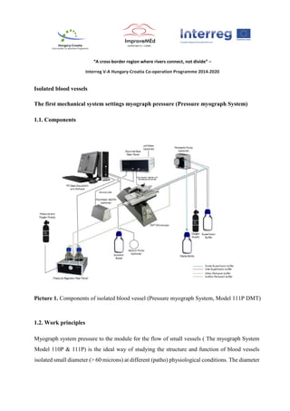
- 1. “A cross-border region where rivers connect, not divide” – Interreg V-A Hungary-Croatia Co-operation Programme 2014-2020 Isolated blood vessels The first mechanical system settings myograph pressure (Pressure myograph System) 1.1. Components Picture 1. Components of isolated blood vessel (Pressure myograph System, Model 111P DMT) 1.2. Work principles Myograph system pressure to the module for the flow of small vessels ( The myograph System Model 110P & 111P) is the ideal way of studying the structure and function of blood vessels isolated small diameter (> 60 microns) at different (patho) physiological conditions. The diameter
- 2. “A cross-border region where rivers connect, not divide” – Interreg V-A Hungary-Croatia Co-operation Programme 2014-2020 of the blood vessels can be measured in response to pharmacological and physiological stimuli. The built-in heating system maintains constant temperature chamber, the cover includes openings for wells superfusion, the quick charging and discharging, the cumulative addition of drugs, and for the oxidation. Chamber is made of stainless steel resistant to acid, which facilitates the cleaning (after use) and maintenance. This system can be tested physiological responses of isolated blood vessels, such as the myogenic response and flow mediated dilation. In our experiments this system we use for the study of small blood vessels such as the rat middle cerebral artery. By combining different vasoactive substances and specific blockers of the enzyme (which were incubated for 20 min in the chamber), it is possible to study the function of endothelial and smooth muscle cells and a variety of vasoconstrictor and vasodilator mechanisms. Also, information may be obtained such as the thickness of the blood vessel wall, the change in vessel diameter, the intravascular pressure of this other set of calculated parameters, such as the shear stress and vascular resistance. The system pressure myograph is designed for use in teaching and research activities. It is not intended for clinical use, nor can it be used in the prevention, diagnosis, treatment or alleviation of disease, injury or handicap. 1.3. Software Manufacturer of our system to study vascular reactivity and endothelial function of isolated small blood vessels is Danish Myo Technology (DMT) (Denmark). The measurements are continuously recorded by computer with software to analyze the dimensions - MyoVIEW. In
- 3. “A cross-border region where rivers connect, not divide” – Interreg V-A Hungary-Croatia Co-operation Programme 2014-2020 addition, Flowmeter - 161FM can be added to the system to measure the size of the flow through the insulated vessel (flow between 15-1500 ml / min). Figure 2. MyoView Second Preparation PSS solution Physiological salt solution (PSS) is a modified Krebs-Henseleit solution, which is commonly used in the study of the physiology of isolated blood vessels. In our experiments we used the PSS solution having the following composition: - To prepare 0.5 L • Distilled water: 450 ml
- 4. “A cross-border region where rivers connect, not divide” – Interreg V-A Hungary-Croatia Co-operation Programme 2014-2020 • Salt stock solution: 25 ml • Buffer stock solution: 25 ml • Glucose: 0.5 g • NaH 2 PER 4: 0.07 g At the end of each experiment, a solution of PSS was replaced with Ca-free PSS solution, in order to determine a maximum dilation of the test vessels. Ca-free PSS chloride solution contains: - To prepare 0.5 L • Distilled water: 450 ml • Salt-free Ca stock solution: 25 ml • Buffer stock Ca-free solution: 25 ml • Glucose: 0.5 g • NaH 2 PER 4: 0.07 g the pH of the PSS and PSS Ca-free should be between 7:35 and 7:45. The third experiment and protocols Each experiment begins preparing PSSotopine, medications you are planning to use this activation system. After start the system and open the source of gas, we have to miss a PSS solution through the whole system of adjusting the pressure P1 ( inflow) to a value of 150 mmHg, and P 2 ( outflow) at 70 mmHg, and certainly have to turn the heater system to a temperature of 37C. 3.1. The process of isolation of middle cerebral artery and experimental protocol
- 5. “A cross-border region where rivers connect, not divide” – Interreg V-A Hungary-Croatia Co-operation Programme 2014-2020 The test animals are weighed and then anesthetized with ketamine 75 mg / kg (Ketanest S 25 mg / ml, 2 ml ampoule, Pfizer) and midazolam 0.5mg / kg (Torrex Midazolam 5 mg / mL, 3 mL, Torrex Chiesi Pharma), followed by decapitation. And using microsurgery accessories operating microscope from the brain was isolated middle cerebral artery, and cleared of surrounding tissue disposed between the glass micropipette (outer diameter = 100-200 microns) located in the chamber that is filled with warm (37 C) physiological salt solution (PSS, pH = 7.4 ± 0.05). The ends of the cerebral arteries are attached to a thin suture (10-0 nylon Black) pipette. The system is continuously oxygenated gas mixture of 21% O2, 5% CO 2 and N balance 2 ( no pressure not exceed 1.0 bar). After placing in a well, followed by incubation of the blood vessel for 60 minutes at Δ80 mmHg (80 mmHg is P1, P2 0 mmHg), to estimate the base (basal) diameter. Vessels all the time recorded by infrared camera and the image displayed on the monitor, and record the change in vessel diameter. After incubation, the blood vessel will then be exposed to the flow of which is achieved by simultaneous changes in input ( inflow) and output ( outflow) pressure (pressure gradients Δ10, Δ20, Δ40, Δ60 and 100 mmHg). Pressure gradient formation will result in the flow of prepared PSS solution flow through the blood vessel. Certainly when setting up the blood vessels in the glass micropipette must be careful that there is no residual air bubbles, which could damage and removed for the endothelium of blood vessels. The role of nitric oxide (NO) in the mechanism of flow induced dilatation ( flow-induced dilation; FID) that dilatation of endothelium-dependent, is determined by study of the FID in the presence of L-NAME-a (Nω-nitro-L-arginine methyl ester) (10- 5 mol / L) - inhibitors of nitric oxide synthase (NOS). The role of COX metabolites is determined in the presence of the
- 6. “A cross-border region where rivers connect, not divide” – Interreg V-A Hungary-Croatia Co-operation Programme 2014-2020 cyclooxygenase inhibitor, indomethacin (INDO), while the role of CYP450 enzymes examined metabolites in the presence of selective inhibitors of the CYP450 epoxidase, PPOH-MS. To test the dilation induced flow that is not dependent on the endothelium uses NO donor direct CDs, sodium nitroprusside (SNP; 10- 6 mol / L), while testing for endothelium-dependent vasodilation uses acetylcholine (ACh, 10 6 mol / L). Reply to ACh and SNP are estimated at a pressure of 80 mmHg (sleep mode no flow). To evaluate the existence and role of oxidative stress in vitro, mediated flow dilation is measured in the presence of a blood vessel "catcher" superoxide radicals TEMPOL times (100 mmol / L) in the chamber. At the end of the experimental protocol, the blood vessels are exposed to a solution of the PSS solution without Ca 2+ in order to measure the maximum diameter of the blood vessel. Subtitle blood vessels that did not show a significant level of the active tone (about 50%), are exempted from the treatment. The active tone (%) was calculated as ((D max - D Bass) / D max) X 100, where D max and D bass and the largest diameter of the blood vessels (the Δ 0 mm Hg, no flow). The diameters of the vessels recorded an infrared camera, and the image is displayed on the monitor. 4. Statistical analysis and data presentation As in our experiments mostly use several groups of experimental animals and to measure response to at several different flow rates (Δ10, Δ20, Δ 40, Δ60, Δ100 mmHg), data processing must use two-way ANOVA repeated measures.
- 7. “A cross-border region where rivers connect, not divide” – Interreg V-A Hungary-Croatia Co-operation Programme 2014-2020 Figure 3. The basic response of resistive arteriolar subcutaneous (SAT) and visceral (VAT) adipose tissue to flow induced dilation (FID), comparison between the two groups