
Recommended
More Related Content
What's hot
What's hot (20)
Similar to Hemiarthroplasty of Hip joint
Similar to Hemiarthroplasty of Hip joint (20)
More from Dr Thouseef Abdul Majeed
Recently uploaded
Recently uploaded (20)
Hemiarthroplasty of Hip joint
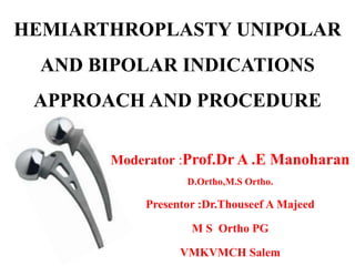
- 1. HEMIARTHROPLASTY UNIPOLAR AND BIPOLAR INDICATIONS APPROACH AND PROCEDURE Moderator :Prof.Dr A .E Manoharan D.Ortho,M.S Ortho. Presentor :Dr.Thouseef A Majeed M S Ortho PG VMKVMCH Salem
- 2. Hemiarthroplasty • Half joint replacement • Hemiarthroplasty involves replacing the femoral head with a prosthesis, while retaining the natural acetabulum (endoprosthesis)
- 3. • Fractures of the neck of femur is the commonest fracture in old aged individuals because of severe osteoporosis and advancing age causing more brittleness of the bone • Non union and avascular necrosis are the two principal complications of this fracture
- 4. • Almost 45yrs ago prosthetic replacement was introduced for solving the unsolved problems of fracture Neck Of Femur • Vitallium intramedullary prosthesis had definite role in its treatment.
- 5. History • 1932: Hey Grooves replaced a femoral head with Ivory • 1938: Smith Peterson first used Vitallium mould arthroplasty in the hip in case of ankylosis as a result of Rheumatoid Arthritis
- 6. • 1944: Judet brothers introduced Acrylic femoral head for the treatment of Osteoarthritis. • 1948: Mc Bride introduced Threaded stem. • 1950: Moore introduced a self locking Cobalt chrome alloy prosthesis
- 7. • 1952 : Thompson worked on a prosthesis at the same time as Moore • In 1947: The Bipolar prosthesis first introduced by James E.Bateman and Gilberty • 1983 : Charnley-Hastings used Bipolar prosthesis
- 8. • Prosthesis should not be used for fresh fractures in preferance to internal fixation unless there are definite indications
- 9. TYPES OF PROSTHESIS • Two types of prosthesis –STEM PROSTHESIS –MEDULLARY PROSTHESIS
- 10. STEM PROSTHESIS • It has a head and a stem • Stem is inserted into the neck and anchored in the cortex of the shaft • They are no more used • E.g– JUDET brothers, R.E.M.THOMSON
- 11. MEDULLARY PROSTHESIS • It has a head and a stem • Anchored in medullary canal • It is either fixed by press fit (inference fit) or by bone cement • Austin Moore 1957 devised intramedullary self locking prosthesisfenestrstion to facilitate bone growth and to increase blood supply
- 12. DIFFERENT PARTS OF UNIPOLAR PROSTHESIS • HEAD: (37mm to 59mm) • NECK • STEM: triangular in shape thin and becomes easy for insertion but chances of breakage of the tip • COLLAR • Fenestrations
- 13. • Collar is transverse in Moores & in Thompsons is acutely angled and wide • FENESTRATION – Moores prosthesis is fenestrated – Bone grows through the fenestrations & – Anchor the prosthesis inside the shaft
- 14. AUSTIN MOORE PROSTHESIS • INDICATIONS – Failure of closed reduction of a displaced intracapsular fracture in an elderly patient >60yrs – Patient with rhematoid disease with minimal arthritis of hip
- 15. – Neglected fractures (>1wk) and when there is no acetabular damage – Fracture associated with pagets disease when there is minimal acetabular damage • Fractures secondary to malignancy • Acute displaced intracapsular Fracture
- 16. • RELATIVE INDICATIONS: • Advanced physiological age • Femoral neck fracture that loose fixation several weeks after operation • Old undiagnosed fractures of femoral neck (3wks) • Pre existing diseases of hip
- 17. • CONTRA INDICATIONS • Pre existing sepsis • Active young patient • Pre existing disese of acetabular cartilage- o Rheumatoid Arthritis o Osteoarthritis
- 18. PREQUESITIES • Traction internal rotation view should be taken • Presence of neck with Calcar femorale
- 19. ADVANTAGES • Allows immediate weight bearing to return the patients to pre-fracture level of activity. • It eliminates Avascular Necrosis and non union. • Hospital stay is cut short by about 30%
- 20. DISADVANTAGES • Salvage procedures are complicated when there is sepsis or mechanical failure • At least 2/3rd of patients treated by internal fixation have functional hips that last the remainder of lifetime, a fact ignored by prosthetic replacement • More extensive surgery
- 21. THOMPSONS PROSTHESIS • Designed for non union of fracture neck of femur when there is no neck available • Designed to rest on the intertrochanteric line
- 22. INDICATION • Absence of neck • Non union fracture neck of femur • Malignancies • Bony Secondaries- Pathological fracture • Osteoporotic fracture with neck
- 23. • Although designed for use without bone cement. • Now frequently used with bone cement due to o Small stem o Difficult to achieve stability within femur
- 24. SELECTION • Not simple because of radiological magnifications in preoperative assessment • Overgrowth of articular cartilage adopts the acetabulam to the size of metal head
- 25. FEMORAL HEAD • Always select the size of femoral head which is removed • If correct size not available, 1 size smaller size is preferred to bigger size • If too large head , equatorial contact occurs, resulting in a tight joint with a decreased motion and pain.
- 26. • If head is too small, polar contact occurs with increased stress over reduced area; leads to erosion, superomedial prosthetic migration & pain.
- 27. FEMORAL NECK LENGTH • If the neck is left excessively long, reduction may be difficult and pressure on acetabular cartilage is increased. • Prostheses should be inserted so that the distance between the greater trochanter and center of the femoral head is restored.
- 28. • Alternatively, attempt to restore the distance between the lesser trochanter and the acetabulum. • This will restore the length of the abductor mechanism and thereby help to prevent postoperative limp.
- 29. FIXATION • Classical fixation is called as Interfernce fit • Obtained by reaming and driving the prosthesis into the shaft of the femur
- 30. POSITION • Fixed in neutral or slightvalgus, • Avoid varus, anteversion or retroversion. • Excessive retroversion causes external rotational deformity and increased risk of dislocation with internal rotation.
- 31. • Excessive anteversion can lead to in-toeing • 10degree of anteversion is ideal to prevent dislocations.
- 33. Gilberty & Bateman in 1974, reported use of bipolar prosthesis. Erosion and protrusion of acetabulum would be less because motion is present between metal head & polyethylene socket (inner bearing). Motion between metallic cup & acetabulum (outer bearing), since cup is not fixed in bone.
- 34. • Because of compound bearing surface, bipolar designs provide greater overall range of motion than either unipolar designs or conventional THR. • Made available with a 22 or 32 mm diameter head
- 35. Recent modifications Axis of metallic and polyethylene cups are now eccentric so that with loading of hip. Metallic cup rotates laterally than medially, and thus avoids fixation in varus position and avoids impingement of head on edge of cup.
- 36. • WIDE RANGE OF MOVEMENTS • STABILITY WILL BE IMPROVED • PREVENTS THE COMPLICATIONS • INCREASED LIFE SPAN OF PROSTHESIS • CAN DO A TOTAL HIP LATER ADVANTAGES
- 37. • WIDE RANGE OF MOVEMENTS – It is due to size and geometry of inner bearing – After a certain arc of abduction-adduction movements and then the further movement occurs between acetabulam and outer metallic cup of prosthesis
- 38. • STABILITY WILL BE IMPROVED – At the degree of movement of the inner bearing, when the joint tends to dislocate, it is prevented by movement of outer bearing in opposite direction.
- 39. • PREVENTS THE COMPLICATIONS LIKE – Acetabular erosion and protrusio acetabulii – Loosening of stem
- 40. • INCREASED LIFE SPAN OF PROSTHESIS – As it is a low friction arthroplasty, the wear and tear is minimal in both implant and the acetabulam – Hence the life span is more when compared to other universal endo prosthesis
- 41. • CAN DO A TOTAL HIP LATER o Bipolar design affords the advantage of low friction arthroplasty without implanting a separate acetabular component.
- 42. o As absence of fixed acetabular cup eliminates the potential complications use of methyl methacrylate for fixation of one acetabular cup, which increases the duration of surgery and complications associated with fixing the cup with cement.
- 43. DISADVANTAGES • INCREASED INCIDENCE OF DISLOCATION REQUIRED IN OPEN REDUCTION • INCREASED COST
- 44. MODULAR PROSTHESIS • Implant of choice for displaced femoral neck of femur fractures • In most cases, inserted as a cemented femoral stem with neck length, offset, and acetabular adjustment • This theoretically decreases the stress on the acetabular cartilage
- 45. • Can be used with a fixed femoral head (unipolar) or bipolar head and provides a relatively easy conversion to a THA, if required in future
- 46. INSERTION OF PROSTHESIS • When an uncemented prosthesis used, it is essential to achieve a firm fit within femoral canal and good seating of the neck of the prosthesis on the calcar. • Prosthesis should be tapped into place fairly gently. If stronger hammer blows used fracture of femur may occur • Valgus rather than a varus position should be borne in mind
- 47. • Reduction of prosthesis is achieved by applying longitudinal traction at the same time gently abducting and externally rotate the leg. • Simultaneously pressure is applied to the femoral head so as to push it distally and medially into the acetabulam. • Confirmation of the reduction is achieved by assessing that the hip has full range of movements.
- 48. Hemiarthroplasty Issues: Unipolar vs. Bipolar • Unipolar – Lower cost – Simpler • Bipolar – Less wear – More modular – More expensive – Can dissociate – Can convert to total hip arthroplasty
- 49. Cement Vs Press fit • Cement (PMMA) – Improved mobility, function, walking aids – Sudden Intra-op cardiac death risk slightly increased: • Non-cemented (Press-fit) – Pain / Loosening higher – Intra-op fracture (theoretical)
- 50. • Patients with a "stove pipe" type of femur (with no tapering of medullary canal) are the best candidates for cemented stems. • Since there will be a higher risk of fracture with press fit stems in these patients. • Risks of cement in hip fractures: methylmethacrylate embolism (leading to death).
- 51. Arthroplasty Issues: Hemiarthroplasty versus THA • Hemi –More revisions • 6-18% –Smaller operation • Less blood loss –More stable • 2-3% dislocation • Total Hip –Fewer revisions • 4% –Better functional outcome –More dislocations • 11% early • 2.5% recurrent [Cabanela, Orthop 1999] [JBJS 1994]
- 52. Approaches
- 53. POSTERIOR APPROACHES Gibsons approach(postero lateral approach) Southern or Mores approach LATERALAPPROACHES Anterolateral approach (Watson Jones approach) Harris lateral approach McFarland & Osborne lateral approach
- 54. Posterior approach(Southern Mores approach POSITION • Lateral decubitus with an axillary roll • All bony prominences are padded
- 55. LANDMARK: Greater trochanter INCISION:10-15cm curved centered on posterior aspect of Greater trochanter • Begin proximally 6-8cms posterosuperior to posterior aspect of Greater trochanter
- 56. • Continue to Greater trochanter • Curve the incision in line with fibers of Greater trochanter • Continue along shaft of femur INTERNERVOUS PLANE: No true plane
- 57. SUPERFICIAL DISSECTION: Incise the fascia lata to expose the Vastus lateralis. Superiorly split the fibers of GM(very important) gently.
- 58. Gluteus Maximus split in line with its fibres Gluteus medius released from crest of trochanter →short rotators exposed DEEP DISSECTION:
- 59. Internally rotate the lower extremity at the hip to aid exposure of external rotator tendons Posterior joint Capsule incised to expose head & neck
- 60. DANGERS • NERVES SCIATIC NERVE – from direct injury or retraction or duing repair of external rotators and capsule when closing FEMORAL NERVE – from retraction and displacement of proximal femur during reaming of the acetabulum or retractor placement OBTURATOR NERVE. – Retractors
- 61. VESSELS INFERIOR GLUTEAL ARTERY – direct injury or retraction MEDIAL FEMORAL CIRCUMFLEX – during takedown of external rotators from bone of posterior proximal femur OBTURATOR ARTERY – retractor in inferior aspect of acetabulum.
- 62. Closure is extremly important with posterior exposure to lessen possibility of dislocation Short rotators are retrieved and are then reattached through bone holes in the posterior margin of trochanter in the region of anatomic attachment
- 63. Gibsons approach (1953) POSITION • Place the patient in lateral position INCISION: • The proximal limb of incision is begin at a point 6-8 cm anterior to posterior superior iliac spine & just distal to iliac crest overlying the anterior border of gluteus maximus muscle.
- 64. • It is extended distally to anterior border of greater trochanter & further distally in line of femur for 15-18 cm. SUPERFICIAL DISSECTION • Iliotibial tract is incised in line with direction of its fibres.
- 65. • Next, gluteus minimus and medius are divided at their insertion to expose the capsule.
- 66. ANTEROIOR -LATERALAPPROACH(Watson Jones approach) POSITION • SUPINE – CLOSE TO EDGE – BUTTOCK HANGS OVER – TILTING THE TABLE TO OPPOSITE SIDE
- 67. INCISION FIGUREOF 4 (FLEX AND ADDUCT SO THAT THE LEG LIES OVER OPPOSITE KNEE) →8-15 cms INCISION CENTERING ACROSS THE POSTERIOR THIRD OF GREATER TROCHANTER
- 68. INTERNERVOUS PLANE NO TRUE INTERNERVOUS PLANE AS BOTH TENSOR FASCIAE LATAE AND GLUTEUS MEDIUS SUPPLIED BY SUPERIOR GLUTEAL NERVE
- 69. Anterior flap consisting of gluteus medius, minimus & vastus lateralis; alternatively this can be done by osteotomy Anterior Capsule exposed & capsulotomy performed release from femoral attachment and a ‘T’ into acetabular rim. Deep dissection
- 70. FEMORAL NERVE o Most laterally placed in femoral triangle. o Not flexing the hip after dissecting up to anterior rim of acetabulum o Placing retractors into substance of iliopsoas Or over exuberant retraction can damage it. VESSELS –FEMORAL ARTERY & VEIN – damaged by acetabular retractors that penetrate iliopsoas substance. DANGERS
- 71. Harris lateral approach Position o Place the patient on unaffected hip,elevate the affected one to 60 degrees and maintain this with a sand bag. Incision o Make a U’shaped incision , o Base at the posterior border of greater trochanter.
- 72. • Begin the incision 5cm proximal to the anterior superior iliac spine. • Curve it distally and posteriorly to the posterio superior corner of the greater trochanter
- 73. • Distally divide the ilio tibial band in line with the skin incision • At the greater trochanter , place a finger deep to the band and feel for gluteus maximus on gluteal tuberosity and guide the incision on fascia latae posteriorly.
- 74. • Make a short oblique incision at the deep surface of the posteriorly reflected fascia latae • Begin the incision at the middle of the greater trochanter extend it medially and proximally into the gluteus maximus muscle
- 75. • Free the abductor muscles by osteotomizing the greater trochanter
- 77. Risks due to trochanteric osteotomy • Trochanteric non-union • Trochanteric bursitis • Heterotopic ossification
- 78. Position: o Lateral with affected hip is above Incision o Mid lateral incision centering greater trochanter. McFarland & Osborne lateral approach
- 79. • Gluteal fascia and iliotibial band are divided in mid lateral line
- 80. • Incision is made to bone obliquely across the trochanter and distally in vastus lateralis
- 81. • Combined mass of gluteus medius & vastus lateralis with their tendinous junction is elevated & retracted anteriorly.
- 82. oTendon of gluteus minimus is split and divided before retraction proximally oCapsule opened to expose joint.
- 84. Radiographic examination • X ray of pelvis with both hip AP view • Anteroposterior and cross –table lateral view of the involved proximal femur.
- 85. Templating • Preoperative templating to determine the appropriate femoral stem and unipolar or bipolar head size.
- 86. • The normal hip is used as a template to duplicate normal leg length and hip offset. • Proper hip offset helps to maintain proper soft tissue tension. • Templating begins anterior posterior view of pelvis that includes proximal femur. • Pelvis should not be rotated
- 87. • 15 degree internal rotation of normal hip eliminates the normal anteversion. • The centre of the head is marked in non injured hip. • A line is drawn down the centre of femoral shaft. • The distance from this lines to the centre of the femoral head is the hip offset
- 88. • Using templates a stem of appropriate size is choosen. • It is also important to check that stem also matches both anteroposterior and lateral views of the injured hip before templating on the normal hip. • For cemented insertion,adequate space must be maintained around the stem to accommodate the cement mantel (usually 2mm)
- 89. • The template is placed over the anterioposterior pelvis film ,dierectly in line with the femoral canal. • It is then slid down the canal until one of the neck length markings matches the offset of normal hip. • The distance from this marking down to the lesser trochanter is measured using the magnified ruler markings on the template.
- 90. • This distance is recorded and later measured intraoperatively to mark the level of desired neck cut. • The distance from the lesser trochanter to the centre of the femoral head is also measured ,to recreate this distance intraoperatively.
- 91. • The neck length marking on the template that most closely matches the offset of the normal is the neck length that will be used first when performing an intra-operative trial & assuming intra-operative stability for the prosthesis itself.
- 92. • Some patients have hips with larger offset than available on the templates. • These patients usally needs a prosthesis of high offset geometry. • If high offset stem is not used ,the soft tissue tension of the hip abducters will be subnormal. • These muscles may function sub optimally and hip stability may be compromised.
- 93. PROCEDURE
- 94. • Position: according to the approach selected for hemi- arthroplasty • Through selected approach hip joint is exposed. • In osteoarthritis hip is dislocated by flexion adduction and inernal rotation and neck is ostetomised in posterior approach • In lateral approach dislocate the hip anteriorly .
- 95. • The neck should be osteotomised approximately 1cm proximal to the lesser trochanter. • Shortening of the limb by excessive femoral neck resection and short femoral neck component may lead to prosthetic dislocation due to soft tissue laxity. • Lengthening of the limb will result in increased pressure on the acetabular cartilage and acetabular erosion.
- 96. • In fracture neck of femur ,head is removed by using cork screw by incising the ligamentum teres. • Femoral head size should be measured by using caliper or template. –Head in smaller diameter will result in assymetric load in acetabulum and lead to protrusio acetabuli. –Head in larger diameter will not fully seat with in acetabulum and leads to increse risk of prosthetic dislocation
- 97. • If pulvinar is excessively large should be trimmed. • Soft tissues from the posterior and lateral aspect of femoral neck to the lesser trochanter is excised.
- 98. • Box osteotome is used to open the femoral canal. • Sequential reaming done with rasp (reamer) until the appropriate size (2size smaller to the template) in appropriate anteversion.
- 99. Anteversion • Orientation of the femoral neck in relation to the femoral condyles at the level of the knee. • In most cases, the femoral neck is oriented anteriorly as compared to the femoral condyles.
- 100. • Femoral anteversion averages between 30-40° at birth, and between 8-14° in adults • Males having a slightly less femoral anteversion than females
- 101. • In the case of posterior orientation, the term femoral retroversion is also applied. • Excessive anteversion result in internal rotation deformity and increased risk of anterior hip dislocation. • Retroversion result in external rotation deformity and increased risk of posterior hip dislocation.
- 102. • Trial femoral component neck and head is placed. • Reduce the hip by traction and external rotation. • Hip stability is assessed through range of motion. – External rotation with hip in full extension. – Flexion and adduction. – Hip in neutral ,straight pull from the foot
- 103. • Trial implant replaced with appropriate prosthesis. • If cementing , the bone plug is inserted and vaccum is created by suction. • Cementing is done through retrograde fashion using a cement gun and good pressurisation technique.(hand packed)
- 104. • Prosthesis is inserted using manual force and light taps with mallet until the fully seated to the level of calcar cut . • Excess cement is removed. • Head is reduced. • Stability is reasessed. • Short external rotators and underlying capsule are repaired. • Suturing done by layers. • Shift the patient in abduction by keeping a pillow between legs.
- 105. OPERATIVE COMPLICATIONS • Erosion of acetabulum • Fracture of stem of prosthesis • Dislocation of Prosthesis • Fracture of femur
- 106. • Retroversion and anteversion of prosthesis • Varus angulation • Neck length variation • Possibility of the sciatic nerve injury
- 107. POST OPERATIVE MANAGEMENT • In case of cemented hemiarthroplasty mobilization will be started on the second day & in uncemented will be after 2weeks • Use of walker • Avoidance of stairs and prevention of excessive hip flexion or adduction • Avoid squatting & sitting cross legged