SlideShare a Scribd company logo
1 of 91
1
Radiographic Projections & Positions
QUESTIONS ANSWERS
The term defined as the path ofcentral ray as it exits the x-ray tube and goes through
the patient to the IR is called a what?
Projection
During this projection a perpendicular central ray enters the anterior body surface
and exits the posterior body surface is known as what projection?
Anteroposterior-
AP
During this projection a perpendicular central ray enters the posterior body and
exiting the anterior body surface.
Posteroanterior-PA
During this projection, there is longitudinal angulation ofthe central ray with the long
axis ofthe body or a specific body part.
Axial projection
This term refers to all projections in which the longitudinal angulation between the
central ray and the long axis ofthe body part is 10 degrees or more.
Axial
Occasionally the central ray is directed toward the outer margin of a curved body
surface to profile a body part just under the surface and project it free of
superimposition during this projection.
Tangential
During this projection, a perpendicular central ray enters one side ofthe body or body
part, passes transversely along the coronal plane and exits on the opposite side.
Lateralprojection
During this projection, the central ray enters the body or body part from a side angle
following an oblique plane.
Oblique projection
If the central ray enters the anterior surface and exits the opposite posterior surface, it
is known as what projection?
AP oblique
projection
If it enters the posterior surface and exits anteriorly, it is known as what projection? PA oblique
projection
The overall posture ofthe patient or the general body position is termed as what? Position
Erect or marked by a vertical position. Upright
Upright position in which the patient is sitting or stool. Seated
Lying on the back Supine
Lying face down Prone
Supine position with the head tilted downward Tredelenburg's
position
Supine position with the head higher than the feet. Fowler's position
General term referring to lying down in any position. Recumbent
A recumbent position with the patient lying on the left anterior side (semiprone) with
the left leg extended and the right knee and thigh partially flexed.
Sim's position
A supine position with the knees and hip flexed and thighs abducted and rotated
externally, supported by ankle supports.
Lithotomy position
This position refers to the side ofthe patient that is placed closest to the IR. Lateralposition
This position is achieved when the entire body or body part is rotated so that the
coronal plane is not parallel with the radiographic table or IR.
Oblique position
Term used to indicate that the patient is lying down and that the central ray is
horizontal and parallel with the floor
Decubitus position
This position is achieved by having the patient lean backward while in the upright
body position so that the shoulders are in contact with the IR.
Lordotic position
This term is used to describe the body part as seen by the IR. View
This term describes the specific radiographic projection that the individual developed. Method
2
Terms & Positioning
QUESTIONS ANSWERS
The lowest level ofstructuaral organization of the human
body is the:
Chemical Level
Four basic types oftisuesin the body: Epithelial, Connective, Muscular, Nervous
The 10 systems ofthe human body: Skeletal, Circulatory, Digestive, Respiratory,
Urinary, Reproductive, Nervous, Muscular,
Endocrine, Integumentary
Eliminates solid waste from the body Digestive System
Regulates fluid and electrolyte balance and volume Urinary System
Maintains posture Muscular System
Regulates body activities with electical impulses Nervous System
Regulates body activities through various hormones Endocrine System
Eliminates carbon dioxide from the blood Respiratory System
Receives stimuli, such as temperature, pressure, and pain Integumentary System
Reproduces the organism Reproductive System
Helps regulate body temperature Circulatory System
Supports and protects many soft tissuesofthe body Skeletal System
True or False: One ofthe six functions ofthe circulatory
system is to protect against disease
True
This body system regulates body temperature: Integumentary System
What is the largest organ system in the body? Integumentary System
List the two divisions ofthe human skeleton Appendicular and Axial
True or False: The adult skeleton system contains 256 bones. False (206)
True or False: The scapula is part ofthe axial skeleton. False (appendicular skeleton)
True or False: The skull is part ofthe axial skeleton. True
True of False: The pelvis is part ofthe appendicular skeleton. True
List the four classifications ofbones Long Bones, Short Bones, Flat Bones,
Irregular Bones
The outer coveringofa long bone, which is composed ofa
dense, fibrous membrane,is called what?
Periosteum
Which aspect oflong bones is responsible for the production
of red blood cells?
Medullary Aspect
Which aspect ofthe long bone is essential for bone growth,
repair, and nutrition?
Periosteum
Identify primary and secondary growth centers for long bones Primary growth center: the body (diaphysis)
Secondary growth center:epiphyses
True of False: Epiphyseal fusion ofthe long bonesis complete
by the age of16 year
False (25 years)
What is the wIder portion ofa long bone in which bone
growth in length occurs
Metaphysis
What are the three functional classifications ofjoints? Synarthrosis, Amphiarthrosis, Diathrosis
What are the three structual classifications ofjoints? Fibrous, Connective, Synovial
First carpometacarpal of thumb Synovial Joint
Roots around teeth Fibrous Joint
Proximal radioulnar joint Synovial Joint
Skull sutures Fibrous Joint
Epiphyses Cartilaginous JoinT
Interphalangeal joints Synovial Joints
3
Distal tibiofibular joint Fibrous Joint
Intervertebral disk space Cartilaginous Joint
Symphysis pubis Cartilaginous Joint
Hip Joint Synovial Joint
What are the seven types of movement for synovial joints? Plane(Gliding) Ginglymus(Hinge)
Trochoid(Pivot) Ellipsoid(Condylar)
Seller(Saddle) Spheroidal(Ball and Socket)
Bicondylar
First carpometacarpal joint Sellar
Elbow joint Ginglymus
Shoulder blade Spheroidal
Intercarpal joint Plane
Wrist joint Ellipsoidal
Temporomandibular joint Bicondylar
First and secnond cervical vertebra joint Trochoidal
Distal radioulnar joint Trochoidal
Second interphalangeal joint Ginglymus
Ankle joint Sellar
Knee joint Bicondylar
Third metacarpophalangeal joint Ellipsoidal
What is an image ofa patient's anatomic parts as porduced by
the actions ofx-rays on an imager receptor?
Radiograph
What is the aspect ofan x-ray beam that has the least
divergence?
Central Ray
Upright position with the arms abducted, palms forward, and
head and feet directed straight ahead
Anatomic Position
Vertical plane that divides the body into equal right and left
parts
Mid-Sagittal Plane
the vertical plane that divides the body into equal anterior and
posterior parts
Mid-Coronal Plane
A plane taken at right angels along any point of the
longitudinal axis ofthe body
Transverse/Axial Plane
True or False: The base plane ofthe skull is a plane located
between the infraorbital margin ofthe orbit and the superior
margin of the external auditory meatus.
True
True or False: The Frankfort horizontal plane is also referred
to as the midcoronal plane.
False
The direction or path of the central ray defines the following
positions term
Projection
The positioning term that describes the general and specific
body position is:
Position
True or False: Oblique and lateral positions are described
according to the side ofthe body closest to the image receptor
True
True or False: Decubitus positions always use a horizontal x-
ray beam
True
What is the name ofthe position in which the body is turned
90 degrees from a true anteriorposterior(AP) or
posterioanterior(PA) projection
LateralPosition
A patient is erect with the back to the image receptor. The left
side ofthe body is turned 45 degrees toward the image
receptor. What is this Position?
Left Posterior Oblique(LPO)
A patient is recumbent facing the image receptor. The right
side ofthe body is turned 15 degrees toward the image
Right Anterior Oblique(RAO)
4
receptor. What is this position?
The patient is lying on his back. The x-ray beam is directed
horizontally and enters the right side ofthe body and exits the
left side ofthe body. An image receptor is placed against the
left side ofthe patient. Which specific position has been used?
Dorsal Decubitus(Left Lateral)
The patient is erect with the right side ofthe body against the
image receptor. The x-ray beam enters the left side and exits
the right side ofthe body. What position has been preformed?
Right Lateral
A patient is lying on the left side ofa cart. The x-ray beam is
directed horizontally and enters the posterior surface and
exits the anterior aspect ofthe body. The image receptor is
against the anterior surface. Which specific position has been
perform
Left LateralDecubitus(PA)
Palm of the hand Palmar
Lying on the back facing upward Supine
An upright position Erect
Lying down in any position Recumbent
Front half of the patient Anterior
Top or anterior surface of the foot Dorsum Pedis
Position in which head is higher than the feet Fowlers
Posterior aspect ofthe foot Plantar
Position in which the head is lower than the feet Trendelenburg
Back half ofthe patient Posterior
What is the name ofthe projection in which the central way
enters the anterior surface and exits the posterior surface
Anteriorposterior
A projection using a CR angle of10 percent or more directed
parallel along the long axis ofhte body or body part is
Axial Projection
The specific position that demonstrates the apices ofteh lungs,
without superimposition ofthe clavicles
Apical Lordotic
True or False: Radiographic "view" is not a correct
positioning term in the United States
True
True or False: The term varus describes the bending ofa port
outward
False(inward, toward midline)
Anteroposterior Projection
Prone Position
Trendelenburg Position
Left posterior oblique Position
Left lateral chest Position
Mediolateral ankle Projection
Tangential Projection
Lordotic Position
Inferosuperior acial Projection
Left lateral decubitus Position
Opposites
Flexion Extension
Ulnar Deviation Radial Deviation
Dorsiflexion Plantarflexion
Eversion Inversion
Lateral(external) Rotation Medial (internal) Rotation
Abduction Adduction
Supination Pronation
Retraction Protraction
5
Depression Elevation
Near the source or beginning Proximal
On the opposite side Contralateral
Toward the center Medial
Toward the head end ofthe body Cephalad or Superior
Away from the source or beginning Distal
Outside or outward Exterior
On the same side Ipsilateral
Near the skin surface Superficial
Away from the head end Caudad or Inferior
Farther from the skin surface Deep
Moving or thrusting the jaw forward form the normal position
is an example of
Protraction
To turn or bend the wrist toward the radius side is called Radial Deviation
Which two types ofinformation should be imprinted on every
radiographic image?
Patient Name and Date,and Anatomic Side
Markers
True or False: a technologist has the right to refuse to perform
an examination on a patient whom he or she finds offensive.
False
True or False: A technologist is responsible for the
professional decisions he or she makesduring care ofa
patient.
True
True or False: The technologistis responsible for
communicating with the patient to obtain pertinent clincal
information.
True
True or False: The technologistis expected to provide a
preliminary interpretation ofradiographic findings to the
referring physician.
False
True or False: The technologistmust reveal confidential
information pertaining to a patient who is less than 18 years of
age to the patient or guardian.
False
What two are rules/principles for determining positioning
routines as they relate to the maximum number ofprojections
required in a basic routine?
A minimum of two projections 90 degrees
from each other, and a minimum of three
projections when the joints are in the prime
interest area
What is the minimum number of projections for the: Foot Three
What is the minimum number of projections for the: Chest Two
What is the minimum number of projections for the: Wrist Three
What is the minimum number of projections for the:
Tibia/Fibula
Two
What is the minimum number of projections for the: Humerus Two
What is the minimum number of projections for the: Fifth Toe Three
What is the minimum number of projections for the:
Postreduction ofwrist
Two
What is the minimum number of projections for the: Left Hip Two
What is the minimum number of projections for the: Knee Three
What is the minimum number of projections for the:
Pelvis(non-hip injury)
One
A young child enters the emergency room with a fractured
forearm. After one projection is completed that confirms a
fracture, the child refuses to move the forearm for any
additional projections. What is the minimum number of
projections needed?
Two
If additional projections are required for a routine forearm Rather than move the forearm for a second
6
series, what should the technologist do with the young patient? projection, place the IR and x-ray tube as
needed for the second projections 90 degrees
from the first projection.
The physical localization oftopographic landmarks on a
patient is called:
Palpation
Which two landmarks may not be palpated because of
institutional policy?
Ischial Tuberosity, and Symphysis Pubis
True or False: Always place a radiograph for viewing as teh
IR "sees" the patient.The patients left is to the viewer's left on
an AP projection)
False, the patients left is to the viewers right
on an AP projection
True or false: Most CT and MRI images are viewed so that
the patient's right is to the viewer's left.
True
The radiographic anolog(film) image is composed of________
on a polyester base.
Metallic Silver
What are the four image quality factors of a radiograph? Density, Spatial Resolution, Contrast,
Exposure Latitude
The range ofexposure over which a film produces an
acceptable image
Exposure Latitude
Which specific exposure factor controls the quality or
pentrationg ability of the x-ray beam?
Kilovoltage(kVp)
Exposure time is usually expressed in units of Milliseconds(ms)
The amount of blackness seen on a prcessed radiograph is
called
Density
The primary controlling factor for the overall blackness on a
radiograph is
mAs
If the distance between the x-ray tube and IR is increased
from 40 to 80 inches, what specific effect will it have on the
radiographic density, ifother fractors are not changed?
Decrease density to 25%
Which term is used to describe a radiograph that has too little
density?
Under Exposure
Doubling the mAs will result in _______ the denisty ofthe IR
image
Doubling
True or False: kVmust be altered to chance the radiographic
density on the IR
False, mAs will change the density, kVp will
change the contrast
When analog images, using manual technique settings, are
underexposed or overexposed, a minimum chance in mAs of
________ is required to make a visible difference in the
radiographic density
25% to 30%
According to the anode heel effect, the x-ray beam is less
intense at the(anode or cathode) end ofthe x-ray tube
Anode
To best use the anode heel effect, the thicker part of the
anatomic structure should be place under the (anode or
cathode) end ofthe x-ray tube
Cathode
What device or method (other than the anode heel effect)may
be used to compensate for the anatomic part thickness
difference and prduce an acceptable density ofthe IR image
Compensating Filters
What are three common types ofcompensating filters Wedge Filter, Trough Filter, Boomerang
Filter
Which type of compensationg filter is used commonly for AP
projections ofthe thorasic spine?
Wedge Filter
Which type of compensating filter permits soft tissue and obny
detail of the shoulder to be equally visualized?
Boomerang Filter
A radiograph produced using conventional analog cassettes 10 mAs
7
resulted in too little density. The origanal exposure was 5 mAs.
What mAs is needed to correct the density(density needs to be
doubled)
The difference in density on adjacent areas ofthe radiograph
defines
Radiographic Contrast
What is the primary controlling factor for radiographic
contrast
kVp
What are the two scales ofradiographic contrast, and identify
which is classified as high contrast and which is lowcontrast.
Long Scale Contrast(low contrast), Short
Scale Contrast(high contrast)
Which scale ofcontrast is produced with a 110-kVtechnique Long Scale Contrast(low contrast)
True or False: A 50-kVtechnique producesa high contrast
image
True
True of False: A low-contrast image demonstrastes more
shades ofgray on the radiograph
True
Which of the following sets ofexposures factors will result in
the least patient exposure and produce long-scale contrast on a
PA chest (50 kV, 800 mAs or 110 kV, to mAs)
110 kV at 10 mAs
A radiograph of a hand is underexposed. The original
technique used was 55kVwith 2.5 mAs. Keeping the mAs and
increasing the kVwhat would the newkVbe to double
density?
8 - 10kV
If an anoatomic part measures greater than _____cm a grid
must be used
10cm
Identify the type ofgrid cuttoff that is created: The central ray
(CR)and face of grid are not perpendicular
Off-LevelGrid Cutoff
Identify the type ofgrid cuttoff that is created: The SID is set
beyond the focal range ofthe grid
Off-Focused Grid Cutoff
Identify the type ofgrid cuttoff that is created: The back of
the grid is facing the x-ray tube
Upside Down Grid Cutoff
The recorded sharpness ofstructures ofobjects on the
radiograph defines
Spatial Resolution/Definiton
The lack ofvisible sharpnessis Blur/Unsharpness
What are the three geometric factors that control or influence
image resolution
Focal Spot Size, Source Image Receptor
Distance(SID),Object Image Receptor
Distance(OID)
The term that describes the unsharp edges ofthe projected
image
Penumbra
True or False: The use ofa small focal spot will entirely
eliminate the problem identified in teh previous question
False
The greatest contributor to image unsharpness as related to
positioning
Motion
Body Movement
QUESTIONS ANSWERS
Movement ofa part away from the central axis ofthe body or body part. abduction
Movement ofpart toward the central axis ofthe body or body part. adduction
Straightening ofa joint; when both elements ofthe joint are in the anatomic position; the
normal position ofa joint.
extension
8
Act of bending a joint; the opposite ofextension. flexion
Forced or excessive extension ofa limb or joints. hyperextension
Forced over flexion ofa limb or joint. hyperflexion
Outward turning of the foot at the ankle. eversion
Inward turning of the foot at the ankle. inversion
Rotation ofthe forearm so that the palm is down. pronate
Rotation ofthe forearm so that palm is up (in the anatomic position). supinate
Turning or rotating ofthe body or body part around its axis. rotate
Circular movement ofa limb. circumduction
Tipping or slanting a body part slightly; in relation to the long axis ofthe body. tilt
A turning away from the regular standard or course. deviation
Body Habitus/Regions
QUESTIONS ANSWERS
Name the 3 superior body regions. Right Hypochondrium, Epigastrium, Left
Hypochondrium
Name the 3 middle body regions Right Lateral, Umbilical, Left Lateral
Name the 3 inferior body regions. Right Inguinal, Hypogastrium, Left
Inguinal
What organs are found in the Right Hypochondrium region? Gallbladder, Liver
What organs are found in the Left Hypochondrium region? Spleen, Stomach
What organs are found in the Right Inguinal region? Appendix
What organs are found in the Hypogastrium region? Bladder, Rectum
What conditions are likely in the Left Inguinal region? Gas pains
What conditions can occur in the Epigastrium region? Heartburn, Ulcer
What organs can be found in the Right Lateral Region? Ascending colon, Kidney
What organs can be found in the Left Lateral Region? Descending colon, Kidney
Name the 4 Quadrants. Left Upper Quandrant, Left Lower
Quandrant, Right Lower Quadrant,Right
Upper Quadrant
What organs are in the LUQ? Stomach, Spleen, Pancreas
What organs are located in the RLQ? Appendix, Cecum, Colon
What organs are located in the RUQ? Gallbladder, Liver, Intestines, Kidney
What organs can be found in the LLQ? Descending colon, Intestines
Name the 4 types ofBody Habitus. Asthenic, Hypersthenic, Hyposthenic, and
Sthenic
Which body habitus has the highest rate ofoccurrence? Sthenic at 50%.
Which body habitus has the lowest rate ofoccurrence? Hypersthenic at 5%
Which body habitus belongs to frail individuals? Asthenic
Asthenic Body Habitus occurs about ___% 10%
Which body habitus is the most difficult to classify? Hyposthenic
This body habitus represents 10%ofthe population,
gallbladder is low and nearer the midline, stomach is lowand
medial. This is the ________ body habitus.
Asthenic
Define body habitus. Common variations in the shape of the
human body.
Body habitus affects the location ofwhich organs? Gallbladder, Stomach, Heart,Lungs,
9
Diaphragm
In which body habitus is the heart nearly vertical at midline? Asthenic
Which body habitus represents 10%ofthe population, the
gallbladder is low and nearer to midline and the stomach is low
and medial?
Asthenic
Which body habitus representing 5% ofthe population, has a
high gallbladder, and a high, transverse stomach?
Hypersthenic
What is the percentage ofoccurrence for hyposthenic body
habitus?
35%
What is the stomach and gallbladder position for sthenic? Stomach: high/upper left; Gallbladder:
center on right
What is the stomach and gallbladder position for asthenic? Stomach: Low/medial; Gallbladder:
low/near midline
What is the stomach and gallbladder position for hypersthenic? Stomach: High/transverse/in middle;
Gallbladder: High & outside
Sthenic organ placement. Stomach: high & left; colon: even spread;
GB: centered right side
Hyposthenic organ placement. No change.
Asthenic organ placement. Stomach: low & medial in pelvis; colon:
low; GB: Low & near midline
Hypersthenic organ placement. Stomach: High & middle; Colon: frames
abdomen; GB: High & outside.
Skeletal Landmarks
C1 MASTOID TIP
C2 - C3 GONION
C3 - C4 HYOID
C5 THYROID
C7 VERTEBRA PROMINENS
T1 APPROXIMATELY 2"ABOVE JUGULAR NOTCH
T2 - T3 JUGULAR NOTCH
T4 - T5 STERNAL ANGLE
T7 INFERIOR ANGLES OF SCAPULA
T9 - T10 XIPHOID PROCESS
L2 - L3 LUMBAR PUNCTURE
L4 - L5 ILIAC CREST
S1 - S2 ASIS
PUBIC SYMPHYSIS/GREATER TROCHANTERS COCCYX
10
Lower Extremities
QUESTIONS ANSWERS
The pelvic girdle consists of 2 hip bones
The Pelvis consists of both hip bones, sacrum,coccyx
The hip is made up ofthe ilium, ischium, and pubic bone
What is the area between the greater and lesser
trochanter called on the ANTERIORaspect ofthe
proximal femur
intertrochanteric line
What is the area between the greater and lesser
trochanter called on the POSTERIOR aspectof
the proximal femur
intertrochanteric crest
A true AP of the hip require howmuch rotation? 15-20 degree internal rotation
kVfor the AP Pelvis, AP Hip, and Lateral Hip is 75-85kV
Center for the AP Pelvis is centered 2" inferior to level of ASIS (crest 1.5" below top
of IR)
Howare you doing? EXCELLENT!
What size IR for a AP Pelvis? 14x17 CW
T/F Lesser trochanters ofthe femur is included in
the AP Pelvis
True
Howdo you detect rotation for Pelvis?
The superior ramus is part ofthe pubis
The inferior ramus is part ofthe Ischium
The Judet method demonstrates the Acetabulum
Center for AP hip (with hardware) 1-2" distal to neck or femur (all of hardware must be
demonstrated)
Lateral of the hip is also called Frog or Modified Cleaves or Lauenstein method
Trauma Hip most often used is called Danelius-Miller or Cross-table lateral or Axiolateral
(inferiorsuperior)
The modified axiolateral trauma hip when both
hips can't be moved. is called
Clements-Nakayama method
Howmuch should the femur be abducted for the
Cleaves method for the hip?
40-45degrees
Howmuch should the femur be abducted for the
Lauenstein method for the hip?
40-45 degrees (with knee flexed 90degrees)
Where is the CR placed for a unilateral frog-leg
projection
mid femoral neck
The AP axial outlet projection for the pelvis
requires the CR to be ______for females and
_______ for males
20-35 and males 30-45degrees
The AP inlet projection for the pelvic ring
requires the CR angle to be
40deg caudad
A male pelvis has an ______ angle while a female
pelvis has a ________
less than 90 degrees acute,female greater than 90 degrees
obtuse
Three differences in a female and male pelvis are males have narrower , deeper and less flared, angle of the
pubic arch is less than 90deg, shape of the inlet is more
narrower and more oval or heart shape
What are some important positioning landmarks
for the pelvis
iliac crest,ASIS, greater trochanter,symphysis pubis,
ischial Tuberosity
The pelvis is separated into ______ superior to the
inlet and ________pelvis is a cavity that is
surrounded by bony structures that is ofgreat
importance during birthing process
greater false pelvis, lesser true pelvis forms birthing canal
11
If the femoral neck is foreshortened and the lesser
trochanters are in profile medially on a
radiograph what is probable cause for positioning
external rotation of the leg and foot
When taking a patient history for a hip x-ray it is
important to ask about a prosthesis or any hip
surgery for what two reasons
so you can position patient without injuring site, and to
make sure you center lower to include all hardware
What pathology is best demonstrated with the
judet method
acetabular fractures
Where is the CR placed for a unilateral frog-leg
projection
mid femoral neck
The ankle joint is formed by what three bones tibia, fibula, talus
A 15deg internal rotated AP oblique projection is
called the
mortise projection
The mortise position demonstrates the joint and
should have even space over entire _____
talar surface
What does the mortise joint do for the body helps stabilize weight
What is the difference between the AP mortise
and AP oblique ankle projections for positioning
internal rotation for mortise is 15-20deg and the ankle is
internal rotation of 45deg
On a true AP of the ankle what is not
demonstrated
entire three part joint space of the ankle mortise
The ankle is what type ofjoint with what type of
movement
synovial joint, sellar or saddle type and movement is
flexion and extension
Which malleolus is longer and is an extension of
the fibula
lateral malleolus
What are the stress views ofthe ankle important shows lack of support, from fractures or tears of ligaments
Before doing a stress viewofthe ankle what
should be ruled out
make sure there is no fracture
What are the two joints are on the tibia proximal and distal tibiofibular joints
What structures are seen in the AP Ankle? 1/3 of tib/fib, ½ of metatarsals,ankle joint with the medial
and upper portion of the joint open.
Name the 3 Ankle positions (routine) AP,AP oblique with medial rotation, Lateral
Positioning for the AP ankle Center to ankle joint, foot dorsiflexed.
Positioning for the AP mortise with medial
rotation
15-20 degrees medial rotation, centered to ankle.
(demonstrates ankle mortise)
Howdo you accurately position for the AP w/
medial rotation?
rotate medially until the malleoli are parallel (equidistant)
to the IR. Rotate the whole leg NOT just the ankle or foot.
What is the visual difference between and AP and
AP Mortise?
the joint space on the lateral side of the Mortise will be
open. In the AP the Fib is superimposed over part of the
talus.
What is the (rarely used) AP oblique with
45degree medial rotation for?
to show tib/fib joint space.
Identify rotation on Lateral ankle talar domes should be superimposed, lateral malleolus
superimposed over posterior half of tibia.
What are Inversion/Eversion viewofthe Ankle
for?
stress views that are used to demonstrate ligament
damage.
What do you do to fit the Tib/Fib on a 14x17? Try it diagonally, then try increasing the SID (44-48in)
T/F There should be partial superimposition of
the Tib and Fib at both proximal AND distal
ends?
TRUE
You are _____? ON FIRE! Someone call 9-1-1!
Describe positioning for the Lateral TIB/FIB Mediolateral, flex knee to 45 degrees,center midshaft and
include both joints. May increase SID.
Identify rotation for the Lateral TIB/FIB Rotation indicated by condyles of femur and ankle joint.
12
Condyles should be superimposed and the proximal head
of FIB superimposed by TIB, distal FIB superimposed
over posterior half of TIB.
Identify rotation for AP TIB/FIB evaluate relationship of the fibula to tibia. Lat. Rot. – fib
shifts toward or under tib, obscuring medial mortise. Med.
Rot – head of fib draws from beneath tib.
.
Name the tarsals ofthe foot Calcaneus, Cuboid, Cuniforms (1 medial, 2 intermediate,
3 lateral), Navicular, Talus
Howmany Tarsals are there? Seven 7
The heel bone is called Calcaneus
The Calcaneus is a Tarsal True
Where would you find Sesamoid Bones in the
foot?
embedded in tendons, near joints, plantar surface
Howmany bones in the foot? 14 (phalanges), 5 (metatarsals),7 (tarsals). 26 total bones.
Name the arches ofthe foot Longitundinal Arch (Lateraland Medial sides of foot)
Transverse arch (across the foot)
Describe the Longitudinal arch of the foot Comprised of lateral and medial, most of the arch is on
the medial side and in the mid aspect of the foot
Describe the Transverse arch ofthe foot primary located along the plantar surface of the distal
tarsals and TMT joints. Made up mostly of the cuniforms
and cuboid (especially 2nd and 3rd cuniforms).
Dorsiflexion is when the foot is raised cephalad
Plantar Flexion is when the foot is extended away from the body (pressing
the gas pedal)
Inversion (varus) ofthe foot is when the bottom of the foot is faced medially
Eversion (valgus) ofthe foot is when the bottom of the foot is faced laterally
Technical factors for the foot 40in SID, 50-70kV, short exp. time, grid if >10cm
Name the Foot positions AP axial, AP oblique, Lateral
Name the Toes positions AP axial, AP oblique, Lateral
Name the Calcaneus positions Axial and Lateral
CR angle for AP axial Toes 15 degrees cephalic
Centering for AP axial Toes MTP joint
Film size for AP axial Toes 8x10 or 10x12 (depends on projections done and if AP
axial FOOT is done as a projection)
Special projection for sesamoid bones tangential of toes – dorsiflex foot 15-20degrees from
vertical, CR perpendicular to IR and centered tangentially
to posterior of 1st MTP
alternative lateral for the foot lateromedial- outside of the foot, CR mid-cuneiform base
of 3rd MT
special projection for the foot to show
longitudinal arches
AP & lateral weight-bearing CR 15deg posterior to base
of MT
Name the Calcaneus projections and centering
point
Axial Plantodorsal –dorsiflexed, CR 40deg cephalic at
base of 3rd MT Lateral-Mediolateral- CR 1in inferior to
medial malleolus
what is gout? form of arthritis, uric acid deposits destuct joint space
Does Lisfranc joint injury requires a decrease or
increase in technique
increase to penetrate tarsalregion
joint effusions are signs of fracture,dislocation,soft tissue damage
what type ofjoints are IP joints hinge (flexion and extension)
what type ofjoints are TMT,intertarsal plane or gliding (limited movement)
what type ofjoints are MTP ellipsoidal or condyloid, (4 movements)
the calcaneal sulcus and a depression on the Talus sinus tarsi
13
form an opening for ligaments to pass through in
the middle ofthe subtalar joint called?
three articular facets appear at the subtalar or
talocalcaneal joint with the Talus through which
the weight ofthe body is transmitted to the
ground in an erect position
posterior, anterior and middle articular
what does the sustentaculum do? provides medial support for weight bearing subtalar or
talocalcaneal joint
.
in what projection is the tuberosity on the 5th MT
demonstrated
oblique-medial of the foot
what is a common trauma site for the foot that
provides attachment ofa tendon
tuberosity of the 5th MT
weight ofthe body is transmitted by this bone
through the important ankle and talocalcaneal
joints
TALUS
what type ofjoint is the ankle synovial-sellar type w/flexion and extension
Longest and strongest bone femur
Four major ligaments for the knee joint posterior cruciate, anterior cruciate, fibular collateral,
tibial collateral
Name three knee positions that are tunnel
projections
BeClere,camp Coventry, homblad
Name two tangential knee projections merchant and sunrise
A distinguishing difference between the lateral
and medial condyle is the presence of
_____________
adductor tubercle on the posterior side of the medial
condyle that receives the tendon of the adductor muscle
What do all tunnel views demonstrate intercondylar fossa
Howdo you position a patient for the camp-
coventry method
patient supine, flex knee 40-50degrees, CR to knee joint
or popliteal depression, CR perpendicular to tib/fib, 40
SID.
What two tunnel projections are PA holmblad and camp Coventry
What one tunnel viewrequires the CRto be
perpendicular to the IR
Homblad method
The settegast method also called the
inferosuperior projection requires the kneesto be
flexed __________ deg and the CR angle
__________ to the lower legs
40-45d, 10-15d
The joints at each end ofthe femur are a frequent
source ofpathology when trauma occurs because
why
The entire weight of the body is transferred through the
femur and associated joints
What do the medial and lateral condyles ofthe
femur articulate with
the tibia
Why must the CR angle for a lateral knee be 5-7
degreescephalad
the medial femoral condyle extends lower than the lateral
femoral condyle when the femoral shaft is vertical
The medial and lateral epicondyles are
attachments for what
the medial and lateral collateral ligaments
What is the largest sesamoid bone in the body the patella
When the leg is extended the patella is where superior to the patellar surface
When the leg is flexed the patella is where downward over the patellar surface
Where is the apex ofthe patella located along the inferior border
Where is the base ofthe patella located the superior border
Does the patella articulate with the tibia no! only with the femur
Where is the femorotibial joint located between the two condyles of the femur and the condyles
14
of the tibia
What is the femorotibial joint classified as a synovial joint, bicondylar and diarthrodial that allows
flexion and extension
Where is the patellofemoral joint located where the patella articulates with the anterior surface of
the distal femur
What is the patellofemoral joint classified as synovial , SELLAR (saddle)
What is the largest joint space ofthe human body cavity of the knee joint
What is the knee joint the knee joint is synovial type enclosed in an articular
capsule or bursa
What are the medial and lateral menisci fibrocartilage disks between the articular facets of the
tibia and the femoral condyles
What projection shows the articular facets in
profile
AP knee
Where do you center for an AP knee parallel to the tibial plateau
Why are the femoral condyles superimposed but
never completely
because of magnification
What is the same for all tunnels ofthe knee CR perpendicular to tib/fib and demonstrates
intercondylar fossa
Why is a PA patella preferred over an AP less OID
What is demonstrated on an AP proximal femur lesser trochanter superimposed and the greater trochanter
in profile
What is demonstrated on an AP Distal femur epicondyles parallel to IR
What is demonstrated on a Lateral proximal
femur
lesser trochanter in profile and the greater trochanter is
superiposed
What is demonstrated on a lateral distal femur condyles are in line with long axis of femur for no
rotation
Beclere method (ap axial) for tunnel knee
requires _____degree knee flexion, CR angle of
____ degreesand the CR centered _______
40-45, 40-45 cephalad, ½ inched distal to apex of patella
Holmblad method (pa axial) for tunnel knee
requires ______degree knee flexion, and the CR
angle of______degrees.
60-70 degree knee flexion and no angle on CR (perp to
IR)
Camp Coventry method (pa axial) for tunnel of
knee requires _____degree knee flexion, and CR
angle of______ degrees.
60-70 degree knee flexion and 40-50 degree caudad angle
on CR
Do you rotate the knee for a true AP? yup, 5 degree internal rotation of anterior knee will align
interepicondylar line parallel to plane of IR.
Howmuch should you flex the knee for a Lateral-
Mediolateral Knee projection?
5-10 degrees additional flexion may cause separation of a
fracture (p.253)
Define Baker Cyst When an excess of knee joint fluid is compressed by the
body weight between the bones of the knee joint, it can
become trapped and separate from the joint to form the
fluid-filled sac in the posterior knee.
The cavity in the hipbone that articulates with the
femoral head is called the
acetabulum
The hip bone consists ofwhat three parts? Ischium, Pubic bone, and Ilium
The ilium and sacrum articulates at the
_________ joint
Iliosacral
The junction of what 2 bones forms the obturator
foramen of the pelvis?
Ischium and Pubic bone
Name the bones that make up the pelvic girdle Right and Left Hip bones
Name the bones that make up the pelvis in an
adult
Sacrum, Coccyx, Right and Left Hip
15
The prominent ridge extending between the
trochanters at the base ofthe neck on the
posterior surface ofthe femur is the
intertrochanteric crest
Name one or more structures that may be helpful
in order to evaluate rotation on an AP pelvis
radiograph (not proximal femur)
Symetry of the Obturator formina or Ischial spines, and
alignment of the Coccyx and Pubis symphisis.
Howmuch do you medially rotate the feet and
lower limbs to place the femoral necks parallel
with the plane ofthe IR on an AP projection of
the pelvis?
15-20 degrees
What position, projection or method is useful in
diagnosing fractures ofthe acetabulum?
Judet (axiolateral)
What is the projection ofthe Modified Cleaves
often called?
Frog leg
Do you see the lesser trochanter with the
Modified Cleaves method?
Yes
What projection/position ofthe hip best
demonstrates the greater trochanter in profile?
AP hip/pelvis
The angulation of the tube for the axiolateral
projection (Danelius-Miller Method) is angled
perpendicular to what structure? (not the film)
Femoral Neck (and IR)
Where is the central directed for the unilateral
frog-leg?
Femoral Neck
The largest sesamoid bone in the body is the patella
The tube angle for the Camp Coventry method
for the PA axial (knee) is
40 degrees
In order to better visualize the joint space in the
AP projection ofthe knee on a large patient, the
central ray should be angled howmany degrees
and in what direction?
3-5 degrees cephalic
In the Be'clere position the patient is placed
(supine, prone,or lateral)?
Supine
The centering point for the AP ofthe knee is 1/2" distal from apex of Patella
This acts as a shock absorber in the knee Meniscus
In the AP projection ofthe proximal femur, the
foot should usually be slightly rotated internally
________ degrees.
15-20
Which projection ofthe patella provides sharper
recorded detail, AP or PA?
PA
What is the name ofthe prominence on the
posterior aspect ofthe femur that forms the
popliteal surface?
Linea Aspera
What is the protrusion on the anterior side of the
proximal tibia called where the patellar ligament
inserts
tibial tuberosity
When looking at a lateral ankle radiograph, how
do you determine ifit is rotated
the talar domes should be superimposed and there should
be superimposition of the posterior tibia
Is the sustentaculum tali on the medial or lateral
side ofthe calcaneus
medial
The lateral malleolus is part ofthis bone fibula
The fibula articulates with the condyles ofthe
femur (T or F?)
False
When doing an oblique ankle that is for the 15-20 degrees medial rotation
16
mortise, howmuch do you rotate the leg and in
which direction
Describe howto position a tib/fib for an AP condyles should be parallel to IR and foot should be AP
Where is the centering point on an AP projection
of the ankle
ankle joint
If an x-ray ofthe toes are requested, howmuch do
you angle your tube on the AP axial projection to
open the joint spaces
15 degrees
If an x-ray ofthe foot is requested, howmuch do
you angle your tube for an AP projection which
opens the joint spaces
10 degrees
On an AP oblique projection ofthe foot, which
oblique and how many degreesobliquity is most
often performed
30 degrees medial oblique
When doing an AP oblique projection ofthe foot
which rotation best demonstrates the sinus tarsi
medial rotation
Where is the central ray directed for the lateral
first toe
IP
Where is the central ray directed for the AP foot base of the 3rd metatarsal
To obtain an axial projection ofthe calcaneus, the
number of the degrees the central ray is
angled____ when the long axis ofthe foot is
perpendicular to the plane ofthe IR
40 degrees
For AP of the toes, the toes/foot are ________ to
the IR and the CR is at the ____ Joint?
PARALLEL and MTP Joint
For AB Oblique ofthe toes, knees are flexed, foot
on IR with toes INTERNALLYrotated are
_______ to the IR with CR TO ____ joint.
30" to 45* Oblique = CR to MTP Joint
For the lateral viewofthe big toe, the foot should
always be in what position
LATERAL
Where is the CR Directed for an AP Dorsoplantar
of the foot?
CR is angled 10* POSTERIORLY toward the heelot
BASE of the 3RD Metatarsal
For and AP OBLIQUE ofthe foot howmany
degreesto the IR ?
30*
For the AP OBLQUE ofthe foot the CR to the
BASE is at?
the 3rd Metatarsal
For a lateral viewofthe foot howshould it be
positioned?
Mediolateral
For the AXIAL PLANTODORSAL position of
the foot and calcaneus (the heel) be positioned to
the IR?
Perpendicular to the IR
In the AXEAL PLANTODORSAL position the
CR should be angled howmany degrees
40* Cephalad toward the id calcaneus
What does CEPHALD mean? Toward the head
What position should the leg be in a lateral
(mediolateral) position
Knee Flexed-Leg rotated externally until lateral side of
foot is against the IR - Ankle is flexed 90*
For and AP OBLIQUE Mortise howfar do you
rotate the ankle?
15* - 20* Oblique
For an AP OBLIQUEhowfar do you rotate the
ankle
45* oblique to IR
When positioning the lower leg the 14"x17" is
placed how?
Diagonally
In the AP ofthe lower leg the lower leg and knee PARALLEL
17
should be _______ to the IR?
When positioning the knee in the AP Viewthe CR
angled should be _____ * cephalad to 1/2" distal
to apex ofpatella
5*
For a lateral knee the knee should be flexed ____*
to ____ *, leg should be rotated _____ until
femoral condyle and patella are ____ to IR
- 20* TO 30* = Externally = PERPENDICULAR to IR
For the lateral knee the CR is _____* to _____*
cephald to _____" distal to medial epicondyle
5* to 1* = 1"
Howmany degrees is the knee flexed for the
TUNNEL VIEW?
Prone with Knee Flexed 40-50* to IR
Howmany degrees for a SUNRISE VIEW? 80*
For the Patella the CR angle is? 15* to 20* Cephalad to APEXof the Patella
What is ASIS? Anterior Superior Iliac Spine
When positioning the femur the 14" x 17" should
be placed __________ with TOP OF IR at level of
_______ for PROXIMAL VIEWS
LONGITUDINALLY - ASIS
When positioning the femur the 14'x17' should be
placed ________ with the BOTTOM ofthe IR
________ belowknee joint for a distal view
Longitudinally - 1" to 2"
For a LATERAL PROXIMAL VIEWthe patient
is turned _______ on side, knees flexed _______
with legs rotated ________ until lateral?
PARTIALLY on side - 30"-45" - Rotated
EXTERNALLY -
For a LATERAL DISTAL VIEWthe patient is
turned on side with ________ leg crossed over
affected leg, knee is flexed ____* with femoral
condyles and patella _______ to the IR
UNAFFECTED LEG - 30*-45* = PERPENDICULAR
When using the bucky to position the hip a 10" x
12" is placed ____________ with TOP OF IR at
level ofASIS
Longitudinally
For an AP Positioning ofthe hip leg is fully
extended with foot and leg rotated ________ *
Internally 15
Name the irregular bones OXCOXAE - SACRUM - COCCYX
The tarsal are what type of bones Short Bones
The phalanges and metatarsals are classified as
what type ofbones?
Long Bones
The tibia and fibula are classified as what type of
bones
Long Bones
The femur is considered what type ofa bone Long Bone
What type ofmovement does all PHANGEAL
JOINTS provide
Hinge Movement
The MTP Joints allowfor what type ofmovement Hinge Movement
The ANKLE (MORTISE) Joint allows for what
type ofmovement
Hinge Movement
The Patella femoral allows for what type of
movement?
Gliding Movement
The hip joint allows for what type of movement Circumduction
Howmany bones are there in the foot? 26
Howmany phalanges (toes)are there in the foot 14
There are ________ tarsals in the foot 7
The FEMUR extends from the _____ to the ____ Hip to the Knee
The proximal end ofthe femur contains what? The head - neck & greater and lesser trochanters
The distal end contains the ______ & _____ with a Medial and the LateralCondyles = Blood vessels and
18
U-shaped notch. This notch lets what pass
through?
nerves
The 1st digit (big toe) contains ____ phalanges 2
What are name of the phalanges found in the big
toe?
Proximal and Distal Phalanx
The foot contians 2 _______ bones near the 1st
metatarsal phalangeal joint
Sesamoid Bone
What are the 3 bones in the Proximal Rowofthe
foot
Navicualr - Talus - Calcaneous (heel0
What are the 2 bones that make up the lower leg Tibia and Fibula
On what disc is the Fibula found LateralSide
The tibia is the larger weight bearing bone
located on the MEDIAL Side
True
Where are the TIBIAL SPINES located Anterior Tibia
The tibial tuberosity is a raised area on Anterior Tibia
The distal tibia contains ____________? Medial Maleolus
The proximal end contains the ______ and
_______ process
Head and Styloid
The POINTAL INFERIOR border is called the? APEX
The ROUNDEDSUPERIOR border is called the Base
The bones that make up the pelvic girdle are the? Right and Left OS COXAE (HIPS)
The hip bones is made up of3 fused bones ..what
are they?
Ilium, Ishium and Pubis
The Pelvis includes the _______ and ______? Pelic Girdle, Sacruml, Coccyx
The ilium has a curved upper portion called the? Iliac Crest
The ilium has a bondy projection called the ASIS Anterior Superior Iliac Spine
In each Os Coxae there are 2 large openings
called the ______ _________ which allows for the
passage ofNERVES and BLOOD VESSELS to
the legs
Obturator Foramen
Howmany IP Joints does the big toe have 1 IP
Digits 2 - 5 have both PIP (Proximal
Interphalangeal) and Distal (DIP)
Interphalangeal Joints
True
The ankle mortise joint seperates the Tibia from
the Lateral Malleolus
FALSE
The Meniscus acts as a ________ helping to
cushion the knee joint.
shock absorber
EC - What are the 2 c-shaped disks between the
femoral condyles and the tibial plateaus called
Meniscus
EC - The medial and laterial condyles have a
Ushaped notch that seperates them and it is called
a ________.Blood vessels and nerves pass
through this notch
Inter Condylar Fossa
Toes (Lateral) Perpendicular, entering IP joint of big toe
Foot (AP Axial) 10 degrees toward heel, entering base of third metatarsal
Calcaneus (Lateral) Perpendicular to calcaneus. Center 1" distal to medial
malleolus.
Calcaneus (Axial) Plantodorsal 40 degrees cephalic to base of 3rd metatarsal
Ankle (AP Oblique) Medial Rotation Perpendicular entering ankle joint midway between
malleoli
Ankle (Lateral) Lateromedial Perpendicular through ankle joint, entering 1/2" superior
to lateral malleolus
19
What is the position ofpart for the Lateral
Femur (with the knee included)?
Distal Femur- draw the patient's uppermost limb forward, true
lateral position, and adjust the position of the Bucky tray so that
the IR projects approximately 2 inches beyond the knee to be
included.
What is the position ofpart for the Lateral
Femur (with the hip included)?
For the proximal femur, place the top of the IR at the level of
the ASIS. Adjust the pelvis so that it is rolled 10 to 15 degrees
from the lateral position to prevent superimposition.
What is the central ray for the Lateral
Femur?
Perpendicular to the midfemur and the center of the IR
What is the structures shown for the Lateral
Femur?
The lateral projection of 3/4ths of the femur and adjacent joint.
If needed, use two IRs for the entire adult femur.
Sesamoids (Tangential) Perpendicular and tangential to 1st metatarsal joint
Knee (AP) Weight-Bearing Horizontal and perpendicular, entering 1/2" below patella
apex
Knee (Lateral) Mediolateral To knee joint 1" distal to medial epicondyle at angle of 5 -
7 degrees cephalic
Patella (Lateral) Mediolateral Perpendicular, entering knee at midpatellofemoral joint
Patella (Tangential) Merchant Method Perpendicular with 40 degree knee flex. Angle CR 30
degrees caudal.
Patella (PA) Perpendicular to midpopliteal, exiting patella
Patella and Patellofemoral Joint (Tangential)
Settegast Method
Perpendicular to perpendicular joint. If not, CR angle will
be 15 - 20 degrees
Patella and Patellofemoral Joint (Tangential)
Hughston Method
Angled 45 degrees cephalic and directed through
patellofemoral joint
Intercondylar Fossa (PA Axial) Camp-Coventry Perpendicular and centered to knee joint. Angled 40
degrees when knee flexed 40 degrees or angled 50 degrees
when knee is 50 degrees
Hip (AP) Line up at ASIS. Go distal 2" and center between ASIS
and pubic symphysis.
Hip (Modified Axiolateral) Clements - Nakayama Directed 15 degrees posteriorly and aligned perpendicular
to femoral neck
Hip (Axiolateral) Danelius-Miller Perpendicular to long axis of femoral neck.
Hip (Lateral) Mediolateral (Lauenstein) Perpendicular through hip joint. Lauenstein: midway bet.
ASIS and Pubic symphysis
Hip (Lateral) Mediolateral (Hickey) Perpendicular through hip joint. Hickey: Cephalic angle is
20 - 25 degrees
Acetabulum (AP Oblique) Judet Method Perpendicular, entering pubic symphysis
Anterior Pelvic Bones (AP Axial) Outlet - Taylor
Method
Male: Directed 20 - 35 degrees cephalic and centered to a
point 2" distal to superior border of pubic symphysis.
Anterior Pelvic Bones (AP Axial) Outlet - Taylor
Method
Female: Directed 30 - 45 degrees cephalic and centered to
point 2" distal to upper border of pubic symphysis.
Anterior Pelvic Bones (Superoinferior Axial)
Inlet - Bridgeman Method
Directed 40 degrees caudal, entering at level of ASIS
SI Joints (AP Oblique) Perpendicular, entering 1" medial to elevated ASIS
SI Joints (PA Oblique) (RAO/LAO) Perpendicular, 1" medial to ASIS
Pelvis (Lateral) Perpendicular to a point at the level just above greater
trochanter
Femoral Necks (AP Oblique) Modified Cleaves Perpendicular, entering at level 1" superior to pubic
symphysis
Femoral Necks (AP) Modified Cleaves Directly to femoral neck
20
What is the evaluation criteria for the
Lateral Femur?
A 2nd radiograph for the other end of femur is
recommended,Any orthopedic,Trabecular detail on the femoral
body, With the knee- Superimposed anterior surface of the
femoral condyles, Patella in profile, opposite thigh, greater
trochanter not prominent.
Added evaluation criteria for the Lateral
Femur?
Patients with the conditions should be examined in the supine
position by placing the IR vertically along the medial or lateral
aspect of the thigh and knee and the directing the centralray
horizontally.
Position ofpatient for the AP Femur? Supine, pelvis is not rotated
Position ofpart for the AP Femur? Center the IR midline, when patient is too tall include the entire
femur, include the joint closest to the area of interest.
Position ofpart for the AP Femur (with hip
included)?
Proximal femur-which must include the hip joint, place the top
of the IR a the level of the ASIS. Rotate limb internally 10to15
degrees to place the femoral neck in profile.
Structures shown and evaluation criteria for
the AP femur?
Projection of the femur, including the knee joint and/or the hip.
Femoral neck not foreshortened on the proximal femur.
What is the position ofthe patient for the
Tangential Projection(Settegast Method)?
Supine or Prone. Use an even, slow flexion to tolerate pain.
Place IR transversely under the knee and center to joint space
b/t the patella and the femoral condyles.
What is the central ray for the Tangential
Projection for the Patella?
Perpendicular to the joint space between the patella and the
femoral condyles when joint is perp. The angulation is 15-
20degress when space is not perp.
What is the evaluation criteria for the
Tangential Settegast?
Patella in profile, open patellofemoral artic., Surfaces of the
femoral condyles, soft tissue, and bony detail of patella and
femoral condyles
What is the SID for Tangential Projection
Merchant Method?
6 feet,to reduce magnification
What is the position ofpart for Tangential
Projection (Merchant Method)?
Adjust the angle of knee flexion to 40 degrees,may varies
30to90 to demonstrate patellofemoral disorders, Place IR
perpendicular to the central ray 1 ft distal to patellae,
What is the structure shown for a
Tangential projection(Merchant Method)?
The right angle alignment of the IR and the central ray, the
patellae are seen as nondistorded, albeit slightly magnified
images.
What is the evaluation criteria for the
Tangential projection(Merchant Method)?
Patellae in profile, Femoral condyles, and intercondylar sulcus,
Open patellofemoral articulations.
What lateral projection is the Patella? Mediolateral
What is the position or patient and part for
the Lateral Patella?
LateralRemcumbent, Flex affected knee approx. 5-
10(Increasing flexion reduces patellofemoral joint; adjust knee
in lateral position so epicondyles are superimposed, center IR to
patella
What is the CR for the Lateral Patella? Perpendicular to the IR entering the knee at the
midpatellofemoral joint. Collimate to the patellar area.
Evaluation Criteria for the Lateral Patella? Knee flexed 5-10, open patellofemoral space,patella in lateral,
close collimation
What is the IR Size for the PA Projection for
the Patella?
8x10 in lengthwise
What is the position ofpart for the PA
Patella?
Center the IR to the Patella, Adjust the leg to place patella
parallel with IR, usually requires that the heel be rotated 5-10
degrees laterally.
What is the CR for the PA Patella? Perpendicular to the midpopliteral area exiting the Patella,
Collimate close to the patellar area.
What is the structures shown for the PA
Patella?
The PA projection of the patella provides sharper recorded
detail than in the AP projection because of the close object to
21
image receptor distance.
What is the position ofpatient in the PA
Axial (Camp-Coventry) for the
Intercondylar Fossa?
Place the patient in prone, and do not rotate.
Position ofPart for the Camp-Coventry? Flex the knee to either 40-50 and rest foot on support, Center
the upper half of the IR to the knee joint the central ray
angulation projects the joint to the center of the IR. Adjust leg
so knee has no medial or lateral rotation.
CR for the Camp-Coventry? Perpendicular to the long axis of the lower leg and centered to
the knee joint. Angled 40 degrees when the knee is flexed 40
degrees and 50 degrees when the knee is flexed 50 degrees.
Structures shown for the Camp-Coventry? Axial image shows unobstructed projection of the
intercondyloid fossa and the medial and lateral intercondylar
tubercles of the intercondylar eminence.
What are the four parts of the lower limb? 1. Hip2. Leg3. Thigh4. Hip
Howmany bones are in the foot? 26 Bones Total14 Phalanges5 Metatarsals7 Tarsals
What are the Forefoot,Midfoot,and
Hindfoot and what do they contain?
Forefoot- The metatarsals and toesMidfoot- Five tarsals,
cuneiforms, navicular, and cuboidHindfoot- Talus and
Calcaneus
What is the largest and strongest tarsal
bone?
Calcaneus
What does the talus articulates with? Tibia, Fibula, Calcaneus, and Navicular
What does the Trochlear surface articulate
with?
Tibia and connects with the foot to the leg.
Where is the cuboid bone located? The cuboid bone lies on the lateral side of the foot between the
calcaneus and the fourth and fifth metatarsals.
Where is the navicular bone located? The navicular bone lies on the medial side of the foot between
the talus and the three cuneiforms.
What does the leg contain? Tibia and Fibula
What is the sharp projection between the
two surfaces ofthe the tibal plateaus?
The intercondylar eminence
What is the distal end ofthe tibia and its
medial surface is prolonged into a large
process?
Medial malleolus
What is the apex? The apex is the lateorposterior aspect of the head that is the
conic projection
What is the enlarged distal end ofthe
fibula?
Lateralmalleolus
Where does the lateral malleolus lie? Axially, the lateral malleolus lies approximally 15-20 degrees
more posterior than the medial malleolus
What is the largest,strongest, and most
heaviest bone in the body
Fibula, convex anteriorly and slants medially from 5to15
degrees.
What is the patella? The patella is the largest and most constant sesamoid bone in
the body. That is flat, triangular anterior surface of the femur
Where is the apex located? Directed inferiorly lies 1/2 inch above the knee joint
What are the three positions for the
Intercondylar Fossa PA Axial (Holmblad
Method)?
1. Standing w/knee of interest in flexed & resting on a stool at
the side of the table 2. Standing at side of table w/affected knee
flexed & placed in contact w/IR. 3. Kneeling on table
w/affected knee over IR. (Patient leans over table for support)
What is the position ofpart for the
Intercondylar Fossa PA Axial (Holmblad
Method)?
Flex knee 70 from full extension (20 difference from the CR)
What is the Position ofpart for the AP Medially rotated the limb, ate elevate the hip of the affected
22
Oblique Knee (Medial)? side enough to rotate the limb 45.
What is the Central Ray for AP Oblique
Knee both Lateral and Medial Rotation?
Directed 1/2 inferior to patellar apex. The angle is variable,
depending on measurement between the ASIS & table:<19cm =
3-5 degrees caudad19-24cm = 0 degrees>24cm= 3-5 degrees
cephalad
What is the position ofpart for the AP
Oblique Knee Lateral?
If necessary,elevate the hip of the unaffected side enough to
rotate the affected limb. Center IR 1/2 below the apex of the
patella. Externally rotate the limb 45 degrees
What is the Central Ray for the AP Weight
Bearing Knees?
Ask the patient to stand straight w/knees fully extended. Center
the IR 1/2 below the apices of the patellae.
What is the position ofpart for the Knee
Lateral?
A flexion of 20-30 is usually preferred bc this position relaxes
the muscles & shows the max. volume of the joint cavity. To
prevent fragment separation in new or unhealed patellar
fractures,the knee should not be flexed more than 10.
What is the Central Ray for the Knee
Lateral?
Directed to the knee joint 1in distal to the medial epicondyles at
an angle of 5-7 cephalad.
What is the Central Ray for the PA Knee? Directed at an angle of 5-7 caudad to exit a point 1/2 inferior to
the patellar apex. B/C the tib/fib are slightly inclined, the CR
will be parallel w/the tibial plateau
What is the position ofpart for the AP
Oblique Leg?
Rotate the limb 45 degrees medially or laterally. For the medial
rotation ensure that the leg is turned inward and not just the
foot.
What is the position ofpart for the Lateral
Leg?
Adjust rotation of the body to place the patella perpendicular to
IR and ensure that a line drawn thru the femoral condyles are
perpendicular. ALT: When pt cannot be turned from supine
position, the lateral projection may be taken cross-table.
What is the position ofpart for the Ankle
AP Oblique?
Place plantar surface of foot in vertical position & laterally
rotate leg and foot 45 degrees.
What is the position ofpart for the Ankle
Mortise Joint (AP Oblique)?
Grasp distal femur area w/one hand and foot w/the other. Assist
pt by internally rotating the entire leg and foot together 15-20
degrees until the intermalleolar plane is parallel w/IR.
Foot, Ankle & Leg
QUESTIONS ANSWERS
FOOT 26 BONES
14 PHALANGES BONES OF TOE
5 METATRSALS BONES OF INSTEP
7 TARSALS BONES OF ANKLE
WALKING, SUPPORTING
BODYWEIGHT
THINGS FOOT DOES
FOREFOOT METATARSALS AND TOES
midfoot 5 tarsals
hindfoot talus and calcaneous
midfoot cuniforms, navicular, cuboid
longitudinal and transverse arches shock absorber that distributes body weight in all directions
superior side offoot dorsum
inferior side offoot plantar surface
big (great) toe medial toe only two phalanx distal and proximal NO MIDDLE
23
metatarsals body and two ends base and head, FIVE HEADS FORM BASE OF
FOOT
1st metatarsal shortest and thickest
2nd metatarsal longest
base of5th metatarsal prominent tuberosity common site for fractures
7 tarsals in foot Calcaneuos,talus, navicular,cuboid, medial cuneiform,intermediate
cuneiform, lateral cuneiform.
begining at the medial side ofthe
foot the cuneiforms are described
as
medial, intermediate, lateral.
the calcaneous largest and strongest tarsal (os Calsis)
calcaneous angle 30 degrees
posterior and inferior parts of the
calcaneous attach to
the Achilles tendon
the talus second largest tarsal bone
talus articulates with four bones( tibia, fibula, calcaneous,and navicular.
cuboid lateral side3 of the foot between calcaneous and 4th and 5th metatarsals
navicular between talus and three cuneiforms
cuniforms central and medial aspect of the foot between the navicular , first second
and third metatarsals
medial cuneiform largest
the intermediadte cunieform smallest
7 tarsal pneumonic CHUBBY ( calcaneous) TWISTED (Talus) Never (Navicular) Could
(cuboid) CHA (Cunieform-medial) CHA ( cunieform -intermediate) CHA
(cunieform-lateral)
sesamoid bones beneath the head of the first metatarsalembedded w/in the tendons
(common site of fracture)
Leg tibia and fibula
tibia second largest bone in body medial side weight bearing bone
fibula lateral side of leg bears no weight
tbia makes up most of the mortise and articulates with the talus
femur longest, strongest and heaviest bone in body
when femur is vertical the medial condyle is lower than the lateral
because the lateral epicondyle on
the femur is lower
the centralray is angled 5m to 7 degrees cephalad to open the joint
the superior portion ofthe femur
articulates with
the acetabulum of the hip joint
patella knee cap
Ankle Tibia & Fibula
QUESTIONS ANSWERS
The ankle joint is formed by what three
bones
tibia, fibula, talus
A 15deg internal rotated AP oblique
projection is called the
mortise projection
The mortise position demonstrates the
joint and should have even space over
talar surface
24
entire
What does the mortise joint do for the
body
helps stabilize weight
What is the difference between the AP
mortise and AP oblique ankle projections
for positioning
internal rotation for mortise is 15-20deg and the ankle is internal
rotation of 45deg
On a true AP of the ankle what is not
demonstrated
entire three part joint space of the ankle mortise
The ankle is what type ofjoint with what
type ofmovement
synovial joint, sellar or saddle type and movement is flexion and
extension
Which malleolus is longer and is an
extension ofthe fibula
lateral malleolus
What are the stress views ofthe ankle
important
shows lack of support, from fractures or tears of ligaments
Before doing a stress viewofthe ankle
what should be ruled out
make sure there is no fracture
What are the two joints are on the tibia proximal and distal tibiofibular joints
Name the 3 Ankle positions (routine) AP,AP oblique with medial rotation, Lateral
Positioning for the AP ankle Center to ankle joint, foot dorsiflexed.
Positioning for the AP with medial
rotation
15-20 degrees medial rotation, centered to ankle. (demonstrates
ankle mortise)
Howdo you accurately position for the
AP w/ medial rotation?
rotate medially until the malleoli are parallel (equidistant) to the
IR. Rotate the whole leg NOT just the ankle or foot.
What is the visual difference between and
AP and AP Mortise?
the joint space on the lateral side of the Mortise will be open. In the
AP the Fib is superimposed over part of the talus.
What is the (rarely used) AP oblique with
45degree medial rotation for?
to show tib/fib joint space.
Identify rotation on Lateral ankle talar domes should be superimposed, lateral malleolus
superimposed over posterior half of tibia.
What are Inversion/Eversion viewofthe
Ankle for?
stress views that are used to demonstrate ligament damage.
What do you do to fit the Tib/Fib on a
14x17?
Try it diagonally, then try increasing the SID (44-48in)
T/F There should be partial
superimposition ofthe Tib and Fib at
both proximal AND distal ends?
TRUE
.
Describe positioning for the Lateral
TIB/FIB
Mediolateral, flex knee to 45 degrees,center midshaft and include
both joints. May increase SID.
Identify rotation for the Lateral TIB/FIB Rotation indicated by condyles of femur and ankle joint. Condyles
should be superimposed and the proximal head of FIB
superimposed by TIB,distal FIB superimposed over posterior half
of TIB.
Identify rotation for AP TIB/FIB evaluate relationship of the fibula to tibia. Lat. Rot. – fib shifts
toward or under tib, obscuring medial mortise. Med. Rot – head of
fib draws from beneath tib.
.
AP stress views for the ankle evaluate
what?
Stability of the mortise joint
What anatomy overlaps on an AP ankle? the distal tibia and fibula overlap eachother and the talus
What is the anterior tubercle? An expanded process at the distal anterior and lateral tibia that
articulates with the superolateral talus and partially overlaps the
fibula anteriorly
25
What is the tibial plafond? The distal tibial joint surface that forms the roof of the ankle.
What does a true lateral ofthe ankle
require?
The lateral malleolus to be about 1 cm posterior to the medial
malleolus.
T/F? The tibia is the weight bearing bone
of the body.
True
The distal tibiofibular joint is classified
as what type of joint?
Fibrous joint and is amphiarthrodial (slightly moveable) of the
syndesmosis type.
The proximal tibiofibular joint is
classified as what type ofjoint?
Synovial joint and is diarthrodial (freely moveable) and is plane
(gliding) type
Where is the fibula located? Laterally and posteriorly to the tibia.
What does an AP ankle need to
demonstrate?
Slight superimposition of the talus and lateral malleolus and slight
superimposition of the distal tibia and fibula.
T/F The entire mortise joint is open on an
AP oblique ankle with medial rotation
(mortise).
True
T/F The intermalleolar line is
perpendicular to the IR on a Mortise
projection.
False. The intermalleolar line is parallel to the IR.
What is demonstrated on a AP oblique
ankle with 45 degree rotation?
The distal tibiofibular joint is open and is in profile.
T/F The intermalleolar line is
perpendicular to the IR on a Mortise
projection.
False. The intermalleolar line is parallel to the IR.
What is demonstrated on a AP oblique
ankle with 45 degree rotation?
The distal tibiofibular joint is open and is in profile.
What needs to be visualized on a lateral
ankle?
The entire talus and calcaneus,lateral malleolus superimposed over
posterior half of tibia and talar domes are superimposed
What do you look for on
inversion/eversion ankle projections?
ligament attachments
T/F An AP tib/fib is done bucky. False. AP tib/fib is done table top
Knee and Femur
QUESTIONS ANSWERS
Longest and strongest bone femur
Four major ligaments for the knee joint posterior cruciate, anterior cruciate, fibular collateral,
tibial collateral
Name three knee positions that are tunnel
projections
BeClere,camp Coventry, homblad
Name two tangential knee projections merchant and sunrise
A distinguishing difference between the lateral
and medial condyle is the presence of
_____________
adductor tubercle on the posterior side of the medial
condyle that receives the tendon of the adductor muscle
What do all tunnel views demonstrate intercondylar fossa
Howdo you position a patient for the camp-
coventry method
patient supine, flex knee 40-50degrees, CR to knee joint or
popliteal depression, CR perpendicular to tib/fib, 40 SID.
What two tunnel projections are PA holmblad and camp Coventry
What one tunnel viewrequires the CRto be
perpendicular to the IR
Homblad method
26
The settegast method also called the
inferosuperior projection requires the kneesto be
flexed __________ deg and the CR angle
__________ to the lower legs
40-45d, 10-15d
The joints at each end ofthe femur are a frequent
source ofpathology when trauma occurs because
why
The entire weight of the body is transferred through the
femur and associated joints
What do the medial and lateral condyles ofthe
femur articulate with
the tibia
Why must the CR angle for a lateral knee be 5-7
degreescephalad
the medial femoral condyle extends lower than the lateral
femoral condyle when the femoral shaft is vertical
The medial and lateral epicondyles are
attachments for what
the medial and lateral collateral ligaments
What is the largest sesmoid bone in the body the patella
When the leg is extended the patella is where superior to the patellar surface
When the leg is flexed the patella is where downward over the patellar surface
Where is the apex ofthe patella located along the inferior border
Where is the base ofthe patella located the superior border
Does the patella articulate with the tibia no! only with the femur
Where is the femorotibial joint located between the two condyles of the femur and the condyles of
the tibia
What is the femorotibial joint classified as a synovial joint, bicondylar and diarthrodial that allows
flexion and extension (and gliding and rotational with
knee partially flexed)
Where is the patellofemoral joint located where the patella articulates with the anterior surface of
the distal femur
What is the patellofemoral joint classified as a synovial joint, sellar/saddle and diarthrodial
What is the largest joint space ofthe human body cavity of the knee joint
What is the knee joint the knee joint is synovial type enclosed in an articular
capsule or bursa
What are the medial and lateral menisci fibrocartilage disks between the articular facets of the tibia
and the femoral condyles
What projection shows the articular facets in
profile
AP knee
Where do you center for an AP knee parallel to the tibial plateau
Why are the femoral condyles superimposed but
never completely
because of magnification
What is the same for all tunnels ofthe knee CR perpendicular to tib/fib and demonstrates intercondylar
fossa
Why is a PA patella preferred over an AP less OID
What is demonstrated on an AP proximal femur lesser trochanter superimposed and the greater trochanter
in profile
What is demonstrated on an AP Distal femur epicondyles parallel to IR
What is demonstrated on a Lateral proximal
femur
lesser trochanter in profile and the greater trochanter is
superiposed
What is demonstrated on a lateral distal femur condyles are in line with long axis of femur for no rotation
Beclere method (ap axial) for tunnel knee
requires _____degree knee flexion, CR angle of
____ degreesand the CR centered _______
40-45, 40-45 cephalad, ½ inched distal to apex of patella
Holmblad method (pa axial) for tunnel knee
requires ______degree knee flexion, and the CR
angle of______degrees.
60-70 degree knee flexion and no angle on CR (perp to
IR)
Camp Coventry method (pa axial) for tunnel of 60-70 degree knee flexion and 40-50 degree caudad angle
27
knee requires _____degree knee flexion, and CR
angle of______ degrees.
on CR
Do you rotate the knee for a true AP? yup, 5 degree internal rotation of anterior knee will align
interepicondylar line parallel to plane of IR.
Howmuch should you flex the knee for a Lateral-
Mediolateral patella projection?
5-10 degrees additional flexion may cause separation of a
fracture (p.253)
Define Baker Cyst When an excess of knee joint fluid is compressed by the
body weight between the bones of the knee joint, it can
become trapped and separate from the joint to form the
fluid-filled sac in the posterior knee.
The largest sesamoid bone in the body is the patella
The tube angle for the Camp Coventry method
for the PA axial (knee) is
40 degrees
In order to better visualize the joint space in the
AP projection ofthe knee on a large patient, the
central ray should be angled howmany degrees
and in what direction?
3-5 degrees cephalic
In the Be'clere position the patient is placed
(supine, prone,or lateral)?
Supine
The centering point for the AP ofthe knee is 1/2" distal from apex of Patella
This acts as a shock absorber in the knee Meniscus
In the AP projection ofthe proximal femur, the
foot should usually be slightly rotated internally
________ degrees.
15-20
Which projection ofthe patella provides sharper
recorded detail, AP or PA?
PA
What is the name ofthe prominence on the
posterior aspect ofthe femur that forms the
popliteal surface?
Linea Aspera
Patella Anatomy
What joints make up the knee? patellofemoral, femorotibial
Where does the ligamentum patellae attach? tibial tuberosity
At what age does the patella form? 3-5 years of age
Howdo the femoral condyles sit in relation to each
other?
the medial condyle sits 5-7 degrees more inferior
What ligaments stabilize the knee joint? posterior cruciate, anterior cruciate, tibial collateral,
fibular collateral.
What are the attachment points for the posterior
cruciate ligament?
medial condyle, posterior intercondylar area
What are the attachment points for the anterior
cruciate ligament?
lateral condyle, anterior intercondylar area
ACL anterior cruciate ligament
PCL posterior cruciate ligament
What are the attachment points for the tibial collateral
ligament?
medial femoral condyle, medial tibial condyle
What are the attachment points for the fibular
collateral ligament?
lateral femoral condyle, lateral fibular head
28
What angle do the tibial plateaus sit at? they slope posteriorly 10-20 degrees
Howis the femur normally situated in the body? it slants medially 5-15 degrees
What type ofbone is the patella? sesamoid
What is the name for the sesamoid bone occasionally
found behind the knee?
flabella
Where does the patella form? in the quadriceps femoris muscle
What is the name ofthe surface the patella articulates
with on the femur?
patellar surface
Where does the patellar apex lie in relation to the knee
joint space?
½ inch superior
Upper Extremities
QUESTIONS ANSWERS
kVfor AP or AP Axial Clavicle 65-75kV
Centering for Clavicle perpendicular to mid clavicle
kVfor AP or Lateral Scapula 70-80kV
AP Axial of Clavicle, the CR is angled
_____?
15-30 degrees cephalad
Bilateral AC joints require what two
positions?
with and without 5-8lbs of weights
Name the three angles ofthe Scapula Superior, Inferior, and Lateralangles
Name the two fossa on the Dorsal Scapula Supraspinous and Infraspinous Fossa
The two views ofthe Scapula AP and Lateral
Criteria for good Scapula image entire scapula, lateral border free of ribs and lungs, optimal
exposure factors
SID for Scapula and Clavicle 40 inches
SID for AC Joints 72 inches
Centering for AC Joints 1 inch above Jugular Notch
True/False: Bilat. AC joints require
markers- R, L, with, without
TRUE
True/False: Bilat. AC Joints can be done
WITHOUT a grid
TRUE
Name the 3 arm positions that can be used
for a lateral scapula
behind back, across chest,over head.
True/False: Humerus should be
superimposed over the scapula
FALSE
Name Criteria for lateral Scapula entire scapula,in profile,separated from ribs, humerous not
superimposed over area of interest.
True/False: Respiration is not important
for a AP Scapula
False - Should be slow respiration
True/False: Respiration is not important
for a Lateral Scapula
False - Should be suspended respiration
Name the Trauma Shoulder positions AP neutral rotation, Transthoracic lateral or the Scapular Y view
Name the Routine Shoulder positions AP with external and internal rotation
Another name for Inferosuperior axial
(Shoulder)
Lawrence method
Another name for Superoinferior axial Hobbs modification
29
(Shoulder)
Another name for Posterior Oblique-
glenoid cavity (Shoulder)
Grashey method
Another name for Tangential projection -
intertubercal groove(Shoulder)
Fisk modification
Another name for Transthoracic lateral
(Shoulder)
Lawrence method
Routine positions for the Humerus are: AP and Lateral
Trauma positions for the Humerus are: Lateralfor distal Humerus, Transthoracic lateral for proximal
Humerus, Y-view for proximal Humerus
Criteria for good AP Humerus entire Humerus, Greater tubercle in profile, epicondyles in
profile, exposure factors.
Criteria for good Lateral Humerus entire Humerus, Lesser tubercle in profile, epicondyles are
superimposed, exp. factors.
Type ofjoint: Scapulohumeral Spheroidal (ball and socket)
Type ofjoint: Sternoclavicular Plane (gliding)
Type ofjoint: Acromioclavicular Plane (gliding)
Describe epicondyles and tubercles with
Shoulder AP External rotation
Epicondyles are parallel to IR, Greater tub in profile laterally,
Lesser tub anterior
Describe epicondyles and tubercles with
Shoulder AP Internal rotation
Epicondyles are perpendicular to IR, Greater tub anterior, Lesser
tub in profile medially
Centering point for AP shoulder? 1" inferior of Coracoid process (Scapulohumeral joint)
Where is the Coranoid Process? The proximal end of the Ulna, articulates with the Trochlea of
the Humerus
Where is the Coracoid Process? Superior border of Scapula and inferior to the Distal end of the
Clavicle
What carpal bone articulates with the
radius?
Scaphoid
What carpal bone articulates with the
radius and the capitate?
Lunate
Which carpal bone is proximal to the first
metacarpal (thumb)?
Trapezium
Which carpal bone is proximal to the 2nd
metacarpal?
Trapezoid
Which carpal bone is proximal to the 3rd
metacarpal?
Capitate
Which carpal bone is proximal to the 4th
and 5th metacarpal?
Hamate
The metacarpals are concave on the
anterior and convex on the posterior.
True
The wrist joint is an ellipsoidal joint which
is the most freely moveable ofsynovial
joints.
True
What is the joint called where the radius
articulates with the scaphoid and the
lunate?
radiocarpal joint
What is the average range ofkVfor the
fingers hand and wrist?
50-65 kV
Where do you center for a PA hand and an
oblique hand?
3rd MCP
Where do you center for a lateral of the
hand?
2nd MCP
What is another name for the Norgaard Ball Catcher's Position - diagnoses rheumatoid arthritis
30
Method and what is it used to diagnose?
Where do you center for a PA and oblique
wrist?
mid carpal area
Where do you center for a lateral wrist? Perpendicular to wrist joint
Howmuch of a CR angle is used for the
Stecher Method (Scaphoid)?
20 degrees up hand centered over the scaphoid
Where do you center on the thumb? At 1st MCP joint
What position is used for an oblique ofthe
thumb?
PA hand
What needs to be demonstrated on an exam
of the thumb?
Entire thumb including the 1st MCP
Where do you center for the 2nd-5th digits? PIP joint
The radial head is proximal/near the elbow
on the lateral or thumb side.
True
The ulnar head is distal/near the wrist on
the medial side.
True
When does the radius cross over the ulna? during pronation
When do the radius and ulna showno
superimposition?
external rotation (oblique with lateral rotation)
What does a true lateral show? The proximal head and neck of the radius, the radial tuberosity,
and the trochlear notch.
Does the forearm need to showboth joints? YES
What exam shows the coronoid process free
of superimposition?
AP oblique (medial rotation)
Acute flexion is also called? Jones method
Technical factors for the Shoulder?
(kV/mAs)
Medium kV (70-80) High mA/low exposure time
The lesser tuberosity ofthe humerus is seen
in profile with the arm in ________ .
Internal rotation
Which part of the scapula does the
humerus articulate with?
glenoid fossa
To demonstrate the shoulder and upper
humerus in anatomical position, the arm
should be rotated __________
Externally
The AP internal rotation of the shoulder
places the humerus _______ in the position
Lateral
What is the centering point for AP shoulder
WITH external rotation?
1" inferior of the coracoid process
Which shoulder position shows the lesser
tubercle in profile?
AP with internal rotation
Another name for inferosuperior, axial
projection ofthe shoulder is?
Lawrence method
In the inferiosuperior, axial projection of
the shoulder, the ______ tubercle is in
profile
Lesser
The AP shoulder with neutral rotation is
done for?
Trauma
When doing the humerus howmany, and
which joints are demonstrated?
2, Scapulohumeral and elbow joint (includes humeralulna,
humeralradial, and proximal radioulnar joints.)
When doing a dislocated shoulder exam,
what positions would be performed?
AP shoulder with neutral rotation and the Y view
What is the centering point for a
transthoracic lateral of the humerus?
surgical neck
31
What is the Grashey method and how
much is the patient rotated?
AP oblique of the shoulder, 35degrees toward the affected side
What is the position ofthe scapula when
doing a Y view?
Lateral
The Grashey method is used to
demonstrate?
profile of the glenoid cavity
For the oblique ofthe Hand, what do you
use to measure your rotation and what is
the degree?
The styloid processes should be at a 45degree angle
Why are the fingers parallel to the IR and
not bent in a hand exam?
to show joint spaces
What should you do with the fingers in a
wrist projection?
curl them, to move the carpals closer to the IR
What is the name ofthe furthest lateral
carpal on the proximal row?
Schapoid
Name the carpals in order, proximal row
first.
Schaphoid, Lunate, Triquetrium, Pisiform, Trapezium,
Trapazoid, Capitate, Hamate
Ok Hotshot, what are the OLD names the
carpals in order ?
Navicular, Semilunar, Triangular, Pisiform, Greater
Multiangular, Lesser Multiangular, Os magnum, Unciform
WOW, you are good! Yeah,I know you know.
In the anatomical position, what is it called
when the hand is moved medially, but the
arm is kept straight?
Ulnar deviation
R______ A_______ is a common pathology
that hand and wrist exams are ordered for.
Rhumatoid Artharitis
Howmany bones are in the hand? 27
Howmany bones in the Phalanges? 14
Howmany carpals? 8
What kind of joint is the 1stMCP? Sellar (saddle)
What kind of joint is the DIP? Ginglymus (hinge)
What kind of joint are the intercarpals? Plane (Gliding)
What kind of joint is the Wrist (carpal to
ulna and radius)?
Ellipsoid (condyloid)
What ind of joint is the proximal and distal
radioulnar joints?
Trochoid (pivot)
What kinda of joint is the elbow? Ginglymus (hinge)
kVAP hand? 50-60
kVLateral hand? 55
kVOblique hand 55-65
kVwrist and trauma wrist? 55-65 and 50-70
Define Subluxion partial dislocation
Define Sprain rupture or tearing of ligaments
Define Contusion bruise without fracture
Define Greenstick incomplete fracture
fx means? fracture
Baseball mallet fx is? fx of distal phalynx
Boxer's fx is? broken knuckle
Name the fat pads of the elbow anterior fat pad, posterior fat pad, supinator fat stripe.
To obtain a lateral forearm: Thumb side must be up & forearm & humerus must be in the
same plane
To clearly see the olecranon process in
profile, which position should be used?
AP Oblique w/medial rotation
32
For some soft tissue injuries the lateral
elbowis only flexed:
30-35 degrees
The proximal radioulnar joint is considered
a:
pivot joint and is diarthrodial
For a lateral viewofthe elbowto be
accurate, what should be superimposed?
epicondyles of the humerus
For a trauma elbow, howmany AP
projections should be taken
2
Which projection ofthe elbow
superimposes the forearm and the
humerus?
AP projection;acute flexion
Are both joints usually visualized when
taking a forearm on an 11 x 14?
YES
Which ligament ofthe wrist extends from
the styloid process ofthe radius to the
lateral aspect ofthe scaphoid & trapezium
bones?
radial collateral ligament
The two important fat stripes around the
wrist joint are:
scaphoid fat stripe & pronator fat stripe
Pathology revealed in a AP forearm? Fractures,dislocations,and pathologic processes such as
osteomyelitis or arthritis.
Describe Positioning for an AP forearm Entire limb in the same planeShoulder at table levelAlign and
centre forearm to long axis of IRSupinate hand (2nd to 5th
metacarpalheads against IR)Elbow fully extendedCheck the
humeral epicondyles are equidistant from the IR
A forearm film is hung from which end? from the fingers...or wrist end.
A shoulder is hung from which end? from the shoulder.
You are _______ Amazing!
Rotation ofthe forearm is shown by ? separation of ulna and radius(lat. rot.) or MORE THAN
SLIGHT superimposition (med. rot.) or pronation- if radius is
rotated across ulna (hand not supinated)
Name wrist fat pads? scaphoid fat stripe and pronator fat stripe
Name Elbowfat pads? Anterior fat pad, posterior fat pad, supinator fat stripe.
Define Bursitis Inflammation of the bursae (fluid filled sacs that enclose joints)
Define Osteroarthritis degenerative joint disease
Define Osteoporosis reduction in quantity of bone or atrophy of skeletal tissue
Define Rheumatoid Arthritis systemic chronic inflammation of connective tissue.
Detect rotation on AP thumb or fingers by? should be symmetric concave sides of phalanges and equal soft
tissue.
Detect rotation ofAP hand. should be symmetric concavity of sides of metacarpals and
phalanges 2 thru 5.
Detect rotation ofOblique hand true 45degree oblique will have some overlap of 3rd, 4th, and
5th metacarpalhead only.
Detect rotation for lateral hand radius and ulna should be superimposed. metacarpals should also
be superimposed.
Detect rotation AP wrist should be equal concavity of proximal metacarpals and near
equal distance between proximal carpals.
Detect rotation ofLateral wrist true lateral ulnar head will be superimposed over distal radius 2-
5 metacarpals aligned and superimposed.
Detect rotation for AP Forearm should be humeral epicondyles in profile. radial head, neck, and
tuberosity slightly superimposed by ulna.
Detect rotation for Lateral Forearm head of ulna and radius SHOULD be superimposed and humeral
epicondyles should be superimposed.
33
the wrist joint is also called the radiocarpal joint
ellipsoidal joints move in howmany
directions
4
cast conversions fiberglass-^25-30%ma or kV^3-4, sm to dry- ^mas 50-60% or
kV^5-7, heavy or wet- ^mas 100% or kV ^8-10
CR for carpal canal-tangential
inferiorsuperior projection for carpal
tunnel syndrome
25-30deg 1 inch distal to base of third metacarpal
Howmany phalanges are there? 14
The largest carpal bone is what? Capitate
Which carpal bone has a "HOOK LIKE" process? Hamate
Which carpal bone is MOONSHAPED Lunate
Howmany carpal bones are in the wrist? 8
The ULNA is located on what dies ofthe forearm Medical (Pinky Side)
The Proximal end ofthe radius contains what? Radial Head,Neck and tuberosity
The Olecranon Fossa is located on? Posterior/Anterior Humerous
The greater and lesser tuberclesare located on the what? Proximal End of the humerous
The shoulder girdle consist ofwhat? Scapula and Clavicle
Which of the following is NOT one ofthe scapula
borders?
Superior - Medial - Lateral
The shoulder joint is formed by the articulation of the
head of the humerous and _____ ofthe scapula?
Glenoid Cavity
The medial end ofthe clavical is called the what? Sternal End
THERE WILL BE MATCHING - KNOWLONG-
SHORT-FLAT BONES (there will be no irregular bones)
Know the LONG - SHORT - FLAT BONES
The Phalanges Bones are what? Long Bones
The Carpal Bones are what? Short Bones
The Radial and Ulna Bones are what? Long Bones
The Metecarpals bones are what? Long Bones
The Scapula are what? Flat Bones
The Clavical Bones are what? Long Bones
The Phalanges (fingers) are called what kind ofjoint? Interphalangeal Joint
All of the Interphalangeal joints allowfor what
movement?
Hinge Movement
The Metacarpal Phalangeal Joints allowfor what
movement?
Circumduction Movement
The radiocarpal joint allows for what movement? Circumduction Movement
Flexion and Extenion ofthe ElbowJoint allows for what
movement?
Hinge Movement
The Proximal RadialUlnar joint allows for what
movement?
Rotational Movement
The Shoulder joint allows for what type ofmovement? Circumduction Movement
The AC (acromioclavicular) joint and the SC
(sternoclavicular) joint allows for what movement?
Gliding Movement
What part of the hand has only 1 Interphalangeal (IP) The THUMP (IP)
What digits Numbers have both PROXIMAL (PIP) and
Distal (DIP)
Digits 2 thru
In the LATERAL PROJECTION ofthe thumb the CR is
directed to what ?
MCP1
For a PA Projection ofthe hand should be in what
position?
LATERAL POSITION
Where is the CR (Central Ray) directed for a PA of the PIP Joint
34
5th Digit
For the lateral of the 2nd digit what projection should you
obtain?
Medial Lateral
(T/F) A medial lateral projection is the correct xray for
the 4th digit?
FALSE (Lateralis correct)
The correct degree ofa PA Oblique ofthe 3rd digit is? 45*
For a LATERAL HAND the fingers should be what? (Karate Chop position) Fingers should be
extended and superimposed-with thumb extended
(T/F) For a PA Projection ofthe hand it is PARRELL to
the IR?
TRUE
Where is the CR drected for a LATERAL Hand? 2nd MCP Joint
The hand should be rotated ____ to obtain an OBLIQUE
Position
EXTERNALLY
For a LATERAL Wrist the CR should be directed to the
Middle ofwhat?
Mid carpals
For a PA Projection ofthe wrist the hand is? Pronated and forms a slight fist. Parallel to the
cassette
For a PA Projection ofthe SCAFOID?
The CR is directed howmany degrees toward the elbow? 20*
(T-F) The CR should be angled 25* to 30* toward the
palm for a tangential viewofthe wrists?
TRUE
To include the wrist and elbowjoint for an adult the IR
should be placed how?
Diagnally
For a LATERAL FOREARM the elbowshould be flexed
how many degrees?
90*
For a LATERAL FOREARM what projection should you
obtain?
LateralMedial
What position or projection ofthe elbowdemonstrates the
radial head/neck ofsuperimpostion
AP External Oblique
What AP Projection ofthe elbowepicondyle should
be_______ to the IR
Parallel
T/F = For the LATERAL Elbowthe CR should be
directed to the Medial Epicondyle?
FALSE (should be LATERAL)
For an AP Humerous Projection a 14" x 17" should be
placed ____________ with the top ofthe IR __________
above the shoulders
1) Longitudinally 2) 1.5" to 2"
For a lateral humerous the hand should be placed on the
what?
HIP
Which position ofprojection demostrates the greater
tubercle in profile laterally
AP with External Rotation
What position or projection is used to demonstrate
"OPEN" joint space with Glenoid Humerous Joints?
AB Oblique (GRASHEY) Glenohumeral View
Where should the CR be directed for AP Shoulder with
External Rotation?
CR 1" Medial and inferior to corticoid process/IR
T/F = A Transthoracic Lateral Shoulder Exam requires a
breathing technique
TRUE
For an AP or PA Oblique Scapular of the Shoulder the
body is rotated howmany degrees?
45*-60*
When X-Raying the Clavical and the AP Projection you
can free from superimposition ofthe shoulder by angling
the CR?
15-30 Cephalad to Mid Clavical
Where should the CR be directed for an AP Clavical? Mid Clavical
T/F= For an AP Scapula the humerous should be TRUE
35
abducted 90* away from the body with the hand
supinated. (Sworn for Trial)
For and AP Projection ofthe lateral scapula the arm
should be placed in what position?
Across the chest or behind the back
For AP Projection ofan AC Joints should be done with a
patient?
Standing or Erect
Is an AP Projection ofan AC Joint done WITH or
WITHOUT weights to demonstrate ligament damamge?
DONE WITH WEIGHTS
What degenrative joint disease is commonly seen on
images ofthe hand and wrist?
Arthritis
T/F= A COLLES Frature of the wrist occurs when a
person falls and extends their hand to break their fall
True
The MOST COMMON Elbowfracture occurs where? At the RADIAL HEAD/NECK
Page 9/10 ID Picture ofDISLOCATED SHOULDER DISLOCATED SHOULDER
Which Carpal Bone most frequently fractured? Navicular or Scapula
What joint has the greatest range ofmovement in the
body?
SHOULDER
What is another name for the AP Oblique? Grashey
What type offracture usually visualizes as a fracture of
the 4th or 5th Metacarpal
BOXERS (Common fracture seen in the hand)
Where is the CR directed on all finger
radiographs?
PIP
Which projection (not routine) will show
the joint interfaces better?
AP
What is the main difference in CR
placement between the thumb vs the
finger?
MCPJ
Why is there a difference in cr placement
between finger and thumb?
No middle phalanx
WHAT ADVANTAGES OR
DISADVANTAGES TO DOING A PA OR
AP PROJECTIONOF THE THUMB?
PA HAS MORE OID BUT IS MORE COMFORTABLE TO PT.
AP HAS LESS OID BUT IS HARDER FOR PT TO ACHIEVE
where is the cr directed for a pa projection
of a hand>
3rd mcp
where is the cr directed for an oblique
projection ofthe hand?
3rd mcp
where is the cr projection for a lateral
projection ofthe hand?
2nd mcp
how many degrees is the hand oblique? 45
what projection does the lateral position of
the hand have?
lateral to medial
what are the variations ofpositioning a
lateral hand? Why are they done?
fan lateral, and lateral-medial finger involvement, fx location mc
how is the pt positioned? seated @ the end of the table
What SID do you use on hand? 40 in sid
what is a bone age film done for adn how
do you do it?
bone ossification, lt PA hand
what projection will demonstrate rheudoid
arthritis? Howdo you do it?
Norgaard/Ballcatchers Midpoint between both hands, Cr 2 3rd
MCP
Name all the bones in the wrist scaphoid, lunate, triquetrium, pisiform, trapezium, trapazoid,
36
capitate, hamate
Howdo you tell the difference between a
PA projection adn a PA oblique ofa wrist?
PA oblique- 3-5th MC heads overlap Well diminstrated trapezoid
and distal scaphoid
On a lateral wirst, how is the hand, thumb,
forearm and elbowpositioned?
Perpendicular to IR, elbow @ 90 degrees
If the radiologist wanted to see the
pisiform in prodile, what projection would
showthat?
AP oblique
Where does the CR go for all wrist
radiographs?
midcarpal
to decrease the distance between the carpal
bones and the film on the PA projection,
what can be done?
Arch hand @ MCPJ
Why does the navicular/scaphoid bone
have a separate routine?
Common fx site
What is the routine discussed in class for
scaphoid?
Ulnar diviation and stetcher (axial)
What will happen to the scaphoid by
diviating the wrist 45 degreestoward the
ulna?
Corrects foreshortening, elongates the scaphoid
Where is the Cr directed on the PA
projection ofthe scaphoid?
perpendicular to scaphoid
What does the term axial mean? Howdoes
it relate to the scaphoid routine?
more than 10 degree angle
Why would we angle the CR on the PA
axial projection? Howmuch and which
direction?
Clear deliniation of scaphoid, 20 degree caudad (toward elbow)
What do you do with your SID when you
angle the CR? Why?
decrease your distance 1 in for every 5 degree,keep oid
radiolgraphically, describe howto tell the
difference betweem the PA projection with
45 degree ulnar diviation and the PA
axial?
Axial- scaphoid projected without self superimposition
What does the term tangential mean? How
does it relate to the Carpal Tunnel
routine?
Skimming, you want to just skim the carpals
Describe howto do the inferosuperior
projection ofthe carpals tunnel?
Hyperextend, CR at the radiostyloid process
Where it the Cr directed on a carpal tunnel
view? which way it angled, howmuch?
radiolstyloid, 25-30 degrees, reduce tube 5-6 inches
Describe howto do the superoinferior
projection on a carpal tunnel view?
dorsoflex and lean forward
What is the only bone to be see free from
superimposition on carpal tunnel views?
pisiform
What is the routine for forearm Ap and Lateral
Where is the Cr directed for a forearm? midpoint to elbow
radiologically, what anatomy must be
included on both forearm projections?
ulna and radium, both elbow and wrist joint
Howis the hand positioned for an AP
projection ofthe wrist? What happens if
the hand is not positioned correctly?
Supinate, radius nad ulna will cross
Howis the lateral wrist positioning done? elbow @ 90 degrees,thumb up, epicondyles perp to IR
When an imaginary line is drawn between AP
37
the humeral epicondyles and it is parallel
to the film, what projection ofthe elbow
will you have?
What is the line that is perp to the film,
and what projection will you have in an
elbow?
Lat
where does the cr go on an ap projection of
the elbow>
Midpoint of the elbow
Howmany degrees do you rotate for a
lat/med oblique ofthe elbow?
45
Howis acute flexion ofthe elbowdone? Elbow fleced, humerus on IR
Why is the angled lateral (axiolateral)
projection ofthe elbowdone? Howis it
done? Howis the CR angled? Which
direction?
Trama view, 45 degree angle toward/away from
how many exposuresmust be done to see
the entire circumference ofthe radial
head?
4, PA,Ap, oblique (lat and med)
what are the Fat pads of the arm? Supinator, anterior and posterior
What is the humerus routine? Ap and Lateral
what breathing instructions do you give the
patient? Why?
Shallow or suspended breathing, blurr motion lines, or decrease
motion
To get and AP projection, what must you
do with the humeral epicondyles? Lateral?
Ap: parallel to IR, Lat:Perp to IR
Where is the CR directed on a humerus? midhumerus
What anatomic structure will be seen
clearly on the humeral head on the AP
projection? Lateral?
Ap: greater trochanter,Lat: lesser trochanter
When would you need to perform a
tranthoracic lateral projection ofthe
humerus?
trauma
Howit a transthoracic donr? film size?
Affected arm? Unaffected arm? Cr?
Breathing instructions?
Affected arm against IR, Cr through surgical neck, midcoronal
plane. Unaffected are up out of the way. Affected arm against the
buckie. 14x17 IR
Lateral Forearm evaluation wrist and distal humerus, superimposed radius and ulna, elbow
flexed 90 degrees
Pa wrist evaluation: carpals, distal rad and ulna, prox. mc. no rotation, Radial and
ulnar joint spaces open. No excessive flexion to obscure mc or
digits.
Pa hand eval: no rotation of hand. = concavity of mc and phalanges. = amount
of soft tissue. =distance between mc heads. open mcp and ipj.
Slight separation w/ soft tissue overlap. All anatomy distal to
radius and ulna
Lat Wrist eval lat projection of mc, c,distal radius and ulna. Ant/post.
displacement of fractures. superimposed distal radius, ulna and
mc
Oblique Hand eval min. overlap of 3rd-4th, 4th-5th mc shafts. Slight overlap of mc
heads and bases. Separation of 2nd and 3rd mc. Open mcp and
ipj. Digits separated w/ no overlap of soft tissue. All anatomy
distal to ulna and radius. Soft tissue and bony trabiculae
Lat hand eval true lat. Superimposition of phalanges, mc and distal rad and ulna
and extended digits. Thumb of superimposition. Each bone
outlined through superimposed shadows of other mc
Norgaard eval both hands from carpalarea to tip of digits. Mc free of
Positioning and-anatomy

More Related Content

What's hot

Radiographic views of proximal femur and pelvis
Radiographic views of proximal femur and pelvisRadiographic views of proximal femur and pelvis
Radiographic views of proximal femur and pelvisChandan Prasad
 
Computed Radiography and digital radiography
Computed Radiography and  digital radiographyComputed Radiography and  digital radiography
Computed Radiography and digital radiographyDurga Singh
 
X ray tube, cassette and screens
X ray tube, cassette and screensX ray tube, cassette and screens
X ray tube, cassette and screensTarun Goyal
 
Reject film Analysis
Reject film AnalysisReject film Analysis
Reject film Analysissunil seksana
 
Recent advancements in modern x ray tube
Recent advancements in modern x ray tubeRecent advancements in modern x ray tube
Recent advancements in modern x ray tubeSantosh Ojha
 
Distortion
DistortionDistortion
Distortionmr_koky
 
Advanced radiographic positions for the lower extremities
Advanced radiographic positions for the lower extremitiesAdvanced radiographic positions for the lower extremities
Advanced radiographic positions for the lower extremitiesmr_koky
 
Radiographic film
Radiographic filmRadiographic film
Radiographic filmRad Tech
 
Computed radiography
Computed radiographyComputed radiography
Computed radiographyLirih Kuu
 
MACRORADIOGRAPHY.pptx
MACRORADIOGRAPHY.pptxMACRORADIOGRAPHY.pptx
MACRORADIOGRAPHY.pptxThejaTej6
 
Radiographic artifacts
Radiographic artifactsRadiographic artifacts
Radiographic artifactsRad Tech
 
Soft tissue radiography.pptx
Soft tissue radiography.pptxSoft tissue radiography.pptx
Soft tissue radiography.pptxRitupanta1
 
Emergency radiography
Emergency radiographyEmergency radiography
Emergency radiographyVignesh Viki
 
Technique 1 inroduction
Technique 1 inroductionTechnique 1 inroduction
Technique 1 inroductionBehzad Ommani
 
Erect Abdomen Final Image Evaluation 2017
Erect Abdomen Final Image Evaluation 2017Erect Abdomen Final Image Evaluation 2017
Erect Abdomen Final Image Evaluation 2017Briana Graber
 
Anode heel effect, line focus principle,
Anode heel effect, line focus principle,Anode heel effect, line focus principle,
Anode heel effect, line focus principle,InosRagan
 

What's hot (20)

Radiographic views of proximal femur and pelvis
Radiographic views of proximal femur and pelvisRadiographic views of proximal femur and pelvis
Radiographic views of proximal femur and pelvis
 
Computed Radiography and digital radiography
Computed Radiography and  digital radiographyComputed Radiography and  digital radiography
Computed Radiography and digital radiography
 
X ray tube, cassette and screens
X ray tube, cassette and screensX ray tube, cassette and screens
X ray tube, cassette and screens
 
Reject film Analysis
Reject film AnalysisReject film Analysis
Reject film Analysis
 
Recent advancements in modern x ray tube
Recent advancements in modern x ray tubeRecent advancements in modern x ray tube
Recent advancements in modern x ray tube
 
Distortion
DistortionDistortion
Distortion
 
Advanced radiographic positions for the lower extremities
Advanced radiographic positions for the lower extremitiesAdvanced radiographic positions for the lower extremities
Advanced radiographic positions for the lower extremities
 
Radiographic film
Radiographic filmRadiographic film
Radiographic film
 
Computed radiography
Computed radiographyComputed radiography
Computed radiography
 
MACRORADIOGRAPHY.pptx
MACRORADIOGRAPHY.pptxMACRORADIOGRAPHY.pptx
MACRORADIOGRAPHY.pptx
 
Trauma Radiography
Trauma RadiographyTrauma Radiography
Trauma Radiography
 
Fluroscopy
FluroscopyFluroscopy
Fluroscopy
 
Radiographic artifacts
Radiographic artifactsRadiographic artifacts
Radiographic artifacts
 
Soft tissue radiography.pptx
Soft tissue radiography.pptxSoft tissue radiography.pptx
Soft tissue radiography.pptx
 
Emergency radiography
Emergency radiographyEmergency radiography
Emergency radiography
 
Technique 1 inroduction
Technique 1 inroductionTechnique 1 inroduction
Technique 1 inroduction
 
Digital Radiography
Digital RadiographyDigital Radiography
Digital Radiography
 
Emergency radiography
Emergency radiography Emergency radiography
Emergency radiography
 
Erect Abdomen Final Image Evaluation 2017
Erect Abdomen Final Image Evaluation 2017Erect Abdomen Final Image Evaluation 2017
Erect Abdomen Final Image Evaluation 2017
 
Anode heel effect, line focus principle,
Anode heel effect, line focus principle,Anode heel effect, line focus principle,
Anode heel effect, line focus principle,
 

Similar to Positioning and-anatomy

Radiographic positioning terminology By A.H Nelson Medical Physicist
Radiographic positioning terminology By A.H Nelson  Medical PhysicistRadiographic positioning terminology By A.H Nelson  Medical Physicist
Radiographic positioning terminology By A.H Nelson Medical PhysicistHarvin Nelson
 
5. Radiology Positioning Terminology_By Anupam Das
5. Radiology Positioning Terminology_By Anupam Das5. Radiology Positioning Terminology_By Anupam Das
5. Radiology Positioning Terminology_By Anupam Dasanupamdas2143
 
Introduction to Human Anatomy
Introduction to Human AnatomyIntroduction to Human Anatomy
Introduction to Human Anatomyssuseraf61fb
 
X-ray imaging planes and terminologies
X-ray imaging planes and terminologies X-ray imaging planes and terminologies
X-ray imaging planes and terminologies MDKR1
 
"Mastering Anatomy: Understanding Anatomic Terminology Simplified"
"Mastering Anatomy: Understanding Anatomic Terminology Simplified""Mastering Anatomy: Understanding Anatomic Terminology Simplified"
"Mastering Anatomy: Understanding Anatomic Terminology Simplified"AyeshaNoor775174
 
Introduction Human Anatomy
Introduction Human AnatomyIntroduction Human Anatomy
Introduction Human AnatomyDr Syed Altaf
 
1.ANAT introduction
1.ANAT introduction1.ANAT introduction
1.ANAT introductionKiran Jose
 
1 introduction to anatomy
1 introduction to anatomy1 introduction to anatomy
1 introduction to anatomyisakwisa willey
 
1. cpp introduction to anatomy 2020 Dr.Gamal
1. cpp introduction to anatomy 2020 Dr.Gamal1. cpp introduction to anatomy 2020 Dr.Gamal
1. cpp introduction to anatomy 2020 Dr.GamalJohn Diggle
 
Introduction to anatomy bmc
Introduction to anatomy   bmcIntroduction to anatomy   bmc
Introduction to anatomy bmcMohamed El Fiky
 
11.20 (dr. nand lal) terminologies anatomical positions anatomical planes ter...
11.20 (dr. nand lal) terminologies anatomical positions anatomical planes ter...11.20 (dr. nand lal) terminologies anatomical positions anatomical planes ter...
11.20 (dr. nand lal) terminologies anatomical positions anatomical planes ter...Fati Naqvi
 
Introduction to Anatomy by Dr. Sami
Introduction to Anatomy by Dr. SamiIntroduction to Anatomy by Dr. Sami
Introduction to Anatomy by Dr. SamiDr Adnan Sami
 
Procedures i chapter 1 bontrager
Procedures i  chapter 1 bontragerProcedures i  chapter 1 bontrager
Procedures i chapter 1 bontragerRad Tech
 
Basics of kinesiology by Dr. Bhanu Pratap
Basics of kinesiology by Dr. Bhanu PratapBasics of kinesiology by Dr. Bhanu Pratap
Basics of kinesiology by Dr. Bhanu PratapDrBhanu1
 
Introduction to General Anatomy I
Introduction to General Anatomy IIntroduction to General Anatomy I
Introduction to General Anatomy IPaolo Zabat
 
Anatomical terminology
Anatomical terminologyAnatomical terminology
Anatomical terminologyLaiba Zahid
 

Similar to Positioning and-anatomy (20)

Radiographic positioning terminology By A.H Nelson Medical Physicist
Radiographic positioning terminology By A.H Nelson  Medical PhysicistRadiographic positioning terminology By A.H Nelson  Medical Physicist
Radiographic positioning terminology By A.H Nelson Medical Physicist
 
5. Radiology Positioning Terminology_By Anupam Das
5. Radiology Positioning Terminology_By Anupam Das5. Radiology Positioning Terminology_By Anupam Das
5. Radiology Positioning Terminology_By Anupam Das
 
Introduction to Human Anatomy
Introduction to Human AnatomyIntroduction to Human Anatomy
Introduction to Human Anatomy
 
X-ray imaging planes and terminologies
X-ray imaging planes and terminologies X-ray imaging planes and terminologies
X-ray imaging planes and terminologies
 
"Mastering Anatomy: Understanding Anatomic Terminology Simplified"
"Mastering Anatomy: Understanding Anatomic Terminology Simplified""Mastering Anatomy: Understanding Anatomic Terminology Simplified"
"Mastering Anatomy: Understanding Anatomic Terminology Simplified"
 
Introduction Human Anatomy
Introduction Human AnatomyIntroduction Human Anatomy
Introduction Human Anatomy
 
1.ANAT introduction
1.ANAT introduction1.ANAT introduction
1.ANAT introduction
 
1 introduction to anatomy
1 introduction to anatomy1 introduction to anatomy
1 introduction to anatomy
 
1. cpp introduction to anatomy 2020 Dr.Gamal
1. cpp introduction to anatomy 2020 Dr.Gamal1. cpp introduction to anatomy 2020 Dr.Gamal
1. cpp introduction to anatomy 2020 Dr.Gamal
 
Human anatomy
Human anatomyHuman anatomy
Human anatomy
 
Introduction to anatomy bmc
Introduction to anatomy   bmcIntroduction to anatomy   bmc
Introduction to anatomy bmc
 
11.20 (dr. nand lal) terminologies anatomical positions anatomical planes ter...
11.20 (dr. nand lal) terminologies anatomical positions anatomical planes ter...11.20 (dr. nand lal) terminologies anatomical positions anatomical planes ter...
11.20 (dr. nand lal) terminologies anatomical positions anatomical planes ter...
 
Introduction to anatomy
Introduction to anatomyIntroduction to anatomy
Introduction to anatomy
 
01_General Anatomy.ppt
01_General Anatomy.ppt01_General Anatomy.ppt
01_General Anatomy.ppt
 
Introduction to Anatomy by Dr. Sami
Introduction to Anatomy by Dr. SamiIntroduction to Anatomy by Dr. Sami
Introduction to Anatomy by Dr. Sami
 
Procedures i chapter 1 bontrager
Procedures i  chapter 1 bontragerProcedures i  chapter 1 bontrager
Procedures i chapter 1 bontrager
 
Basics of kinesiology by Dr. Bhanu Pratap
Basics of kinesiology by Dr. Bhanu PratapBasics of kinesiology by Dr. Bhanu Pratap
Basics of kinesiology by Dr. Bhanu Pratap
 
Introduction to General Anatomy I
Introduction to General Anatomy IIntroduction to General Anatomy I
Introduction to General Anatomy I
 
Medical terminology
Medical terminologyMedical terminology
Medical terminology
 
Anatomical terminology
Anatomical terminologyAnatomical terminology
Anatomical terminology
 

Recently uploaded

Call Girls Service Pune Vaishnavi 9907093804 Short 1500 Night 6000 Best call ...
Call Girls Service Pune Vaishnavi 9907093804 Short 1500 Night 6000 Best call ...Call Girls Service Pune Vaishnavi 9907093804 Short 1500 Night 6000 Best call ...
Call Girls Service Pune Vaishnavi 9907093804 Short 1500 Night 6000 Best call ...Miss joya
 
VIP Call Girls Indore Kirti 💚😋 9256729539 🚀 Indore Escorts
VIP Call Girls Indore Kirti 💚😋  9256729539 🚀 Indore EscortsVIP Call Girls Indore Kirti 💚😋  9256729539 🚀 Indore Escorts
VIP Call Girls Indore Kirti 💚😋 9256729539 🚀 Indore Escortsaditipandeya
 
Call Girls Service Chennai Jiya 7001305949 Independent Escort Service Chennai
Call Girls Service Chennai Jiya 7001305949 Independent Escort Service ChennaiCall Girls Service Chennai Jiya 7001305949 Independent Escort Service Chennai
Call Girls Service Chennai Jiya 7001305949 Independent Escort Service ChennaiNehru place Escorts
 
Bangalore Call Girls Majestic 📞 9907093804 High Profile Service 100% Safe
Bangalore Call Girls Majestic 📞 9907093804 High Profile Service 100% SafeBangalore Call Girls Majestic 📞 9907093804 High Profile Service 100% Safe
Bangalore Call Girls Majestic 📞 9907093804 High Profile Service 100% Safenarwatsonia7
 
Bangalore Call Girls Hebbal Kempapura Number 7001035870 Meetin With Bangalor...
Bangalore Call Girls Hebbal Kempapura Number 7001035870  Meetin With Bangalor...Bangalore Call Girls Hebbal Kempapura Number 7001035870  Meetin With Bangalor...
Bangalore Call Girls Hebbal Kempapura Number 7001035870 Meetin With Bangalor...narwatsonia7
 
Russian Escorts Girls Nehru Place ZINATHI 🔝9711199012 ☪ 24/7 Call Girls Delhi
Russian Escorts Girls  Nehru Place ZINATHI 🔝9711199012 ☪ 24/7 Call Girls DelhiRussian Escorts Girls  Nehru Place ZINATHI 🔝9711199012 ☪ 24/7 Call Girls Delhi
Russian Escorts Girls Nehru Place ZINATHI 🔝9711199012 ☪ 24/7 Call Girls DelhiAlinaDevecerski
 
Call Girls Horamavu WhatsApp Number 7001035870 Meeting With Bangalore Escorts
Call Girls Horamavu WhatsApp Number 7001035870 Meeting With Bangalore EscortsCall Girls Horamavu WhatsApp Number 7001035870 Meeting With Bangalore Escorts
Call Girls Horamavu WhatsApp Number 7001035870 Meeting With Bangalore Escortsvidya singh
 
Call Girls In Andheri East Call 9920874524 Book Hot And Sexy Girls
Call Girls In Andheri East Call 9920874524 Book Hot And Sexy GirlsCall Girls In Andheri East Call 9920874524 Book Hot And Sexy Girls
Call Girls In Andheri East Call 9920874524 Book Hot And Sexy Girlsnehamumbai
 
Call Girls Service Surat Samaira ❤️🍑 8250192130 👄 Independent Escort Service ...
Call Girls Service Surat Samaira ❤️🍑 8250192130 👄 Independent Escort Service ...Call Girls Service Surat Samaira ❤️🍑 8250192130 👄 Independent Escort Service ...
Call Girls Service Surat Samaira ❤️🍑 8250192130 👄 Independent Escort Service ...CALL GIRLS
 
Call Girls Yelahanka Bangalore 📲 9907093804 💞 Full Night Enjoy
Call Girls Yelahanka Bangalore 📲 9907093804 💞 Full Night EnjoyCall Girls Yelahanka Bangalore 📲 9907093804 💞 Full Night Enjoy
Call Girls Yelahanka Bangalore 📲 9907093804 💞 Full Night Enjoynarwatsonia7
 
Artifacts in Nuclear Medicine with Identifying and resolving artifacts.
Artifacts in Nuclear Medicine with Identifying and resolving artifacts.Artifacts in Nuclear Medicine with Identifying and resolving artifacts.
Artifacts in Nuclear Medicine with Identifying and resolving artifacts.MiadAlsulami
 
💎VVIP Kolkata Call Girls Parganas🩱7001035870🩱Independent Girl ( Ac Rooms Avai...
💎VVIP Kolkata Call Girls Parganas🩱7001035870🩱Independent Girl ( Ac Rooms Avai...💎VVIP Kolkata Call Girls Parganas🩱7001035870🩱Independent Girl ( Ac Rooms Avai...
💎VVIP Kolkata Call Girls Parganas🩱7001035870🩱Independent Girl ( Ac Rooms Avai...Taniya Sharma
 
Call Girl Number in Vashi Mumbai📲 9833363713 💞 Full Night Enjoy
Call Girl Number in Vashi Mumbai📲 9833363713 💞 Full Night EnjoyCall Girl Number in Vashi Mumbai📲 9833363713 💞 Full Night Enjoy
Call Girl Number in Vashi Mumbai📲 9833363713 💞 Full Night Enjoybabeytanya
 
College Call Girls Pune Mira 9907093804 Short 1500 Night 6000 Best call girls...
College Call Girls Pune Mira 9907093804 Short 1500 Night 6000 Best call girls...College Call Girls Pune Mira 9907093804 Short 1500 Night 6000 Best call girls...
College Call Girls Pune Mira 9907093804 Short 1500 Night 6000 Best call girls...Miss joya
 
Call Girls Service Navi Mumbai Samaira 8617697112 Independent Escort Service ...
Call Girls Service Navi Mumbai Samaira 8617697112 Independent Escort Service ...Call Girls Service Navi Mumbai Samaira 8617697112 Independent Escort Service ...
Call Girls Service Navi Mumbai Samaira 8617697112 Independent Escort Service ...Call girls in Ahmedabad High profile
 
(Rocky) Jaipur Call Girl - 9001626015 Escorts Service 50% Off with Cash ON De...
(Rocky) Jaipur Call Girl - 9001626015 Escorts Service 50% Off with Cash ON De...(Rocky) Jaipur Call Girl - 9001626015 Escorts Service 50% Off with Cash ON De...
(Rocky) Jaipur Call Girl - 9001626015 Escorts Service 50% Off with Cash ON De...indiancallgirl4rent
 
(👑VVIP ISHAAN ) Russian Call Girls Service Navi Mumbai🖕9920874524🖕Independent...
(👑VVIP ISHAAN ) Russian Call Girls Service Navi Mumbai🖕9920874524🖕Independent...(👑VVIP ISHAAN ) Russian Call Girls Service Navi Mumbai🖕9920874524🖕Independent...
(👑VVIP ISHAAN ) Russian Call Girls Service Navi Mumbai🖕9920874524🖕Independent...Taniya Sharma
 
Call Girls Service In Shyam Nagar Whatsapp 8445551418 Independent Escort Service
Call Girls Service In Shyam Nagar Whatsapp 8445551418 Independent Escort ServiceCall Girls Service In Shyam Nagar Whatsapp 8445551418 Independent Escort Service
Call Girls Service In Shyam Nagar Whatsapp 8445551418 Independent Escort Serviceparulsinha
 

Recently uploaded (20)

Call Girls Service Pune Vaishnavi 9907093804 Short 1500 Night 6000 Best call ...
Call Girls Service Pune Vaishnavi 9907093804 Short 1500 Night 6000 Best call ...Call Girls Service Pune Vaishnavi 9907093804 Short 1500 Night 6000 Best call ...
Call Girls Service Pune Vaishnavi 9907093804 Short 1500 Night 6000 Best call ...
 
VIP Call Girls Indore Kirti 💚😋 9256729539 🚀 Indore Escorts
VIP Call Girls Indore Kirti 💚😋  9256729539 🚀 Indore EscortsVIP Call Girls Indore Kirti 💚😋  9256729539 🚀 Indore Escorts
VIP Call Girls Indore Kirti 💚😋 9256729539 🚀 Indore Escorts
 
Call Girls Service Chennai Jiya 7001305949 Independent Escort Service Chennai
Call Girls Service Chennai Jiya 7001305949 Independent Escort Service ChennaiCall Girls Service Chennai Jiya 7001305949 Independent Escort Service Chennai
Call Girls Service Chennai Jiya 7001305949 Independent Escort Service Chennai
 
Bangalore Call Girls Majestic 📞 9907093804 High Profile Service 100% Safe
Bangalore Call Girls Majestic 📞 9907093804 High Profile Service 100% SafeBangalore Call Girls Majestic 📞 9907093804 High Profile Service 100% Safe
Bangalore Call Girls Majestic 📞 9907093804 High Profile Service 100% Safe
 
Bangalore Call Girls Hebbal Kempapura Number 7001035870 Meetin With Bangalor...
Bangalore Call Girls Hebbal Kempapura Number 7001035870  Meetin With Bangalor...Bangalore Call Girls Hebbal Kempapura Number 7001035870  Meetin With Bangalor...
Bangalore Call Girls Hebbal Kempapura Number 7001035870 Meetin With Bangalor...
 
Russian Escorts Girls Nehru Place ZINATHI 🔝9711199012 ☪ 24/7 Call Girls Delhi
Russian Escorts Girls  Nehru Place ZINATHI 🔝9711199012 ☪ 24/7 Call Girls DelhiRussian Escorts Girls  Nehru Place ZINATHI 🔝9711199012 ☪ 24/7 Call Girls Delhi
Russian Escorts Girls Nehru Place ZINATHI 🔝9711199012 ☪ 24/7 Call Girls Delhi
 
Call Girls Horamavu WhatsApp Number 7001035870 Meeting With Bangalore Escorts
Call Girls Horamavu WhatsApp Number 7001035870 Meeting With Bangalore EscortsCall Girls Horamavu WhatsApp Number 7001035870 Meeting With Bangalore Escorts
Call Girls Horamavu WhatsApp Number 7001035870 Meeting With Bangalore Escorts
 
Call Girls In Andheri East Call 9920874524 Book Hot And Sexy Girls
Call Girls In Andheri East Call 9920874524 Book Hot And Sexy GirlsCall Girls In Andheri East Call 9920874524 Book Hot And Sexy Girls
Call Girls In Andheri East Call 9920874524 Book Hot And Sexy Girls
 
Call Girls Service Surat Samaira ❤️🍑 8250192130 👄 Independent Escort Service ...
Call Girls Service Surat Samaira ❤️🍑 8250192130 👄 Independent Escort Service ...Call Girls Service Surat Samaira ❤️🍑 8250192130 👄 Independent Escort Service ...
Call Girls Service Surat Samaira ❤️🍑 8250192130 👄 Independent Escort Service ...
 
Call Girls Yelahanka Bangalore 📲 9907093804 💞 Full Night Enjoy
Call Girls Yelahanka Bangalore 📲 9907093804 💞 Full Night EnjoyCall Girls Yelahanka Bangalore 📲 9907093804 💞 Full Night Enjoy
Call Girls Yelahanka Bangalore 📲 9907093804 💞 Full Night Enjoy
 
Escort Service Call Girls In Sarita Vihar,, 99530°56974 Delhi NCR
Escort Service Call Girls In Sarita Vihar,, 99530°56974 Delhi NCREscort Service Call Girls In Sarita Vihar,, 99530°56974 Delhi NCR
Escort Service Call Girls In Sarita Vihar,, 99530°56974 Delhi NCR
 
Russian Call Girls in Delhi Tanvi ➡️ 9711199012 💋📞 Independent Escort Service...
Russian Call Girls in Delhi Tanvi ➡️ 9711199012 💋📞 Independent Escort Service...Russian Call Girls in Delhi Tanvi ➡️ 9711199012 💋📞 Independent Escort Service...
Russian Call Girls in Delhi Tanvi ➡️ 9711199012 💋📞 Independent Escort Service...
 
Artifacts in Nuclear Medicine with Identifying and resolving artifacts.
Artifacts in Nuclear Medicine with Identifying and resolving artifacts.Artifacts in Nuclear Medicine with Identifying and resolving artifacts.
Artifacts in Nuclear Medicine with Identifying and resolving artifacts.
 
💎VVIP Kolkata Call Girls Parganas🩱7001035870🩱Independent Girl ( Ac Rooms Avai...
💎VVIP Kolkata Call Girls Parganas🩱7001035870🩱Independent Girl ( Ac Rooms Avai...💎VVIP Kolkata Call Girls Parganas🩱7001035870🩱Independent Girl ( Ac Rooms Avai...
💎VVIP Kolkata Call Girls Parganas🩱7001035870🩱Independent Girl ( Ac Rooms Avai...
 
Call Girl Number in Vashi Mumbai📲 9833363713 💞 Full Night Enjoy
Call Girl Number in Vashi Mumbai📲 9833363713 💞 Full Night EnjoyCall Girl Number in Vashi Mumbai📲 9833363713 💞 Full Night Enjoy
Call Girl Number in Vashi Mumbai📲 9833363713 💞 Full Night Enjoy
 
College Call Girls Pune Mira 9907093804 Short 1500 Night 6000 Best call girls...
College Call Girls Pune Mira 9907093804 Short 1500 Night 6000 Best call girls...College Call Girls Pune Mira 9907093804 Short 1500 Night 6000 Best call girls...
College Call Girls Pune Mira 9907093804 Short 1500 Night 6000 Best call girls...
 
Call Girls Service Navi Mumbai Samaira 8617697112 Independent Escort Service ...
Call Girls Service Navi Mumbai Samaira 8617697112 Independent Escort Service ...Call Girls Service Navi Mumbai Samaira 8617697112 Independent Escort Service ...
Call Girls Service Navi Mumbai Samaira 8617697112 Independent Escort Service ...
 
(Rocky) Jaipur Call Girl - 9001626015 Escorts Service 50% Off with Cash ON De...
(Rocky) Jaipur Call Girl - 9001626015 Escorts Service 50% Off with Cash ON De...(Rocky) Jaipur Call Girl - 9001626015 Escorts Service 50% Off with Cash ON De...
(Rocky) Jaipur Call Girl - 9001626015 Escorts Service 50% Off with Cash ON De...
 
(👑VVIP ISHAAN ) Russian Call Girls Service Navi Mumbai🖕9920874524🖕Independent...
(👑VVIP ISHAAN ) Russian Call Girls Service Navi Mumbai🖕9920874524🖕Independent...(👑VVIP ISHAAN ) Russian Call Girls Service Navi Mumbai🖕9920874524🖕Independent...
(👑VVIP ISHAAN ) Russian Call Girls Service Navi Mumbai🖕9920874524🖕Independent...
 
Call Girls Service In Shyam Nagar Whatsapp 8445551418 Independent Escort Service
Call Girls Service In Shyam Nagar Whatsapp 8445551418 Independent Escort ServiceCall Girls Service In Shyam Nagar Whatsapp 8445551418 Independent Escort Service
Call Girls Service In Shyam Nagar Whatsapp 8445551418 Independent Escort Service
 

Positioning and-anatomy

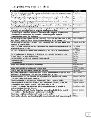
  • 1. 1 Radiographic Projections & Positions QUESTIONS ANSWERS The term defined as the path ofcentral ray as it exits the x-ray tube and goes through the patient to the IR is called a what? Projection During this projection a perpendicular central ray enters the anterior body surface and exits the posterior body surface is known as what projection? Anteroposterior- AP During this projection a perpendicular central ray enters the posterior body and exiting the anterior body surface. Posteroanterior-PA During this projection, there is longitudinal angulation ofthe central ray with the long axis ofthe body or a specific body part. Axial projection This term refers to all projections in which the longitudinal angulation between the central ray and the long axis ofthe body part is 10 degrees or more. Axial Occasionally the central ray is directed toward the outer margin of a curved body surface to profile a body part just under the surface and project it free of superimposition during this projection. Tangential During this projection, a perpendicular central ray enters one side ofthe body or body part, passes transversely along the coronal plane and exits on the opposite side. Lateralprojection During this projection, the central ray enters the body or body part from a side angle following an oblique plane. Oblique projection If the central ray enters the anterior surface and exits the opposite posterior surface, it is known as what projection? AP oblique projection If it enters the posterior surface and exits anteriorly, it is known as what projection? PA oblique projection The overall posture ofthe patient or the general body position is termed as what? Position Erect or marked by a vertical position. Upright Upright position in which the patient is sitting or stool. Seated Lying on the back Supine Lying face down Prone Supine position with the head tilted downward Tredelenburg's position Supine position with the head higher than the feet. Fowler's position General term referring to lying down in any position. Recumbent A recumbent position with the patient lying on the left anterior side (semiprone) with the left leg extended and the right knee and thigh partially flexed. Sim's position A supine position with the knees and hip flexed and thighs abducted and rotated externally, supported by ankle supports. Lithotomy position This position refers to the side ofthe patient that is placed closest to the IR. Lateralposition This position is achieved when the entire body or body part is rotated so that the coronal plane is not parallel with the radiographic table or IR. Oblique position Term used to indicate that the patient is lying down and that the central ray is horizontal and parallel with the floor Decubitus position This position is achieved by having the patient lean backward while in the upright body position so that the shoulders are in contact with the IR. Lordotic position This term is used to describe the body part as seen by the IR. View This term describes the specific radiographic projection that the individual developed. Method
  • 2. 2 Terms & Positioning QUESTIONS ANSWERS The lowest level ofstructuaral organization of the human body is the: Chemical Level Four basic types oftisuesin the body: Epithelial, Connective, Muscular, Nervous The 10 systems ofthe human body: Skeletal, Circulatory, Digestive, Respiratory, Urinary, Reproductive, Nervous, Muscular, Endocrine, Integumentary Eliminates solid waste from the body Digestive System Regulates fluid and electrolyte balance and volume Urinary System Maintains posture Muscular System Regulates body activities with electical impulses Nervous System Regulates body activities through various hormones Endocrine System Eliminates carbon dioxide from the blood Respiratory System Receives stimuli, such as temperature, pressure, and pain Integumentary System Reproduces the organism Reproductive System Helps regulate body temperature Circulatory System Supports and protects many soft tissuesofthe body Skeletal System True or False: One ofthe six functions ofthe circulatory system is to protect against disease True This body system regulates body temperature: Integumentary System What is the largest organ system in the body? Integumentary System List the two divisions ofthe human skeleton Appendicular and Axial True or False: The adult skeleton system contains 256 bones. False (206) True or False: The scapula is part ofthe axial skeleton. False (appendicular skeleton) True or False: The skull is part ofthe axial skeleton. True True of False: The pelvis is part ofthe appendicular skeleton. True List the four classifications ofbones Long Bones, Short Bones, Flat Bones, Irregular Bones The outer coveringofa long bone, which is composed ofa dense, fibrous membrane,is called what? Periosteum Which aspect oflong bones is responsible for the production of red blood cells? Medullary Aspect Which aspect ofthe long bone is essential for bone growth, repair, and nutrition? Periosteum Identify primary and secondary growth centers for long bones Primary growth center: the body (diaphysis) Secondary growth center:epiphyses True of False: Epiphyseal fusion ofthe long bonesis complete by the age of16 year False (25 years) What is the wIder portion ofa long bone in which bone growth in length occurs Metaphysis What are the three functional classifications ofjoints? Synarthrosis, Amphiarthrosis, Diathrosis What are the three structual classifications ofjoints? Fibrous, Connective, Synovial First carpometacarpal of thumb Synovial Joint Roots around teeth Fibrous Joint Proximal radioulnar joint Synovial Joint Skull sutures Fibrous Joint Epiphyses Cartilaginous JoinT Interphalangeal joints Synovial Joints
  • 3. 3 Distal tibiofibular joint Fibrous Joint Intervertebral disk space Cartilaginous Joint Symphysis pubis Cartilaginous Joint Hip Joint Synovial Joint What are the seven types of movement for synovial joints? Plane(Gliding) Ginglymus(Hinge) Trochoid(Pivot) Ellipsoid(Condylar) Seller(Saddle) Spheroidal(Ball and Socket) Bicondylar First carpometacarpal joint Sellar Elbow joint Ginglymus Shoulder blade Spheroidal Intercarpal joint Plane Wrist joint Ellipsoidal Temporomandibular joint Bicondylar First and secnond cervical vertebra joint Trochoidal Distal radioulnar joint Trochoidal Second interphalangeal joint Ginglymus Ankle joint Sellar Knee joint Bicondylar Third metacarpophalangeal joint Ellipsoidal What is an image ofa patient's anatomic parts as porduced by the actions ofx-rays on an imager receptor? Radiograph What is the aspect ofan x-ray beam that has the least divergence? Central Ray Upright position with the arms abducted, palms forward, and head and feet directed straight ahead Anatomic Position Vertical plane that divides the body into equal right and left parts Mid-Sagittal Plane the vertical plane that divides the body into equal anterior and posterior parts Mid-Coronal Plane A plane taken at right angels along any point of the longitudinal axis ofthe body Transverse/Axial Plane True or False: The base plane ofthe skull is a plane located between the infraorbital margin ofthe orbit and the superior margin of the external auditory meatus. True True or False: The Frankfort horizontal plane is also referred to as the midcoronal plane. False The direction or path of the central ray defines the following positions term Projection The positioning term that describes the general and specific body position is: Position True or False: Oblique and lateral positions are described according to the side ofthe body closest to the image receptor True True or False: Decubitus positions always use a horizontal x- ray beam True What is the name ofthe position in which the body is turned 90 degrees from a true anteriorposterior(AP) or posterioanterior(PA) projection LateralPosition A patient is erect with the back to the image receptor. The left side ofthe body is turned 45 degrees toward the image receptor. What is this Position? Left Posterior Oblique(LPO) A patient is recumbent facing the image receptor. The right side ofthe body is turned 15 degrees toward the image Right Anterior Oblique(RAO)
  • 4. 4 receptor. What is this position? The patient is lying on his back. The x-ray beam is directed horizontally and enters the right side ofthe body and exits the left side ofthe body. An image receptor is placed against the left side ofthe patient. Which specific position has been used? Dorsal Decubitus(Left Lateral) The patient is erect with the right side ofthe body against the image receptor. The x-ray beam enters the left side and exits the right side ofthe body. What position has been preformed? Right Lateral A patient is lying on the left side ofa cart. The x-ray beam is directed horizontally and enters the posterior surface and exits the anterior aspect ofthe body. The image receptor is against the anterior surface. Which specific position has been perform Left LateralDecubitus(PA) Palm of the hand Palmar Lying on the back facing upward Supine An upright position Erect Lying down in any position Recumbent Front half of the patient Anterior Top or anterior surface of the foot Dorsum Pedis Position in which head is higher than the feet Fowlers Posterior aspect ofthe foot Plantar Position in which the head is lower than the feet Trendelenburg Back half ofthe patient Posterior What is the name ofthe projection in which the central way enters the anterior surface and exits the posterior surface Anteriorposterior A projection using a CR angle of10 percent or more directed parallel along the long axis ofhte body or body part is Axial Projection The specific position that demonstrates the apices ofteh lungs, without superimposition ofthe clavicles Apical Lordotic True or False: Radiographic "view" is not a correct positioning term in the United States True True or False: The term varus describes the bending ofa port outward False(inward, toward midline) Anteroposterior Projection Prone Position Trendelenburg Position Left posterior oblique Position Left lateral chest Position Mediolateral ankle Projection Tangential Projection Lordotic Position Inferosuperior acial Projection Left lateral decubitus Position Opposites Flexion Extension Ulnar Deviation Radial Deviation Dorsiflexion Plantarflexion Eversion Inversion Lateral(external) Rotation Medial (internal) Rotation Abduction Adduction Supination Pronation Retraction Protraction
  • 5. 5 Depression Elevation Near the source or beginning Proximal On the opposite side Contralateral Toward the center Medial Toward the head end ofthe body Cephalad or Superior Away from the source or beginning Distal Outside or outward Exterior On the same side Ipsilateral Near the skin surface Superficial Away from the head end Caudad or Inferior Farther from the skin surface Deep Moving or thrusting the jaw forward form the normal position is an example of Protraction To turn or bend the wrist toward the radius side is called Radial Deviation Which two types ofinformation should be imprinted on every radiographic image? Patient Name and Date,and Anatomic Side Markers True or False: a technologist has the right to refuse to perform an examination on a patient whom he or she finds offensive. False True or False: A technologist is responsible for the professional decisions he or she makesduring care ofa patient. True True or False: The technologistis responsible for communicating with the patient to obtain pertinent clincal information. True True or False: The technologistis expected to provide a preliminary interpretation ofradiographic findings to the referring physician. False True or False: The technologistmust reveal confidential information pertaining to a patient who is less than 18 years of age to the patient or guardian. False What two are rules/principles for determining positioning routines as they relate to the maximum number ofprojections required in a basic routine? A minimum of two projections 90 degrees from each other, and a minimum of three projections when the joints are in the prime interest area What is the minimum number of projections for the: Foot Three What is the minimum number of projections for the: Chest Two What is the minimum number of projections for the: Wrist Three What is the minimum number of projections for the: Tibia/Fibula Two What is the minimum number of projections for the: Humerus Two What is the minimum number of projections for the: Fifth Toe Three What is the minimum number of projections for the: Postreduction ofwrist Two What is the minimum number of projections for the: Left Hip Two What is the minimum number of projections for the: Knee Three What is the minimum number of projections for the: Pelvis(non-hip injury) One A young child enters the emergency room with a fractured forearm. After one projection is completed that confirms a fracture, the child refuses to move the forearm for any additional projections. What is the minimum number of projections needed? Two If additional projections are required for a routine forearm Rather than move the forearm for a second
  • 6. 6 series, what should the technologist do with the young patient? projection, place the IR and x-ray tube as needed for the second projections 90 degrees from the first projection. The physical localization oftopographic landmarks on a patient is called: Palpation Which two landmarks may not be palpated because of institutional policy? Ischial Tuberosity, and Symphysis Pubis True or False: Always place a radiograph for viewing as teh IR "sees" the patient.The patients left is to the viewer's left on an AP projection) False, the patients left is to the viewers right on an AP projection True or false: Most CT and MRI images are viewed so that the patient's right is to the viewer's left. True The radiographic anolog(film) image is composed of________ on a polyester base. Metallic Silver What are the four image quality factors of a radiograph? Density, Spatial Resolution, Contrast, Exposure Latitude The range ofexposure over which a film produces an acceptable image Exposure Latitude Which specific exposure factor controls the quality or pentrationg ability of the x-ray beam? Kilovoltage(kVp) Exposure time is usually expressed in units of Milliseconds(ms) The amount of blackness seen on a prcessed radiograph is called Density The primary controlling factor for the overall blackness on a radiograph is mAs If the distance between the x-ray tube and IR is increased from 40 to 80 inches, what specific effect will it have on the radiographic density, ifother fractors are not changed? Decrease density to 25% Which term is used to describe a radiograph that has too little density? Under Exposure Doubling the mAs will result in _______ the denisty ofthe IR image Doubling True or False: kVmust be altered to chance the radiographic density on the IR False, mAs will change the density, kVp will change the contrast When analog images, using manual technique settings, are underexposed or overexposed, a minimum chance in mAs of ________ is required to make a visible difference in the radiographic density 25% to 30% According to the anode heel effect, the x-ray beam is less intense at the(anode or cathode) end ofthe x-ray tube Anode To best use the anode heel effect, the thicker part of the anatomic structure should be place under the (anode or cathode) end ofthe x-ray tube Cathode What device or method (other than the anode heel effect)may be used to compensate for the anatomic part thickness difference and prduce an acceptable density ofthe IR image Compensating Filters What are three common types ofcompensating filters Wedge Filter, Trough Filter, Boomerang Filter Which type of compensationg filter is used commonly for AP projections ofthe thorasic spine? Wedge Filter Which type of compensating filter permits soft tissue and obny detail of the shoulder to be equally visualized? Boomerang Filter A radiograph produced using conventional analog cassettes 10 mAs
  • 7. 7 resulted in too little density. The origanal exposure was 5 mAs. What mAs is needed to correct the density(density needs to be doubled) The difference in density on adjacent areas ofthe radiograph defines Radiographic Contrast What is the primary controlling factor for radiographic contrast kVp What are the two scales ofradiographic contrast, and identify which is classified as high contrast and which is lowcontrast. Long Scale Contrast(low contrast), Short Scale Contrast(high contrast) Which scale ofcontrast is produced with a 110-kVtechnique Long Scale Contrast(low contrast) True or False: A 50-kVtechnique producesa high contrast image True True of False: A low-contrast image demonstrastes more shades ofgray on the radiograph True Which of the following sets ofexposures factors will result in the least patient exposure and produce long-scale contrast on a PA chest (50 kV, 800 mAs or 110 kV, to mAs) 110 kV at 10 mAs A radiograph of a hand is underexposed. The original technique used was 55kVwith 2.5 mAs. Keeping the mAs and increasing the kVwhat would the newkVbe to double density? 8 - 10kV If an anoatomic part measures greater than _____cm a grid must be used 10cm Identify the type ofgrid cuttoff that is created: The central ray (CR)and face of grid are not perpendicular Off-LevelGrid Cutoff Identify the type ofgrid cuttoff that is created: The SID is set beyond the focal range ofthe grid Off-Focused Grid Cutoff Identify the type ofgrid cuttoff that is created: The back of the grid is facing the x-ray tube Upside Down Grid Cutoff The recorded sharpness ofstructures ofobjects on the radiograph defines Spatial Resolution/Definiton The lack ofvisible sharpnessis Blur/Unsharpness What are the three geometric factors that control or influence image resolution Focal Spot Size, Source Image Receptor Distance(SID),Object Image Receptor Distance(OID) The term that describes the unsharp edges ofthe projected image Penumbra True or False: The use ofa small focal spot will entirely eliminate the problem identified in teh previous question False The greatest contributor to image unsharpness as related to positioning Motion Body Movement QUESTIONS ANSWERS Movement ofa part away from the central axis ofthe body or body part. abduction Movement ofpart toward the central axis ofthe body or body part. adduction Straightening ofa joint; when both elements ofthe joint are in the anatomic position; the normal position ofa joint. extension
  • 8. 8 Act of bending a joint; the opposite ofextension. flexion Forced or excessive extension ofa limb or joints. hyperextension Forced over flexion ofa limb or joint. hyperflexion Outward turning of the foot at the ankle. eversion Inward turning of the foot at the ankle. inversion Rotation ofthe forearm so that the palm is down. pronate Rotation ofthe forearm so that palm is up (in the anatomic position). supinate Turning or rotating ofthe body or body part around its axis. rotate Circular movement ofa limb. circumduction Tipping or slanting a body part slightly; in relation to the long axis ofthe body. tilt A turning away from the regular standard or course. deviation Body Habitus/Regions QUESTIONS ANSWERS Name the 3 superior body regions. Right Hypochondrium, Epigastrium, Left Hypochondrium Name the 3 middle body regions Right Lateral, Umbilical, Left Lateral Name the 3 inferior body regions. Right Inguinal, Hypogastrium, Left Inguinal What organs are found in the Right Hypochondrium region? Gallbladder, Liver What organs are found in the Left Hypochondrium region? Spleen, Stomach What organs are found in the Right Inguinal region? Appendix What organs are found in the Hypogastrium region? Bladder, Rectum What conditions are likely in the Left Inguinal region? Gas pains What conditions can occur in the Epigastrium region? Heartburn, Ulcer What organs can be found in the Right Lateral Region? Ascending colon, Kidney What organs can be found in the Left Lateral Region? Descending colon, Kidney Name the 4 Quadrants. Left Upper Quandrant, Left Lower Quandrant, Right Lower Quadrant,Right Upper Quadrant What organs are in the LUQ? Stomach, Spleen, Pancreas What organs are located in the RLQ? Appendix, Cecum, Colon What organs are located in the RUQ? Gallbladder, Liver, Intestines, Kidney What organs can be found in the LLQ? Descending colon, Intestines Name the 4 types ofBody Habitus. Asthenic, Hypersthenic, Hyposthenic, and Sthenic Which body habitus has the highest rate ofoccurrence? Sthenic at 50%. Which body habitus has the lowest rate ofoccurrence? Hypersthenic at 5% Which body habitus belongs to frail individuals? Asthenic Asthenic Body Habitus occurs about ___% 10% Which body habitus is the most difficult to classify? Hyposthenic This body habitus represents 10%ofthe population, gallbladder is low and nearer the midline, stomach is lowand medial. This is the ________ body habitus. Asthenic Define body habitus. Common variations in the shape of the human body. Body habitus affects the location ofwhich organs? Gallbladder, Stomach, Heart,Lungs,
  • 9. 9 Diaphragm In which body habitus is the heart nearly vertical at midline? Asthenic Which body habitus represents 10%ofthe population, the gallbladder is low and nearer to midline and the stomach is low and medial? Asthenic Which body habitus representing 5% ofthe population, has a high gallbladder, and a high, transverse stomach? Hypersthenic What is the percentage ofoccurrence for hyposthenic body habitus? 35% What is the stomach and gallbladder position for sthenic? Stomach: high/upper left; Gallbladder: center on right What is the stomach and gallbladder position for asthenic? Stomach: Low/medial; Gallbladder: low/near midline What is the stomach and gallbladder position for hypersthenic? Stomach: High/transverse/in middle; Gallbladder: High & outside Sthenic organ placement. Stomach: high & left; colon: even spread; GB: centered right side Hyposthenic organ placement. No change. Asthenic organ placement. Stomach: low & medial in pelvis; colon: low; GB: Low & near midline Hypersthenic organ placement. Stomach: High & middle; Colon: frames abdomen; GB: High & outside. Skeletal Landmarks C1 MASTOID TIP C2 - C3 GONION C3 - C4 HYOID C5 THYROID C7 VERTEBRA PROMINENS T1 APPROXIMATELY 2"ABOVE JUGULAR NOTCH T2 - T3 JUGULAR NOTCH T4 - T5 STERNAL ANGLE T7 INFERIOR ANGLES OF SCAPULA T9 - T10 XIPHOID PROCESS L2 - L3 LUMBAR PUNCTURE L4 - L5 ILIAC CREST S1 - S2 ASIS PUBIC SYMPHYSIS/GREATER TROCHANTERS COCCYX
  • 10. 10 Lower Extremities QUESTIONS ANSWERS The pelvic girdle consists of 2 hip bones The Pelvis consists of both hip bones, sacrum,coccyx The hip is made up ofthe ilium, ischium, and pubic bone What is the area between the greater and lesser trochanter called on the ANTERIORaspect ofthe proximal femur intertrochanteric line What is the area between the greater and lesser trochanter called on the POSTERIOR aspectof the proximal femur intertrochanteric crest A true AP of the hip require howmuch rotation? 15-20 degree internal rotation kVfor the AP Pelvis, AP Hip, and Lateral Hip is 75-85kV Center for the AP Pelvis is centered 2" inferior to level of ASIS (crest 1.5" below top of IR) Howare you doing? EXCELLENT! What size IR for a AP Pelvis? 14x17 CW T/F Lesser trochanters ofthe femur is included in the AP Pelvis True Howdo you detect rotation for Pelvis? The superior ramus is part ofthe pubis The inferior ramus is part ofthe Ischium The Judet method demonstrates the Acetabulum Center for AP hip (with hardware) 1-2" distal to neck or femur (all of hardware must be demonstrated) Lateral of the hip is also called Frog or Modified Cleaves or Lauenstein method Trauma Hip most often used is called Danelius-Miller or Cross-table lateral or Axiolateral (inferiorsuperior) The modified axiolateral trauma hip when both hips can't be moved. is called Clements-Nakayama method Howmuch should the femur be abducted for the Cleaves method for the hip? 40-45degrees Howmuch should the femur be abducted for the Lauenstein method for the hip? 40-45 degrees (with knee flexed 90degrees) Where is the CR placed for a unilateral frog-leg projection mid femoral neck The AP axial outlet projection for the pelvis requires the CR to be ______for females and _______ for males 20-35 and males 30-45degrees The AP inlet projection for the pelvic ring requires the CR angle to be 40deg caudad A male pelvis has an ______ angle while a female pelvis has a ________ less than 90 degrees acute,female greater than 90 degrees obtuse Three differences in a female and male pelvis are males have narrower , deeper and less flared, angle of the pubic arch is less than 90deg, shape of the inlet is more narrower and more oval or heart shape What are some important positioning landmarks for the pelvis iliac crest,ASIS, greater trochanter,symphysis pubis, ischial Tuberosity The pelvis is separated into ______ superior to the inlet and ________pelvis is a cavity that is surrounded by bony structures that is ofgreat importance during birthing process greater false pelvis, lesser true pelvis forms birthing canal
  • 11. 11 If the femoral neck is foreshortened and the lesser trochanters are in profile medially on a radiograph what is probable cause for positioning external rotation of the leg and foot When taking a patient history for a hip x-ray it is important to ask about a prosthesis or any hip surgery for what two reasons so you can position patient without injuring site, and to make sure you center lower to include all hardware What pathology is best demonstrated with the judet method acetabular fractures Where is the CR placed for a unilateral frog-leg projection mid femoral neck The ankle joint is formed by what three bones tibia, fibula, talus A 15deg internal rotated AP oblique projection is called the mortise projection The mortise position demonstrates the joint and should have even space over entire _____ talar surface What does the mortise joint do for the body helps stabilize weight What is the difference between the AP mortise and AP oblique ankle projections for positioning internal rotation for mortise is 15-20deg and the ankle is internal rotation of 45deg On a true AP of the ankle what is not demonstrated entire three part joint space of the ankle mortise The ankle is what type ofjoint with what type of movement synovial joint, sellar or saddle type and movement is flexion and extension Which malleolus is longer and is an extension of the fibula lateral malleolus What are the stress views ofthe ankle important shows lack of support, from fractures or tears of ligaments Before doing a stress viewofthe ankle what should be ruled out make sure there is no fracture What are the two joints are on the tibia proximal and distal tibiofibular joints What structures are seen in the AP Ankle? 1/3 of tib/fib, ½ of metatarsals,ankle joint with the medial and upper portion of the joint open. Name the 3 Ankle positions (routine) AP,AP oblique with medial rotation, Lateral Positioning for the AP ankle Center to ankle joint, foot dorsiflexed. Positioning for the AP mortise with medial rotation 15-20 degrees medial rotation, centered to ankle. (demonstrates ankle mortise) Howdo you accurately position for the AP w/ medial rotation? rotate medially until the malleoli are parallel (equidistant) to the IR. Rotate the whole leg NOT just the ankle or foot. What is the visual difference between and AP and AP Mortise? the joint space on the lateral side of the Mortise will be open. In the AP the Fib is superimposed over part of the talus. What is the (rarely used) AP oblique with 45degree medial rotation for? to show tib/fib joint space. Identify rotation on Lateral ankle talar domes should be superimposed, lateral malleolus superimposed over posterior half of tibia. What are Inversion/Eversion viewofthe Ankle for? stress views that are used to demonstrate ligament damage. What do you do to fit the Tib/Fib on a 14x17? Try it diagonally, then try increasing the SID (44-48in) T/F There should be partial superimposition of the Tib and Fib at both proximal AND distal ends? TRUE You are _____? ON FIRE! Someone call 9-1-1! Describe positioning for the Lateral TIB/FIB Mediolateral, flex knee to 45 degrees,center midshaft and include both joints. May increase SID. Identify rotation for the Lateral TIB/FIB Rotation indicated by condyles of femur and ankle joint.
  • 12. 12 Condyles should be superimposed and the proximal head of FIB superimposed by TIB, distal FIB superimposed over posterior half of TIB. Identify rotation for AP TIB/FIB evaluate relationship of the fibula to tibia. Lat. Rot. – fib shifts toward or under tib, obscuring medial mortise. Med. Rot – head of fib draws from beneath tib. . Name the tarsals ofthe foot Calcaneus, Cuboid, Cuniforms (1 medial, 2 intermediate, 3 lateral), Navicular, Talus Howmany Tarsals are there? Seven 7 The heel bone is called Calcaneus The Calcaneus is a Tarsal True Where would you find Sesamoid Bones in the foot? embedded in tendons, near joints, plantar surface Howmany bones in the foot? 14 (phalanges), 5 (metatarsals),7 (tarsals). 26 total bones. Name the arches ofthe foot Longitundinal Arch (Lateraland Medial sides of foot) Transverse arch (across the foot) Describe the Longitudinal arch of the foot Comprised of lateral and medial, most of the arch is on the medial side and in the mid aspect of the foot Describe the Transverse arch ofthe foot primary located along the plantar surface of the distal tarsals and TMT joints. Made up mostly of the cuniforms and cuboid (especially 2nd and 3rd cuniforms). Dorsiflexion is when the foot is raised cephalad Plantar Flexion is when the foot is extended away from the body (pressing the gas pedal) Inversion (varus) ofthe foot is when the bottom of the foot is faced medially Eversion (valgus) ofthe foot is when the bottom of the foot is faced laterally Technical factors for the foot 40in SID, 50-70kV, short exp. time, grid if >10cm Name the Foot positions AP axial, AP oblique, Lateral Name the Toes positions AP axial, AP oblique, Lateral Name the Calcaneus positions Axial and Lateral CR angle for AP axial Toes 15 degrees cephalic Centering for AP axial Toes MTP joint Film size for AP axial Toes 8x10 or 10x12 (depends on projections done and if AP axial FOOT is done as a projection) Special projection for sesamoid bones tangential of toes – dorsiflex foot 15-20degrees from vertical, CR perpendicular to IR and centered tangentially to posterior of 1st MTP alternative lateral for the foot lateromedial- outside of the foot, CR mid-cuneiform base of 3rd MT special projection for the foot to show longitudinal arches AP & lateral weight-bearing CR 15deg posterior to base of MT Name the Calcaneus projections and centering point Axial Plantodorsal –dorsiflexed, CR 40deg cephalic at base of 3rd MT Lateral-Mediolateral- CR 1in inferior to medial malleolus what is gout? form of arthritis, uric acid deposits destuct joint space Does Lisfranc joint injury requires a decrease or increase in technique increase to penetrate tarsalregion joint effusions are signs of fracture,dislocation,soft tissue damage what type ofjoints are IP joints hinge (flexion and extension) what type ofjoints are TMT,intertarsal plane or gliding (limited movement) what type ofjoints are MTP ellipsoidal or condyloid, (4 movements) the calcaneal sulcus and a depression on the Talus sinus tarsi
  • 13. 13 form an opening for ligaments to pass through in the middle ofthe subtalar joint called? three articular facets appear at the subtalar or talocalcaneal joint with the Talus through which the weight ofthe body is transmitted to the ground in an erect position posterior, anterior and middle articular what does the sustentaculum do? provides medial support for weight bearing subtalar or talocalcaneal joint . in what projection is the tuberosity on the 5th MT demonstrated oblique-medial of the foot what is a common trauma site for the foot that provides attachment ofa tendon tuberosity of the 5th MT weight ofthe body is transmitted by this bone through the important ankle and talocalcaneal joints TALUS what type ofjoint is the ankle synovial-sellar type w/flexion and extension Longest and strongest bone femur Four major ligaments for the knee joint posterior cruciate, anterior cruciate, fibular collateral, tibial collateral Name three knee positions that are tunnel projections BeClere,camp Coventry, homblad Name two tangential knee projections merchant and sunrise A distinguishing difference between the lateral and medial condyle is the presence of _____________ adductor tubercle on the posterior side of the medial condyle that receives the tendon of the adductor muscle What do all tunnel views demonstrate intercondylar fossa Howdo you position a patient for the camp- coventry method patient supine, flex knee 40-50degrees, CR to knee joint or popliteal depression, CR perpendicular to tib/fib, 40 SID. What two tunnel projections are PA holmblad and camp Coventry What one tunnel viewrequires the CRto be perpendicular to the IR Homblad method The settegast method also called the inferosuperior projection requires the kneesto be flexed __________ deg and the CR angle __________ to the lower legs 40-45d, 10-15d The joints at each end ofthe femur are a frequent source ofpathology when trauma occurs because why The entire weight of the body is transferred through the femur and associated joints What do the medial and lateral condyles ofthe femur articulate with the tibia Why must the CR angle for a lateral knee be 5-7 degreescephalad the medial femoral condyle extends lower than the lateral femoral condyle when the femoral shaft is vertical The medial and lateral epicondyles are attachments for what the medial and lateral collateral ligaments What is the largest sesamoid bone in the body the patella When the leg is extended the patella is where superior to the patellar surface When the leg is flexed the patella is where downward over the patellar surface Where is the apex ofthe patella located along the inferior border Where is the base ofthe patella located the superior border Does the patella articulate with the tibia no! only with the femur Where is the femorotibial joint located between the two condyles of the femur and the condyles
  • 14. 14 of the tibia What is the femorotibial joint classified as a synovial joint, bicondylar and diarthrodial that allows flexion and extension Where is the patellofemoral joint located where the patella articulates with the anterior surface of the distal femur What is the patellofemoral joint classified as synovial , SELLAR (saddle) What is the largest joint space ofthe human body cavity of the knee joint What is the knee joint the knee joint is synovial type enclosed in an articular capsule or bursa What are the medial and lateral menisci fibrocartilage disks between the articular facets of the tibia and the femoral condyles What projection shows the articular facets in profile AP knee Where do you center for an AP knee parallel to the tibial plateau Why are the femoral condyles superimposed but never completely because of magnification What is the same for all tunnels ofthe knee CR perpendicular to tib/fib and demonstrates intercondylar fossa Why is a PA patella preferred over an AP less OID What is demonstrated on an AP proximal femur lesser trochanter superimposed and the greater trochanter in profile What is demonstrated on an AP Distal femur epicondyles parallel to IR What is demonstrated on a Lateral proximal femur lesser trochanter in profile and the greater trochanter is superiposed What is demonstrated on a lateral distal femur condyles are in line with long axis of femur for no rotation Beclere method (ap axial) for tunnel knee requires _____degree knee flexion, CR angle of ____ degreesand the CR centered _______ 40-45, 40-45 cephalad, ½ inched distal to apex of patella Holmblad method (pa axial) for tunnel knee requires ______degree knee flexion, and the CR angle of______degrees. 60-70 degree knee flexion and no angle on CR (perp to IR) Camp Coventry method (pa axial) for tunnel of knee requires _____degree knee flexion, and CR angle of______ degrees. 60-70 degree knee flexion and 40-50 degree caudad angle on CR Do you rotate the knee for a true AP? yup, 5 degree internal rotation of anterior knee will align interepicondylar line parallel to plane of IR. Howmuch should you flex the knee for a Lateral- Mediolateral Knee projection? 5-10 degrees additional flexion may cause separation of a fracture (p.253) Define Baker Cyst When an excess of knee joint fluid is compressed by the body weight between the bones of the knee joint, it can become trapped and separate from the joint to form the fluid-filled sac in the posterior knee. The cavity in the hipbone that articulates with the femoral head is called the acetabulum The hip bone consists ofwhat three parts? Ischium, Pubic bone, and Ilium The ilium and sacrum articulates at the _________ joint Iliosacral The junction of what 2 bones forms the obturator foramen of the pelvis? Ischium and Pubic bone Name the bones that make up the pelvic girdle Right and Left Hip bones Name the bones that make up the pelvis in an adult Sacrum, Coccyx, Right and Left Hip
  • 15. 15 The prominent ridge extending between the trochanters at the base ofthe neck on the posterior surface ofthe femur is the intertrochanteric crest Name one or more structures that may be helpful in order to evaluate rotation on an AP pelvis radiograph (not proximal femur) Symetry of the Obturator formina or Ischial spines, and alignment of the Coccyx and Pubis symphisis. Howmuch do you medially rotate the feet and lower limbs to place the femoral necks parallel with the plane ofthe IR on an AP projection of the pelvis? 15-20 degrees What position, projection or method is useful in diagnosing fractures ofthe acetabulum? Judet (axiolateral) What is the projection ofthe Modified Cleaves often called? Frog leg Do you see the lesser trochanter with the Modified Cleaves method? Yes What projection/position ofthe hip best demonstrates the greater trochanter in profile? AP hip/pelvis The angulation of the tube for the axiolateral projection (Danelius-Miller Method) is angled perpendicular to what structure? (not the film) Femoral Neck (and IR) Where is the central directed for the unilateral frog-leg? Femoral Neck The largest sesamoid bone in the body is the patella The tube angle for the Camp Coventry method for the PA axial (knee) is 40 degrees In order to better visualize the joint space in the AP projection ofthe knee on a large patient, the central ray should be angled howmany degrees and in what direction? 3-5 degrees cephalic In the Be'clere position the patient is placed (supine, prone,or lateral)? Supine The centering point for the AP ofthe knee is 1/2" distal from apex of Patella This acts as a shock absorber in the knee Meniscus In the AP projection ofthe proximal femur, the foot should usually be slightly rotated internally ________ degrees. 15-20 Which projection ofthe patella provides sharper recorded detail, AP or PA? PA What is the name ofthe prominence on the posterior aspect ofthe femur that forms the popliteal surface? Linea Aspera What is the protrusion on the anterior side of the proximal tibia called where the patellar ligament inserts tibial tuberosity When looking at a lateral ankle radiograph, how do you determine ifit is rotated the talar domes should be superimposed and there should be superimposition of the posterior tibia Is the sustentaculum tali on the medial or lateral side ofthe calcaneus medial The lateral malleolus is part ofthis bone fibula The fibula articulates with the condyles ofthe femur (T or F?) False When doing an oblique ankle that is for the 15-20 degrees medial rotation
  • 16. 16 mortise, howmuch do you rotate the leg and in which direction Describe howto position a tib/fib for an AP condyles should be parallel to IR and foot should be AP Where is the centering point on an AP projection of the ankle ankle joint If an x-ray ofthe toes are requested, howmuch do you angle your tube on the AP axial projection to open the joint spaces 15 degrees If an x-ray ofthe foot is requested, howmuch do you angle your tube for an AP projection which opens the joint spaces 10 degrees On an AP oblique projection ofthe foot, which oblique and how many degreesobliquity is most often performed 30 degrees medial oblique When doing an AP oblique projection ofthe foot which rotation best demonstrates the sinus tarsi medial rotation Where is the central ray directed for the lateral first toe IP Where is the central ray directed for the AP foot base of the 3rd metatarsal To obtain an axial projection ofthe calcaneus, the number of the degrees the central ray is angled____ when the long axis ofthe foot is perpendicular to the plane ofthe IR 40 degrees For AP of the toes, the toes/foot are ________ to the IR and the CR is at the ____ Joint? PARALLEL and MTP Joint For AB Oblique ofthe toes, knees are flexed, foot on IR with toes INTERNALLYrotated are _______ to the IR with CR TO ____ joint. 30" to 45* Oblique = CR to MTP Joint For the lateral viewofthe big toe, the foot should always be in what position LATERAL Where is the CR Directed for an AP Dorsoplantar of the foot? CR is angled 10* POSTERIORLY toward the heelot BASE of the 3RD Metatarsal For and AP OBLIQUE ofthe foot howmany degreesto the IR ? 30* For the AP OBLQUE ofthe foot the CR to the BASE is at? the 3rd Metatarsal For a lateral viewofthe foot howshould it be positioned? Mediolateral For the AXIAL PLANTODORSAL position of the foot and calcaneus (the heel) be positioned to the IR? Perpendicular to the IR In the AXEAL PLANTODORSAL position the CR should be angled howmany degrees 40* Cephalad toward the id calcaneus What does CEPHALD mean? Toward the head What position should the leg be in a lateral (mediolateral) position Knee Flexed-Leg rotated externally until lateral side of foot is against the IR - Ankle is flexed 90* For and AP OBLIQUE Mortise howfar do you rotate the ankle? 15* - 20* Oblique For an AP OBLIQUEhowfar do you rotate the ankle 45* oblique to IR When positioning the lower leg the 14"x17" is placed how? Diagonally In the AP ofthe lower leg the lower leg and knee PARALLEL
  • 17. 17 should be _______ to the IR? When positioning the knee in the AP Viewthe CR angled should be _____ * cephalad to 1/2" distal to apex ofpatella 5* For a lateral knee the knee should be flexed ____* to ____ *, leg should be rotated _____ until femoral condyle and patella are ____ to IR - 20* TO 30* = Externally = PERPENDICULAR to IR For the lateral knee the CR is _____* to _____* cephald to _____" distal to medial epicondyle 5* to 1* = 1" Howmany degrees is the knee flexed for the TUNNEL VIEW? Prone with Knee Flexed 40-50* to IR Howmany degrees for a SUNRISE VIEW? 80* For the Patella the CR angle is? 15* to 20* Cephalad to APEXof the Patella What is ASIS? Anterior Superior Iliac Spine When positioning the femur the 14" x 17" should be placed __________ with TOP OF IR at level of _______ for PROXIMAL VIEWS LONGITUDINALLY - ASIS When positioning the femur the 14'x17' should be placed ________ with the BOTTOM ofthe IR ________ belowknee joint for a distal view Longitudinally - 1" to 2" For a LATERAL PROXIMAL VIEWthe patient is turned _______ on side, knees flexed _______ with legs rotated ________ until lateral? PARTIALLY on side - 30"-45" - Rotated EXTERNALLY - For a LATERAL DISTAL VIEWthe patient is turned on side with ________ leg crossed over affected leg, knee is flexed ____* with femoral condyles and patella _______ to the IR UNAFFECTED LEG - 30*-45* = PERPENDICULAR When using the bucky to position the hip a 10" x 12" is placed ____________ with TOP OF IR at level ofASIS Longitudinally For an AP Positioning ofthe hip leg is fully extended with foot and leg rotated ________ * Internally 15 Name the irregular bones OXCOXAE - SACRUM - COCCYX The tarsal are what type of bones Short Bones The phalanges and metatarsals are classified as what type ofbones? Long Bones The tibia and fibula are classified as what type of bones Long Bones The femur is considered what type ofa bone Long Bone What type ofmovement does all PHANGEAL JOINTS provide Hinge Movement The MTP Joints allowfor what type ofmovement Hinge Movement The ANKLE (MORTISE) Joint allows for what type ofmovement Hinge Movement The Patella femoral allows for what type of movement? Gliding Movement The hip joint allows for what type of movement Circumduction Howmany bones are there in the foot? 26 Howmany phalanges (toes)are there in the foot 14 There are ________ tarsals in the foot 7 The FEMUR extends from the _____ to the ____ Hip to the Knee The proximal end ofthe femur contains what? The head - neck & greater and lesser trochanters The distal end contains the ______ & _____ with a Medial and the LateralCondyles = Blood vessels and
  • 18. 18 U-shaped notch. This notch lets what pass through? nerves The 1st digit (big toe) contains ____ phalanges 2 What are name of the phalanges found in the big toe? Proximal and Distal Phalanx The foot contians 2 _______ bones near the 1st metatarsal phalangeal joint Sesamoid Bone What are the 3 bones in the Proximal Rowofthe foot Navicualr - Talus - Calcaneous (heel0 What are the 2 bones that make up the lower leg Tibia and Fibula On what disc is the Fibula found LateralSide The tibia is the larger weight bearing bone located on the MEDIAL Side True Where are the TIBIAL SPINES located Anterior Tibia The tibial tuberosity is a raised area on Anterior Tibia The distal tibia contains ____________? Medial Maleolus The proximal end contains the ______ and _______ process Head and Styloid The POINTAL INFERIOR border is called the? APEX The ROUNDEDSUPERIOR border is called the Base The bones that make up the pelvic girdle are the? Right and Left OS COXAE (HIPS) The hip bones is made up of3 fused bones ..what are they? Ilium, Ishium and Pubis The Pelvis includes the _______ and ______? Pelic Girdle, Sacruml, Coccyx The ilium has a curved upper portion called the? Iliac Crest The ilium has a bondy projection called the ASIS Anterior Superior Iliac Spine In each Os Coxae there are 2 large openings called the ______ _________ which allows for the passage ofNERVES and BLOOD VESSELS to the legs Obturator Foramen Howmany IP Joints does the big toe have 1 IP Digits 2 - 5 have both PIP (Proximal Interphalangeal) and Distal (DIP) Interphalangeal Joints True The ankle mortise joint seperates the Tibia from the Lateral Malleolus FALSE The Meniscus acts as a ________ helping to cushion the knee joint. shock absorber EC - What are the 2 c-shaped disks between the femoral condyles and the tibial plateaus called Meniscus EC - The medial and laterial condyles have a Ushaped notch that seperates them and it is called a ________.Blood vessels and nerves pass through this notch Inter Condylar Fossa Toes (Lateral) Perpendicular, entering IP joint of big toe Foot (AP Axial) 10 degrees toward heel, entering base of third metatarsal Calcaneus (Lateral) Perpendicular to calcaneus. Center 1" distal to medial malleolus. Calcaneus (Axial) Plantodorsal 40 degrees cephalic to base of 3rd metatarsal Ankle (AP Oblique) Medial Rotation Perpendicular entering ankle joint midway between malleoli Ankle (Lateral) Lateromedial Perpendicular through ankle joint, entering 1/2" superior to lateral malleolus
  • 19. 19 What is the position ofpart for the Lateral Femur (with the knee included)? Distal Femur- draw the patient's uppermost limb forward, true lateral position, and adjust the position of the Bucky tray so that the IR projects approximately 2 inches beyond the knee to be included. What is the position ofpart for the Lateral Femur (with the hip included)? For the proximal femur, place the top of the IR at the level of the ASIS. Adjust the pelvis so that it is rolled 10 to 15 degrees from the lateral position to prevent superimposition. What is the central ray for the Lateral Femur? Perpendicular to the midfemur and the center of the IR What is the structures shown for the Lateral Femur? The lateral projection of 3/4ths of the femur and adjacent joint. If needed, use two IRs for the entire adult femur. Sesamoids (Tangential) Perpendicular and tangential to 1st metatarsal joint Knee (AP) Weight-Bearing Horizontal and perpendicular, entering 1/2" below patella apex Knee (Lateral) Mediolateral To knee joint 1" distal to medial epicondyle at angle of 5 - 7 degrees cephalic Patella (Lateral) Mediolateral Perpendicular, entering knee at midpatellofemoral joint Patella (Tangential) Merchant Method Perpendicular with 40 degree knee flex. Angle CR 30 degrees caudal. Patella (PA) Perpendicular to midpopliteal, exiting patella Patella and Patellofemoral Joint (Tangential) Settegast Method Perpendicular to perpendicular joint. If not, CR angle will be 15 - 20 degrees Patella and Patellofemoral Joint (Tangential) Hughston Method Angled 45 degrees cephalic and directed through patellofemoral joint Intercondylar Fossa (PA Axial) Camp-Coventry Perpendicular and centered to knee joint. Angled 40 degrees when knee flexed 40 degrees or angled 50 degrees when knee is 50 degrees Hip (AP) Line up at ASIS. Go distal 2" and center between ASIS and pubic symphysis. Hip (Modified Axiolateral) Clements - Nakayama Directed 15 degrees posteriorly and aligned perpendicular to femoral neck Hip (Axiolateral) Danelius-Miller Perpendicular to long axis of femoral neck. Hip (Lateral) Mediolateral (Lauenstein) Perpendicular through hip joint. Lauenstein: midway bet. ASIS and Pubic symphysis Hip (Lateral) Mediolateral (Hickey) Perpendicular through hip joint. Hickey: Cephalic angle is 20 - 25 degrees Acetabulum (AP Oblique) Judet Method Perpendicular, entering pubic symphysis Anterior Pelvic Bones (AP Axial) Outlet - Taylor Method Male: Directed 20 - 35 degrees cephalic and centered to a point 2" distal to superior border of pubic symphysis. Anterior Pelvic Bones (AP Axial) Outlet - Taylor Method Female: Directed 30 - 45 degrees cephalic and centered to point 2" distal to upper border of pubic symphysis. Anterior Pelvic Bones (Superoinferior Axial) Inlet - Bridgeman Method Directed 40 degrees caudal, entering at level of ASIS SI Joints (AP Oblique) Perpendicular, entering 1" medial to elevated ASIS SI Joints (PA Oblique) (RAO/LAO) Perpendicular, 1" medial to ASIS Pelvis (Lateral) Perpendicular to a point at the level just above greater trochanter Femoral Necks (AP Oblique) Modified Cleaves Perpendicular, entering at level 1" superior to pubic symphysis Femoral Necks (AP) Modified Cleaves Directly to femoral neck
  • 20. 20 What is the evaluation criteria for the Lateral Femur? A 2nd radiograph for the other end of femur is recommended,Any orthopedic,Trabecular detail on the femoral body, With the knee- Superimposed anterior surface of the femoral condyles, Patella in profile, opposite thigh, greater trochanter not prominent. Added evaluation criteria for the Lateral Femur? Patients with the conditions should be examined in the supine position by placing the IR vertically along the medial or lateral aspect of the thigh and knee and the directing the centralray horizontally. Position ofpatient for the AP Femur? Supine, pelvis is not rotated Position ofpart for the AP Femur? Center the IR midline, when patient is too tall include the entire femur, include the joint closest to the area of interest. Position ofpart for the AP Femur (with hip included)? Proximal femur-which must include the hip joint, place the top of the IR a the level of the ASIS. Rotate limb internally 10to15 degrees to place the femoral neck in profile. Structures shown and evaluation criteria for the AP femur? Projection of the femur, including the knee joint and/or the hip. Femoral neck not foreshortened on the proximal femur. What is the position ofthe patient for the Tangential Projection(Settegast Method)? Supine or Prone. Use an even, slow flexion to tolerate pain. Place IR transversely under the knee and center to joint space b/t the patella and the femoral condyles. What is the central ray for the Tangential Projection for the Patella? Perpendicular to the joint space between the patella and the femoral condyles when joint is perp. The angulation is 15- 20degress when space is not perp. What is the evaluation criteria for the Tangential Settegast? Patella in profile, open patellofemoral artic., Surfaces of the femoral condyles, soft tissue, and bony detail of patella and femoral condyles What is the SID for Tangential Projection Merchant Method? 6 feet,to reduce magnification What is the position ofpart for Tangential Projection (Merchant Method)? Adjust the angle of knee flexion to 40 degrees,may varies 30to90 to demonstrate patellofemoral disorders, Place IR perpendicular to the central ray 1 ft distal to patellae, What is the structure shown for a Tangential projection(Merchant Method)? The right angle alignment of the IR and the central ray, the patellae are seen as nondistorded, albeit slightly magnified images. What is the evaluation criteria for the Tangential projection(Merchant Method)? Patellae in profile, Femoral condyles, and intercondylar sulcus, Open patellofemoral articulations. What lateral projection is the Patella? Mediolateral What is the position or patient and part for the Lateral Patella? LateralRemcumbent, Flex affected knee approx. 5- 10(Increasing flexion reduces patellofemoral joint; adjust knee in lateral position so epicondyles are superimposed, center IR to patella What is the CR for the Lateral Patella? Perpendicular to the IR entering the knee at the midpatellofemoral joint. Collimate to the patellar area. Evaluation Criteria for the Lateral Patella? Knee flexed 5-10, open patellofemoral space,patella in lateral, close collimation What is the IR Size for the PA Projection for the Patella? 8x10 in lengthwise What is the position ofpart for the PA Patella? Center the IR to the Patella, Adjust the leg to place patella parallel with IR, usually requires that the heel be rotated 5-10 degrees laterally. What is the CR for the PA Patella? Perpendicular to the midpopliteral area exiting the Patella, Collimate close to the patellar area. What is the structures shown for the PA Patella? The PA projection of the patella provides sharper recorded detail than in the AP projection because of the close object to
  • 21. 21 image receptor distance. What is the position ofpatient in the PA Axial (Camp-Coventry) for the Intercondylar Fossa? Place the patient in prone, and do not rotate. Position ofPart for the Camp-Coventry? Flex the knee to either 40-50 and rest foot on support, Center the upper half of the IR to the knee joint the central ray angulation projects the joint to the center of the IR. Adjust leg so knee has no medial or lateral rotation. CR for the Camp-Coventry? Perpendicular to the long axis of the lower leg and centered to the knee joint. Angled 40 degrees when the knee is flexed 40 degrees and 50 degrees when the knee is flexed 50 degrees. Structures shown for the Camp-Coventry? Axial image shows unobstructed projection of the intercondyloid fossa and the medial and lateral intercondylar tubercles of the intercondylar eminence. What are the four parts of the lower limb? 1. Hip2. Leg3. Thigh4. Hip Howmany bones are in the foot? 26 Bones Total14 Phalanges5 Metatarsals7 Tarsals What are the Forefoot,Midfoot,and Hindfoot and what do they contain? Forefoot- The metatarsals and toesMidfoot- Five tarsals, cuneiforms, navicular, and cuboidHindfoot- Talus and Calcaneus What is the largest and strongest tarsal bone? Calcaneus What does the talus articulates with? Tibia, Fibula, Calcaneus, and Navicular What does the Trochlear surface articulate with? Tibia and connects with the foot to the leg. Where is the cuboid bone located? The cuboid bone lies on the lateral side of the foot between the calcaneus and the fourth and fifth metatarsals. Where is the navicular bone located? The navicular bone lies on the medial side of the foot between the talus and the three cuneiforms. What does the leg contain? Tibia and Fibula What is the sharp projection between the two surfaces ofthe the tibal plateaus? The intercondylar eminence What is the distal end ofthe tibia and its medial surface is prolonged into a large process? Medial malleolus What is the apex? The apex is the lateorposterior aspect of the head that is the conic projection What is the enlarged distal end ofthe fibula? Lateralmalleolus Where does the lateral malleolus lie? Axially, the lateral malleolus lies approximally 15-20 degrees more posterior than the medial malleolus What is the largest,strongest, and most heaviest bone in the body Fibula, convex anteriorly and slants medially from 5to15 degrees. What is the patella? The patella is the largest and most constant sesamoid bone in the body. That is flat, triangular anterior surface of the femur Where is the apex located? Directed inferiorly lies 1/2 inch above the knee joint What are the three positions for the Intercondylar Fossa PA Axial (Holmblad Method)? 1. Standing w/knee of interest in flexed & resting on a stool at the side of the table 2. Standing at side of table w/affected knee flexed & placed in contact w/IR. 3. Kneeling on table w/affected knee over IR. (Patient leans over table for support) What is the position ofpart for the Intercondylar Fossa PA Axial (Holmblad Method)? Flex knee 70 from full extension (20 difference from the CR) What is the Position ofpart for the AP Medially rotated the limb, ate elevate the hip of the affected
  • 22. 22 Oblique Knee (Medial)? side enough to rotate the limb 45. What is the Central Ray for AP Oblique Knee both Lateral and Medial Rotation? Directed 1/2 inferior to patellar apex. The angle is variable, depending on measurement between the ASIS & table:<19cm = 3-5 degrees caudad19-24cm = 0 degrees>24cm= 3-5 degrees cephalad What is the position ofpart for the AP Oblique Knee Lateral? If necessary,elevate the hip of the unaffected side enough to rotate the affected limb. Center IR 1/2 below the apex of the patella. Externally rotate the limb 45 degrees What is the Central Ray for the AP Weight Bearing Knees? Ask the patient to stand straight w/knees fully extended. Center the IR 1/2 below the apices of the patellae. What is the position ofpart for the Knee Lateral? A flexion of 20-30 is usually preferred bc this position relaxes the muscles & shows the max. volume of the joint cavity. To prevent fragment separation in new or unhealed patellar fractures,the knee should not be flexed more than 10. What is the Central Ray for the Knee Lateral? Directed to the knee joint 1in distal to the medial epicondyles at an angle of 5-7 cephalad. What is the Central Ray for the PA Knee? Directed at an angle of 5-7 caudad to exit a point 1/2 inferior to the patellar apex. B/C the tib/fib are slightly inclined, the CR will be parallel w/the tibial plateau What is the position ofpart for the AP Oblique Leg? Rotate the limb 45 degrees medially or laterally. For the medial rotation ensure that the leg is turned inward and not just the foot. What is the position ofpart for the Lateral Leg? Adjust rotation of the body to place the patella perpendicular to IR and ensure that a line drawn thru the femoral condyles are perpendicular. ALT: When pt cannot be turned from supine position, the lateral projection may be taken cross-table. What is the position ofpart for the Ankle AP Oblique? Place plantar surface of foot in vertical position & laterally rotate leg and foot 45 degrees. What is the position ofpart for the Ankle Mortise Joint (AP Oblique)? Grasp distal femur area w/one hand and foot w/the other. Assist pt by internally rotating the entire leg and foot together 15-20 degrees until the intermalleolar plane is parallel w/IR. Foot, Ankle & Leg QUESTIONS ANSWERS FOOT 26 BONES 14 PHALANGES BONES OF TOE 5 METATRSALS BONES OF INSTEP 7 TARSALS BONES OF ANKLE WALKING, SUPPORTING BODYWEIGHT THINGS FOOT DOES FOREFOOT METATARSALS AND TOES midfoot 5 tarsals hindfoot talus and calcaneous midfoot cuniforms, navicular, cuboid longitudinal and transverse arches shock absorber that distributes body weight in all directions superior side offoot dorsum inferior side offoot plantar surface big (great) toe medial toe only two phalanx distal and proximal NO MIDDLE
  • 23. 23 metatarsals body and two ends base and head, FIVE HEADS FORM BASE OF FOOT 1st metatarsal shortest and thickest 2nd metatarsal longest base of5th metatarsal prominent tuberosity common site for fractures 7 tarsals in foot Calcaneuos,talus, navicular,cuboid, medial cuneiform,intermediate cuneiform, lateral cuneiform. begining at the medial side ofthe foot the cuneiforms are described as medial, intermediate, lateral. the calcaneous largest and strongest tarsal (os Calsis) calcaneous angle 30 degrees posterior and inferior parts of the calcaneous attach to the Achilles tendon the talus second largest tarsal bone talus articulates with four bones( tibia, fibula, calcaneous,and navicular. cuboid lateral side3 of the foot between calcaneous and 4th and 5th metatarsals navicular between talus and three cuneiforms cuniforms central and medial aspect of the foot between the navicular , first second and third metatarsals medial cuneiform largest the intermediadte cunieform smallest 7 tarsal pneumonic CHUBBY ( calcaneous) TWISTED (Talus) Never (Navicular) Could (cuboid) CHA (Cunieform-medial) CHA ( cunieform -intermediate) CHA (cunieform-lateral) sesamoid bones beneath the head of the first metatarsalembedded w/in the tendons (common site of fracture) Leg tibia and fibula tibia second largest bone in body medial side weight bearing bone fibula lateral side of leg bears no weight tbia makes up most of the mortise and articulates with the talus femur longest, strongest and heaviest bone in body when femur is vertical the medial condyle is lower than the lateral because the lateral epicondyle on the femur is lower the centralray is angled 5m to 7 degrees cephalad to open the joint the superior portion ofthe femur articulates with the acetabulum of the hip joint patella knee cap Ankle Tibia & Fibula QUESTIONS ANSWERS The ankle joint is formed by what three bones tibia, fibula, talus A 15deg internal rotated AP oblique projection is called the mortise projection The mortise position demonstrates the joint and should have even space over talar surface
  • 24. 24 entire What does the mortise joint do for the body helps stabilize weight What is the difference between the AP mortise and AP oblique ankle projections for positioning internal rotation for mortise is 15-20deg and the ankle is internal rotation of 45deg On a true AP of the ankle what is not demonstrated entire three part joint space of the ankle mortise The ankle is what type ofjoint with what type ofmovement synovial joint, sellar or saddle type and movement is flexion and extension Which malleolus is longer and is an extension ofthe fibula lateral malleolus What are the stress views ofthe ankle important shows lack of support, from fractures or tears of ligaments Before doing a stress viewofthe ankle what should be ruled out make sure there is no fracture What are the two joints are on the tibia proximal and distal tibiofibular joints Name the 3 Ankle positions (routine) AP,AP oblique with medial rotation, Lateral Positioning for the AP ankle Center to ankle joint, foot dorsiflexed. Positioning for the AP with medial rotation 15-20 degrees medial rotation, centered to ankle. (demonstrates ankle mortise) Howdo you accurately position for the AP w/ medial rotation? rotate medially until the malleoli are parallel (equidistant) to the IR. Rotate the whole leg NOT just the ankle or foot. What is the visual difference between and AP and AP Mortise? the joint space on the lateral side of the Mortise will be open. In the AP the Fib is superimposed over part of the talus. What is the (rarely used) AP oblique with 45degree medial rotation for? to show tib/fib joint space. Identify rotation on Lateral ankle talar domes should be superimposed, lateral malleolus superimposed over posterior half of tibia. What are Inversion/Eversion viewofthe Ankle for? stress views that are used to demonstrate ligament damage. What do you do to fit the Tib/Fib on a 14x17? Try it diagonally, then try increasing the SID (44-48in) T/F There should be partial superimposition ofthe Tib and Fib at both proximal AND distal ends? TRUE . Describe positioning for the Lateral TIB/FIB Mediolateral, flex knee to 45 degrees,center midshaft and include both joints. May increase SID. Identify rotation for the Lateral TIB/FIB Rotation indicated by condyles of femur and ankle joint. Condyles should be superimposed and the proximal head of FIB superimposed by TIB,distal FIB superimposed over posterior half of TIB. Identify rotation for AP TIB/FIB evaluate relationship of the fibula to tibia. Lat. Rot. – fib shifts toward or under tib, obscuring medial mortise. Med. Rot – head of fib draws from beneath tib. . AP stress views for the ankle evaluate what? Stability of the mortise joint What anatomy overlaps on an AP ankle? the distal tibia and fibula overlap eachother and the talus What is the anterior tubercle? An expanded process at the distal anterior and lateral tibia that articulates with the superolateral talus and partially overlaps the fibula anteriorly
  • 25. 25 What is the tibial plafond? The distal tibial joint surface that forms the roof of the ankle. What does a true lateral ofthe ankle require? The lateral malleolus to be about 1 cm posterior to the medial malleolus. T/F? The tibia is the weight bearing bone of the body. True The distal tibiofibular joint is classified as what type of joint? Fibrous joint and is amphiarthrodial (slightly moveable) of the syndesmosis type. The proximal tibiofibular joint is classified as what type ofjoint? Synovial joint and is diarthrodial (freely moveable) and is plane (gliding) type Where is the fibula located? Laterally and posteriorly to the tibia. What does an AP ankle need to demonstrate? Slight superimposition of the talus and lateral malleolus and slight superimposition of the distal tibia and fibula. T/F The entire mortise joint is open on an AP oblique ankle with medial rotation (mortise). True T/F The intermalleolar line is perpendicular to the IR on a Mortise projection. False. The intermalleolar line is parallel to the IR. What is demonstrated on a AP oblique ankle with 45 degree rotation? The distal tibiofibular joint is open and is in profile. T/F The intermalleolar line is perpendicular to the IR on a Mortise projection. False. The intermalleolar line is parallel to the IR. What is demonstrated on a AP oblique ankle with 45 degree rotation? The distal tibiofibular joint is open and is in profile. What needs to be visualized on a lateral ankle? The entire talus and calcaneus,lateral malleolus superimposed over posterior half of tibia and talar domes are superimposed What do you look for on inversion/eversion ankle projections? ligament attachments T/F An AP tib/fib is done bucky. False. AP tib/fib is done table top Knee and Femur QUESTIONS ANSWERS Longest and strongest bone femur Four major ligaments for the knee joint posterior cruciate, anterior cruciate, fibular collateral, tibial collateral Name three knee positions that are tunnel projections BeClere,camp Coventry, homblad Name two tangential knee projections merchant and sunrise A distinguishing difference between the lateral and medial condyle is the presence of _____________ adductor tubercle on the posterior side of the medial condyle that receives the tendon of the adductor muscle What do all tunnel views demonstrate intercondylar fossa Howdo you position a patient for the camp- coventry method patient supine, flex knee 40-50degrees, CR to knee joint or popliteal depression, CR perpendicular to tib/fib, 40 SID. What two tunnel projections are PA holmblad and camp Coventry What one tunnel viewrequires the CRto be perpendicular to the IR Homblad method
  • 26. 26 The settegast method also called the inferosuperior projection requires the kneesto be flexed __________ deg and the CR angle __________ to the lower legs 40-45d, 10-15d The joints at each end ofthe femur are a frequent source ofpathology when trauma occurs because why The entire weight of the body is transferred through the femur and associated joints What do the medial and lateral condyles ofthe femur articulate with the tibia Why must the CR angle for a lateral knee be 5-7 degreescephalad the medial femoral condyle extends lower than the lateral femoral condyle when the femoral shaft is vertical The medial and lateral epicondyles are attachments for what the medial and lateral collateral ligaments What is the largest sesmoid bone in the body the patella When the leg is extended the patella is where superior to the patellar surface When the leg is flexed the patella is where downward over the patellar surface Where is the apex ofthe patella located along the inferior border Where is the base ofthe patella located the superior border Does the patella articulate with the tibia no! only with the femur Where is the femorotibial joint located between the two condyles of the femur and the condyles of the tibia What is the femorotibial joint classified as a synovial joint, bicondylar and diarthrodial that allows flexion and extension (and gliding and rotational with knee partially flexed) Where is the patellofemoral joint located where the patella articulates with the anterior surface of the distal femur What is the patellofemoral joint classified as a synovial joint, sellar/saddle and diarthrodial What is the largest joint space ofthe human body cavity of the knee joint What is the knee joint the knee joint is synovial type enclosed in an articular capsule or bursa What are the medial and lateral menisci fibrocartilage disks between the articular facets of the tibia and the femoral condyles What projection shows the articular facets in profile AP knee Where do you center for an AP knee parallel to the tibial plateau Why are the femoral condyles superimposed but never completely because of magnification What is the same for all tunnels ofthe knee CR perpendicular to tib/fib and demonstrates intercondylar fossa Why is a PA patella preferred over an AP less OID What is demonstrated on an AP proximal femur lesser trochanter superimposed and the greater trochanter in profile What is demonstrated on an AP Distal femur epicondyles parallel to IR What is demonstrated on a Lateral proximal femur lesser trochanter in profile and the greater trochanter is superiposed What is demonstrated on a lateral distal femur condyles are in line with long axis of femur for no rotation Beclere method (ap axial) for tunnel knee requires _____degree knee flexion, CR angle of ____ degreesand the CR centered _______ 40-45, 40-45 cephalad, ½ inched distal to apex of patella Holmblad method (pa axial) for tunnel knee requires ______degree knee flexion, and the CR angle of______degrees. 60-70 degree knee flexion and no angle on CR (perp to IR) Camp Coventry method (pa axial) for tunnel of 60-70 degree knee flexion and 40-50 degree caudad angle
  • 27. 27 knee requires _____degree knee flexion, and CR angle of______ degrees. on CR Do you rotate the knee for a true AP? yup, 5 degree internal rotation of anterior knee will align interepicondylar line parallel to plane of IR. Howmuch should you flex the knee for a Lateral- Mediolateral patella projection? 5-10 degrees additional flexion may cause separation of a fracture (p.253) Define Baker Cyst When an excess of knee joint fluid is compressed by the body weight between the bones of the knee joint, it can become trapped and separate from the joint to form the fluid-filled sac in the posterior knee. The largest sesamoid bone in the body is the patella The tube angle for the Camp Coventry method for the PA axial (knee) is 40 degrees In order to better visualize the joint space in the AP projection ofthe knee on a large patient, the central ray should be angled howmany degrees and in what direction? 3-5 degrees cephalic In the Be'clere position the patient is placed (supine, prone,or lateral)? Supine The centering point for the AP ofthe knee is 1/2" distal from apex of Patella This acts as a shock absorber in the knee Meniscus In the AP projection ofthe proximal femur, the foot should usually be slightly rotated internally ________ degrees. 15-20 Which projection ofthe patella provides sharper recorded detail, AP or PA? PA What is the name ofthe prominence on the posterior aspect ofthe femur that forms the popliteal surface? Linea Aspera Patella Anatomy What joints make up the knee? patellofemoral, femorotibial Where does the ligamentum patellae attach? tibial tuberosity At what age does the patella form? 3-5 years of age Howdo the femoral condyles sit in relation to each other? the medial condyle sits 5-7 degrees more inferior What ligaments stabilize the knee joint? posterior cruciate, anterior cruciate, tibial collateral, fibular collateral. What are the attachment points for the posterior cruciate ligament? medial condyle, posterior intercondylar area What are the attachment points for the anterior cruciate ligament? lateral condyle, anterior intercondylar area ACL anterior cruciate ligament PCL posterior cruciate ligament What are the attachment points for the tibial collateral ligament? medial femoral condyle, medial tibial condyle What are the attachment points for the fibular collateral ligament? lateral femoral condyle, lateral fibular head
  • 28. 28 What angle do the tibial plateaus sit at? they slope posteriorly 10-20 degrees Howis the femur normally situated in the body? it slants medially 5-15 degrees What type ofbone is the patella? sesamoid What is the name for the sesamoid bone occasionally found behind the knee? flabella Where does the patella form? in the quadriceps femoris muscle What is the name ofthe surface the patella articulates with on the femur? patellar surface Where does the patellar apex lie in relation to the knee joint space? ½ inch superior Upper Extremities QUESTIONS ANSWERS kVfor AP or AP Axial Clavicle 65-75kV Centering for Clavicle perpendicular to mid clavicle kVfor AP or Lateral Scapula 70-80kV AP Axial of Clavicle, the CR is angled _____? 15-30 degrees cephalad Bilateral AC joints require what two positions? with and without 5-8lbs of weights Name the three angles ofthe Scapula Superior, Inferior, and Lateralangles Name the two fossa on the Dorsal Scapula Supraspinous and Infraspinous Fossa The two views ofthe Scapula AP and Lateral Criteria for good Scapula image entire scapula, lateral border free of ribs and lungs, optimal exposure factors SID for Scapula and Clavicle 40 inches SID for AC Joints 72 inches Centering for AC Joints 1 inch above Jugular Notch True/False: Bilat. AC joints require markers- R, L, with, without TRUE True/False: Bilat. AC Joints can be done WITHOUT a grid TRUE Name the 3 arm positions that can be used for a lateral scapula behind back, across chest,over head. True/False: Humerus should be superimposed over the scapula FALSE Name Criteria for lateral Scapula entire scapula,in profile,separated from ribs, humerous not superimposed over area of interest. True/False: Respiration is not important for a AP Scapula False - Should be slow respiration True/False: Respiration is not important for a Lateral Scapula False - Should be suspended respiration Name the Trauma Shoulder positions AP neutral rotation, Transthoracic lateral or the Scapular Y view Name the Routine Shoulder positions AP with external and internal rotation Another name for Inferosuperior axial (Shoulder) Lawrence method Another name for Superoinferior axial Hobbs modification
  • 29. 29 (Shoulder) Another name for Posterior Oblique- glenoid cavity (Shoulder) Grashey method Another name for Tangential projection - intertubercal groove(Shoulder) Fisk modification Another name for Transthoracic lateral (Shoulder) Lawrence method Routine positions for the Humerus are: AP and Lateral Trauma positions for the Humerus are: Lateralfor distal Humerus, Transthoracic lateral for proximal Humerus, Y-view for proximal Humerus Criteria for good AP Humerus entire Humerus, Greater tubercle in profile, epicondyles in profile, exposure factors. Criteria for good Lateral Humerus entire Humerus, Lesser tubercle in profile, epicondyles are superimposed, exp. factors. Type ofjoint: Scapulohumeral Spheroidal (ball and socket) Type ofjoint: Sternoclavicular Plane (gliding) Type ofjoint: Acromioclavicular Plane (gliding) Describe epicondyles and tubercles with Shoulder AP External rotation Epicondyles are parallel to IR, Greater tub in profile laterally, Lesser tub anterior Describe epicondyles and tubercles with Shoulder AP Internal rotation Epicondyles are perpendicular to IR, Greater tub anterior, Lesser tub in profile medially Centering point for AP shoulder? 1" inferior of Coracoid process (Scapulohumeral joint) Where is the Coranoid Process? The proximal end of the Ulna, articulates with the Trochlea of the Humerus Where is the Coracoid Process? Superior border of Scapula and inferior to the Distal end of the Clavicle What carpal bone articulates with the radius? Scaphoid What carpal bone articulates with the radius and the capitate? Lunate Which carpal bone is proximal to the first metacarpal (thumb)? Trapezium Which carpal bone is proximal to the 2nd metacarpal? Trapezoid Which carpal bone is proximal to the 3rd metacarpal? Capitate Which carpal bone is proximal to the 4th and 5th metacarpal? Hamate The metacarpals are concave on the anterior and convex on the posterior. True The wrist joint is an ellipsoidal joint which is the most freely moveable ofsynovial joints. True What is the joint called where the radius articulates with the scaphoid and the lunate? radiocarpal joint What is the average range ofkVfor the fingers hand and wrist? 50-65 kV Where do you center for a PA hand and an oblique hand? 3rd MCP Where do you center for a lateral of the hand? 2nd MCP What is another name for the Norgaard Ball Catcher's Position - diagnoses rheumatoid arthritis
  • 30. 30 Method and what is it used to diagnose? Where do you center for a PA and oblique wrist? mid carpal area Where do you center for a lateral wrist? Perpendicular to wrist joint Howmuch of a CR angle is used for the Stecher Method (Scaphoid)? 20 degrees up hand centered over the scaphoid Where do you center on the thumb? At 1st MCP joint What position is used for an oblique ofthe thumb? PA hand What needs to be demonstrated on an exam of the thumb? Entire thumb including the 1st MCP Where do you center for the 2nd-5th digits? PIP joint The radial head is proximal/near the elbow on the lateral or thumb side. True The ulnar head is distal/near the wrist on the medial side. True When does the radius cross over the ulna? during pronation When do the radius and ulna showno superimposition? external rotation (oblique with lateral rotation) What does a true lateral show? The proximal head and neck of the radius, the radial tuberosity, and the trochlear notch. Does the forearm need to showboth joints? YES What exam shows the coronoid process free of superimposition? AP oblique (medial rotation) Acute flexion is also called? Jones method Technical factors for the Shoulder? (kV/mAs) Medium kV (70-80) High mA/low exposure time The lesser tuberosity ofthe humerus is seen in profile with the arm in ________ . Internal rotation Which part of the scapula does the humerus articulate with? glenoid fossa To demonstrate the shoulder and upper humerus in anatomical position, the arm should be rotated __________ Externally The AP internal rotation of the shoulder places the humerus _______ in the position Lateral What is the centering point for AP shoulder WITH external rotation? 1" inferior of the coracoid process Which shoulder position shows the lesser tubercle in profile? AP with internal rotation Another name for inferosuperior, axial projection ofthe shoulder is? Lawrence method In the inferiosuperior, axial projection of the shoulder, the ______ tubercle is in profile Lesser The AP shoulder with neutral rotation is done for? Trauma When doing the humerus howmany, and which joints are demonstrated? 2, Scapulohumeral and elbow joint (includes humeralulna, humeralradial, and proximal radioulnar joints.) When doing a dislocated shoulder exam, what positions would be performed? AP shoulder with neutral rotation and the Y view What is the centering point for a transthoracic lateral of the humerus? surgical neck
  • 31. 31 What is the Grashey method and how much is the patient rotated? AP oblique of the shoulder, 35degrees toward the affected side What is the position ofthe scapula when doing a Y view? Lateral The Grashey method is used to demonstrate? profile of the glenoid cavity For the oblique ofthe Hand, what do you use to measure your rotation and what is the degree? The styloid processes should be at a 45degree angle Why are the fingers parallel to the IR and not bent in a hand exam? to show joint spaces What should you do with the fingers in a wrist projection? curl them, to move the carpals closer to the IR What is the name ofthe furthest lateral carpal on the proximal row? Schapoid Name the carpals in order, proximal row first. Schaphoid, Lunate, Triquetrium, Pisiform, Trapezium, Trapazoid, Capitate, Hamate Ok Hotshot, what are the OLD names the carpals in order ? Navicular, Semilunar, Triangular, Pisiform, Greater Multiangular, Lesser Multiangular, Os magnum, Unciform WOW, you are good! Yeah,I know you know. In the anatomical position, what is it called when the hand is moved medially, but the arm is kept straight? Ulnar deviation R______ A_______ is a common pathology that hand and wrist exams are ordered for. Rhumatoid Artharitis Howmany bones are in the hand? 27 Howmany bones in the Phalanges? 14 Howmany carpals? 8 What kind of joint is the 1stMCP? Sellar (saddle) What kind of joint is the DIP? Ginglymus (hinge) What kind of joint are the intercarpals? Plane (Gliding) What kind of joint is the Wrist (carpal to ulna and radius)? Ellipsoid (condyloid) What ind of joint is the proximal and distal radioulnar joints? Trochoid (pivot) What kinda of joint is the elbow? Ginglymus (hinge) kVAP hand? 50-60 kVLateral hand? 55 kVOblique hand 55-65 kVwrist and trauma wrist? 55-65 and 50-70 Define Subluxion partial dislocation Define Sprain rupture or tearing of ligaments Define Contusion bruise without fracture Define Greenstick incomplete fracture fx means? fracture Baseball mallet fx is? fx of distal phalynx Boxer's fx is? broken knuckle Name the fat pads of the elbow anterior fat pad, posterior fat pad, supinator fat stripe. To obtain a lateral forearm: Thumb side must be up & forearm & humerus must be in the same plane To clearly see the olecranon process in profile, which position should be used? AP Oblique w/medial rotation
  • 32. 32 For some soft tissue injuries the lateral elbowis only flexed: 30-35 degrees The proximal radioulnar joint is considered a: pivot joint and is diarthrodial For a lateral viewofthe elbowto be accurate, what should be superimposed? epicondyles of the humerus For a trauma elbow, howmany AP projections should be taken 2 Which projection ofthe elbow superimposes the forearm and the humerus? AP projection;acute flexion Are both joints usually visualized when taking a forearm on an 11 x 14? YES Which ligament ofthe wrist extends from the styloid process ofthe radius to the lateral aspect ofthe scaphoid & trapezium bones? radial collateral ligament The two important fat stripes around the wrist joint are: scaphoid fat stripe & pronator fat stripe Pathology revealed in a AP forearm? Fractures,dislocations,and pathologic processes such as osteomyelitis or arthritis. Describe Positioning for an AP forearm Entire limb in the same planeShoulder at table levelAlign and centre forearm to long axis of IRSupinate hand (2nd to 5th metacarpalheads against IR)Elbow fully extendedCheck the humeral epicondyles are equidistant from the IR A forearm film is hung from which end? from the fingers...or wrist end. A shoulder is hung from which end? from the shoulder. You are _______ Amazing! Rotation ofthe forearm is shown by ? separation of ulna and radius(lat. rot.) or MORE THAN SLIGHT superimposition (med. rot.) or pronation- if radius is rotated across ulna (hand not supinated) Name wrist fat pads? scaphoid fat stripe and pronator fat stripe Name Elbowfat pads? Anterior fat pad, posterior fat pad, supinator fat stripe. Define Bursitis Inflammation of the bursae (fluid filled sacs that enclose joints) Define Osteroarthritis degenerative joint disease Define Osteoporosis reduction in quantity of bone or atrophy of skeletal tissue Define Rheumatoid Arthritis systemic chronic inflammation of connective tissue. Detect rotation on AP thumb or fingers by? should be symmetric concave sides of phalanges and equal soft tissue. Detect rotation ofAP hand. should be symmetric concavity of sides of metacarpals and phalanges 2 thru 5. Detect rotation ofOblique hand true 45degree oblique will have some overlap of 3rd, 4th, and 5th metacarpalhead only. Detect rotation for lateral hand radius and ulna should be superimposed. metacarpals should also be superimposed. Detect rotation AP wrist should be equal concavity of proximal metacarpals and near equal distance between proximal carpals. Detect rotation ofLateral wrist true lateral ulnar head will be superimposed over distal radius 2- 5 metacarpals aligned and superimposed. Detect rotation for AP Forearm should be humeral epicondyles in profile. radial head, neck, and tuberosity slightly superimposed by ulna. Detect rotation for Lateral Forearm head of ulna and radius SHOULD be superimposed and humeral epicondyles should be superimposed.
  • 33. 33 the wrist joint is also called the radiocarpal joint ellipsoidal joints move in howmany directions 4 cast conversions fiberglass-^25-30%ma or kV^3-4, sm to dry- ^mas 50-60% or kV^5-7, heavy or wet- ^mas 100% or kV ^8-10 CR for carpal canal-tangential inferiorsuperior projection for carpal tunnel syndrome 25-30deg 1 inch distal to base of third metacarpal Howmany phalanges are there? 14 The largest carpal bone is what? Capitate Which carpal bone has a "HOOK LIKE" process? Hamate Which carpal bone is MOONSHAPED Lunate Howmany carpal bones are in the wrist? 8 The ULNA is located on what dies ofthe forearm Medical (Pinky Side) The Proximal end ofthe radius contains what? Radial Head,Neck and tuberosity The Olecranon Fossa is located on? Posterior/Anterior Humerous The greater and lesser tuberclesare located on the what? Proximal End of the humerous The shoulder girdle consist ofwhat? Scapula and Clavicle Which of the following is NOT one ofthe scapula borders? Superior - Medial - Lateral The shoulder joint is formed by the articulation of the head of the humerous and _____ ofthe scapula? Glenoid Cavity The medial end ofthe clavical is called the what? Sternal End THERE WILL BE MATCHING - KNOWLONG- SHORT-FLAT BONES (there will be no irregular bones) Know the LONG - SHORT - FLAT BONES The Phalanges Bones are what? Long Bones The Carpal Bones are what? Short Bones The Radial and Ulna Bones are what? Long Bones The Metecarpals bones are what? Long Bones The Scapula are what? Flat Bones The Clavical Bones are what? Long Bones The Phalanges (fingers) are called what kind ofjoint? Interphalangeal Joint All of the Interphalangeal joints allowfor what movement? Hinge Movement The Metacarpal Phalangeal Joints allowfor what movement? Circumduction Movement The radiocarpal joint allows for what movement? Circumduction Movement Flexion and Extenion ofthe ElbowJoint allows for what movement? Hinge Movement The Proximal RadialUlnar joint allows for what movement? Rotational Movement The Shoulder joint allows for what type ofmovement? Circumduction Movement The AC (acromioclavicular) joint and the SC (sternoclavicular) joint allows for what movement? Gliding Movement What part of the hand has only 1 Interphalangeal (IP) The THUMP (IP) What digits Numbers have both PROXIMAL (PIP) and Distal (DIP) Digits 2 thru In the LATERAL PROJECTION ofthe thumb the CR is directed to what ? MCP1 For a PA Projection ofthe hand should be in what position? LATERAL POSITION Where is the CR (Central Ray) directed for a PA of the PIP Joint
  • 34. 34 5th Digit For the lateral of the 2nd digit what projection should you obtain? Medial Lateral (T/F) A medial lateral projection is the correct xray for the 4th digit? FALSE (Lateralis correct) The correct degree ofa PA Oblique ofthe 3rd digit is? 45* For a LATERAL HAND the fingers should be what? (Karate Chop position) Fingers should be extended and superimposed-with thumb extended (T/F) For a PA Projection ofthe hand it is PARRELL to the IR? TRUE Where is the CR drected for a LATERAL Hand? 2nd MCP Joint The hand should be rotated ____ to obtain an OBLIQUE Position EXTERNALLY For a LATERAL Wrist the CR should be directed to the Middle ofwhat? Mid carpals For a PA Projection ofthe wrist the hand is? Pronated and forms a slight fist. Parallel to the cassette For a PA Projection ofthe SCAFOID? The CR is directed howmany degrees toward the elbow? 20* (T-F) The CR should be angled 25* to 30* toward the palm for a tangential viewofthe wrists? TRUE To include the wrist and elbowjoint for an adult the IR should be placed how? Diagnally For a LATERAL FOREARM the elbowshould be flexed how many degrees? 90* For a LATERAL FOREARM what projection should you obtain? LateralMedial What position or projection ofthe elbowdemonstrates the radial head/neck ofsuperimpostion AP External Oblique What AP Projection ofthe elbowepicondyle should be_______ to the IR Parallel T/F = For the LATERAL Elbowthe CR should be directed to the Medial Epicondyle? FALSE (should be LATERAL) For an AP Humerous Projection a 14" x 17" should be placed ____________ with the top ofthe IR __________ above the shoulders 1) Longitudinally 2) 1.5" to 2" For a lateral humerous the hand should be placed on the what? HIP Which position ofprojection demostrates the greater tubercle in profile laterally AP with External Rotation What position or projection is used to demonstrate "OPEN" joint space with Glenoid Humerous Joints? AB Oblique (GRASHEY) Glenohumeral View Where should the CR be directed for AP Shoulder with External Rotation? CR 1" Medial and inferior to corticoid process/IR T/F = A Transthoracic Lateral Shoulder Exam requires a breathing technique TRUE For an AP or PA Oblique Scapular of the Shoulder the body is rotated howmany degrees? 45*-60* When X-Raying the Clavical and the AP Projection you can free from superimposition ofthe shoulder by angling the CR? 15-30 Cephalad to Mid Clavical Where should the CR be directed for an AP Clavical? Mid Clavical T/F= For an AP Scapula the humerous should be TRUE
  • 35. 35 abducted 90* away from the body with the hand supinated. (Sworn for Trial) For and AP Projection ofthe lateral scapula the arm should be placed in what position? Across the chest or behind the back For AP Projection ofan AC Joints should be done with a patient? Standing or Erect Is an AP Projection ofan AC Joint done WITH or WITHOUT weights to demonstrate ligament damamge? DONE WITH WEIGHTS What degenrative joint disease is commonly seen on images ofthe hand and wrist? Arthritis T/F= A COLLES Frature of the wrist occurs when a person falls and extends their hand to break their fall True The MOST COMMON Elbowfracture occurs where? At the RADIAL HEAD/NECK Page 9/10 ID Picture ofDISLOCATED SHOULDER DISLOCATED SHOULDER Which Carpal Bone most frequently fractured? Navicular or Scapula What joint has the greatest range ofmovement in the body? SHOULDER What is another name for the AP Oblique? Grashey What type offracture usually visualizes as a fracture of the 4th or 5th Metacarpal BOXERS (Common fracture seen in the hand) Where is the CR directed on all finger radiographs? PIP Which projection (not routine) will show the joint interfaces better? AP What is the main difference in CR placement between the thumb vs the finger? MCPJ Why is there a difference in cr placement between finger and thumb? No middle phalanx WHAT ADVANTAGES OR DISADVANTAGES TO DOING A PA OR AP PROJECTIONOF THE THUMB? PA HAS MORE OID BUT IS MORE COMFORTABLE TO PT. AP HAS LESS OID BUT IS HARDER FOR PT TO ACHIEVE where is the cr directed for a pa projection of a hand> 3rd mcp where is the cr directed for an oblique projection ofthe hand? 3rd mcp where is the cr projection for a lateral projection ofthe hand? 2nd mcp how many degrees is the hand oblique? 45 what projection does the lateral position of the hand have? lateral to medial what are the variations ofpositioning a lateral hand? Why are they done? fan lateral, and lateral-medial finger involvement, fx location mc how is the pt positioned? seated @ the end of the table What SID do you use on hand? 40 in sid what is a bone age film done for adn how do you do it? bone ossification, lt PA hand what projection will demonstrate rheudoid arthritis? Howdo you do it? Norgaard/Ballcatchers Midpoint between both hands, Cr 2 3rd MCP Name all the bones in the wrist scaphoid, lunate, triquetrium, pisiform, trapezium, trapazoid,
  • 36. 36 capitate, hamate Howdo you tell the difference between a PA projection adn a PA oblique ofa wrist? PA oblique- 3-5th MC heads overlap Well diminstrated trapezoid and distal scaphoid On a lateral wirst, how is the hand, thumb, forearm and elbowpositioned? Perpendicular to IR, elbow @ 90 degrees If the radiologist wanted to see the pisiform in prodile, what projection would showthat? AP oblique Where does the CR go for all wrist radiographs? midcarpal to decrease the distance between the carpal bones and the film on the PA projection, what can be done? Arch hand @ MCPJ Why does the navicular/scaphoid bone have a separate routine? Common fx site What is the routine discussed in class for scaphoid? Ulnar diviation and stetcher (axial) What will happen to the scaphoid by diviating the wrist 45 degreestoward the ulna? Corrects foreshortening, elongates the scaphoid Where is the Cr directed on the PA projection ofthe scaphoid? perpendicular to scaphoid What does the term axial mean? Howdoes it relate to the scaphoid routine? more than 10 degree angle Why would we angle the CR on the PA axial projection? Howmuch and which direction? Clear deliniation of scaphoid, 20 degree caudad (toward elbow) What do you do with your SID when you angle the CR? Why? decrease your distance 1 in for every 5 degree,keep oid radiolgraphically, describe howto tell the difference betweem the PA projection with 45 degree ulnar diviation and the PA axial? Axial- scaphoid projected without self superimposition What does the term tangential mean? How does it relate to the Carpal Tunnel routine? Skimming, you want to just skim the carpals Describe howto do the inferosuperior projection ofthe carpals tunnel? Hyperextend, CR at the radiostyloid process Where it the Cr directed on a carpal tunnel view? which way it angled, howmuch? radiolstyloid, 25-30 degrees, reduce tube 5-6 inches Describe howto do the superoinferior projection on a carpal tunnel view? dorsoflex and lean forward What is the only bone to be see free from superimposition on carpal tunnel views? pisiform What is the routine for forearm Ap and Lateral Where is the Cr directed for a forearm? midpoint to elbow radiologically, what anatomy must be included on both forearm projections? ulna and radium, both elbow and wrist joint Howis the hand positioned for an AP projection ofthe wrist? What happens if the hand is not positioned correctly? Supinate, radius nad ulna will cross Howis the lateral wrist positioning done? elbow @ 90 degrees,thumb up, epicondyles perp to IR When an imaginary line is drawn between AP
  • 37. 37 the humeral epicondyles and it is parallel to the film, what projection ofthe elbow will you have? What is the line that is perp to the film, and what projection will you have in an elbow? Lat where does the cr go on an ap projection of the elbow> Midpoint of the elbow Howmany degrees do you rotate for a lat/med oblique ofthe elbow? 45 Howis acute flexion ofthe elbowdone? Elbow fleced, humerus on IR Why is the angled lateral (axiolateral) projection ofthe elbowdone? Howis it done? Howis the CR angled? Which direction? Trama view, 45 degree angle toward/away from how many exposuresmust be done to see the entire circumference ofthe radial head? 4, PA,Ap, oblique (lat and med) what are the Fat pads of the arm? Supinator, anterior and posterior What is the humerus routine? Ap and Lateral what breathing instructions do you give the patient? Why? Shallow or suspended breathing, blurr motion lines, or decrease motion To get and AP projection, what must you do with the humeral epicondyles? Lateral? Ap: parallel to IR, Lat:Perp to IR Where is the CR directed on a humerus? midhumerus What anatomic structure will be seen clearly on the humeral head on the AP projection? Lateral? Ap: greater trochanter,Lat: lesser trochanter When would you need to perform a tranthoracic lateral projection ofthe humerus? trauma Howit a transthoracic donr? film size? Affected arm? Unaffected arm? Cr? Breathing instructions? Affected arm against IR, Cr through surgical neck, midcoronal plane. Unaffected are up out of the way. Affected arm against the buckie. 14x17 IR Lateral Forearm evaluation wrist and distal humerus, superimposed radius and ulna, elbow flexed 90 degrees Pa wrist evaluation: carpals, distal rad and ulna, prox. mc. no rotation, Radial and ulnar joint spaces open. No excessive flexion to obscure mc or digits. Pa hand eval: no rotation of hand. = concavity of mc and phalanges. = amount of soft tissue. =distance between mc heads. open mcp and ipj. Slight separation w/ soft tissue overlap. All anatomy distal to radius and ulna Lat Wrist eval lat projection of mc, c,distal radius and ulna. Ant/post. displacement of fractures. superimposed distal radius, ulna and mc Oblique Hand eval min. overlap of 3rd-4th, 4th-5th mc shafts. Slight overlap of mc heads and bases. Separation of 2nd and 3rd mc. Open mcp and ipj. Digits separated w/ no overlap of soft tissue. All anatomy distal to ulna and radius. Soft tissue and bony trabiculae Lat hand eval true lat. Superimposition of phalanges, mc and distal rad and ulna and extended digits. Thumb of superimposition. Each bone outlined through superimposed shadows of other mc Norgaard eval both hands from carpalarea to tip of digits. Mc free of