
Recommended
More Related Content
What's hot
What's hot (20)
Similar to Cardiac Output and Its Regulation
Similar to Cardiac Output and Its Regulation (20)
Recently uploaded
Recently uploaded (20)
Cardiac Output and Its Regulation
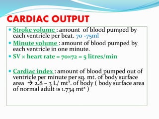
- 1. CARDIAC OUTPUT Stroke volume : amount of blood pumped by each ventricle per beat. 70 -75ml Minute volume : amount of blood pumped by each ventricle in one minute. SV × heart rate = 70×72 = 5 litres/min Cardiac index : amount of blood pumped out of ventricle per minute per sq. mt. of body surface area 2.8 – 3 L/ mt2. of body ( body surface area of normal adult is 1.734 mt2 )
- 3. CARDIAC RESERVE maximum increase in cardiac output above normal value. e.g. excercise 300 – 400 % in adults In cardiac disease : reserve decrease or no reserve
- 4. FACTORS AFFECTING CARDIAC OUTPUT Physiological: 1. Age 2. Gender 3. Diurnal variation 4. Environmental temperature 5. Emotions 6. Exercise, high altitude 7. Posture 8. Pregnancy 9. Sleep
- 5. PATHOLOGICAL FACTORS Increased CO: Fever Anemia Hyperthyroidism Decreased CO: Hypothyroidism Heart block Heart failure Shock Hemorrhage
- 6. DISTRIBUTION OF CARDIAC OUTPUT Liver : 1400 ml - 25% Kidneys : 1300 ml - 25% Skin, Subcutaneous tissue, Skeletal muscles : 1200ml - 25% Heart, lungs, brain : 1300 ml - 25%
- 7. REGULATION / FACTORS AFFECTING / DETERMITANTS OF CO CO = SV x HR I. Venous return II. Peripheral resistance control of SV (Intrinsic R) III. Force of contraction IV. Frequency of heart rate (Extrinsic R)
- 8. VENOUS RETURN (preload) Frank Starling’s law Increased venous return, increases cardiac output Respiratory pump : during inspiration, venous return increased, CO increases. Muscle pump : during exercise, venous return increases, CO increases Venomotor tone : sympathetic stimulation decrease volume of capacitance vessels increased VR increase CO. Gravity – upright posture decrease VR Cardiac pump -
- 9. PERIPHERAL RESISTANCE Resistance offered by vessel wall Load (after load) against which heart has to pump the blood. Afterload for the left ventricle is determined by aortic pressure Afterload for the right ventricle is determined by pulmonary artery pressure. Increased peripheral resistance (in old age & atherosclerosis) decreases cardiac output. (by decreasing SV)
- 10. FORCE OF CONTRACTION Increase myocardial contractility increase CO. Sympathetic stimulation & increase circulating catecholamines increase force of contraction of myocardium. (Positive ionotropic effect)
- 11. FREQUENCY OF HEART BEAT CO directly proportional to HR (SV x HR) Adult HR is normally 80-100 beats per minute (bpm.) Heart rate is modified by autonomic, immune, and local factors. For example: a. An increase in parasympathetic activity via M2 cholinergic receptors in the heart will decrease the heart rate. b. An increase in sympathetic activity via B1 adrenergic receptors throughout the heart will increase the heart rate. But In extreme tachycardia, stroke volume decreased (less diastolic filling), cardiac output decrease.
- 13. MEASUREMENT OF CARDIAC OUTPUT A) Direct methods (animals): Cardiometer, flowmeter B) Indirect methods : 1. Fick’s principle 2. Dye dilution method 3. Thermodilution method 4. Echocardiography
- 14. FICK’S PRINCIPLE Adolf Eugen Fick ( 1829 – 1901) Amount of substance taken per minute = (A-V) diff. of substance blood flow/min Blood flow/min = amount of subst. taken per min (A-V) diff. of that substance
- 15. 1) OXYGEN CONSUMPTION 2) CO2 EVOLVED 1)CARDIAC = O2 CONSUMED (ml/min) OUTPUT ARTERIOVENOUS DIFF. O2 CONSUMED = BENEDICT ROTH APPARATUS O2 CONTENT IN ARTERIAL BLD = FROM ARTERY O2 CONTENT IN VENOUS BLOOD = CARDIAC CATHETERISATION CARDIAC OUTPUT = 250 * 100 = 5000ml/ min 20-15/100 ml 5
- 17. DYE DILUTION METHOD Based on how fast the flowing blood can dilute the substances introduced into the circulation BLOOD FLOW = q C (t2-t1) q = CONCENTRATION OF DYE INJECTED C = MEAN CONC. OF DYE t1 = APPEARANCE OF DYE t2 = DISAPEARANCE OF DYE
- 19. THERMAL DILUTION METHOD Indicator : cold saline injected in to RA Temp. change in blood measure in aorta by thermostate Temperature change in blood is inversely related to blood flow from aorta. (depend on extent to which cold saline diluted)
- 20. ECHOCARDIOGRAPHY Pulses of ultrasonic wave are used frequency 2.25MHz Ultrasonic waves are emitted from a transdusers which also act as receiver to detect waves echo from different parts of heart. Echoes are displaced against time on osciloscope. Recording of movements of ventricular wall, septum, heart valves. Measurement of EDV, ESV, SV & ejection fraction.
- 21. BLOOD PRESSURE “ Lateral pressure exerted by column of blood on wall of blood vessels ” Systolic pressure : maximum pressure during systole = 100 to 140 mmHg Diastolic pressure : minimum pressure during diastole = 70 to 90 mmHg Pulse pressure = SBP - DBP Mean BP = DP + 1/3 PP
- 22. Functions : 1. To maintain sufficient pressure to keep blood flowing through the blood vessels. 2. To provide force of filtration at the capillaries.
- 23. Normal blood pressure in different portion of circulatory system
- 24. VARIATIONS IN BP Physiological factors affecting : 1. Age – SBP & DBP increase with age 2. Gender – male SBP more than female 3. Obesity – increase SBP & DBP 4. Diurnal variation – peak value in the evening 5. After meals – increase SBP 6. Sleep – decrease SBP 7. Exercise, emotions – increase SBP • Pathological : Hypertension : increased BP Hypotension : decreased BP
- 25. FACTORS AFFECTING BP 1. Cardiac output 2. Heart rate 3. Peripheral resistance 4. Blood volume 5. Elasticity of blood vessels 6. Velocity of blood Flow 7. Diameter of blood Vessel 8. Viscosity of blood
- 26. BP = CO PR Cardiac output : Increased co, increases SBP Peripheral resistance: Resistance offered in arterioles Increase SBP Elasticity of blood Vessels : inversely proportional Blood volume : directly proportional Venous return : directly proportional Velocity : inversely proportional (Bernoulli’s principle – in tube or blood vessel some of kinetic energy of flow & pressure energy is constant) Diameter of blood vessel : inversely prop. Viscosity : inversely proportional.
- 27. REGULATION OF BP Short term regulation : nervous regulation Intermediate regulation Long term regulation : renal mechanism Hormonal regulation Local regulation
- 28. SHORT TERM REGULATION Nervous regulation can increase arterial BP to double within 5-10 S & reduce to half within 10-40s Vasomotor center (VMC) : in medulla (anterolateral part) sympathetic outflow to CVS (vasoconstrictor area-upper/ vasodilator area-lower ) Cardiac vagal centre : in medulla in neucleus ambigus parasympathetic outflow to CVS (via vagus nerve) Nucleus of tractus solitarius : receiving sensory information from aortic & carotid baroreceptors & chemoreceptors.
- 32. Continuous Partial Constriction of the Blood Vessels Is Normally Caused by Sympathetic Vasoconstrictor Tone.
- 33. FACTORS AFFECTING VMC 1. BARORECEPTOR REFLEX 2. CHEMORECEPTORS REFLEX 3. CNS ISCHEMIC REFLEX 4. HIGHER CENTERS
- 34. BARORECEPTOR REFLEX Spray type nerve endings Stretch receptors at bifurcation of common carotid A & arch of aorta. Respond to change in mean BP Carotid sinus : IX cranial nerve from CCA BR Arch of aorta : X cranial nerve from AA BR Function: Increased BP -> baroreceptors activated -> suppresses VMC -> Stimulates cardioinhibitory center -> vasodilatation -> decreased BP Response mainly to rapidly changing pressure than to a stationery Pressure
- 37. CHEMORECEPTORS Carotid body & aortic body. Nerve supply via IX and X nerves. Respond to change in chemicals in blood (PaO2 , PaCO2 , H+ ION concentration) Decrease BP (<80mmHg) decrease blood supply decrease O2 & increase CO2 stimulate chemoreceptors that excites VMC increase BP
- 38. CNS ISCHEMIC REFLEX Emergency pressure control system operates between 15-50 mmHg SBP Cerebral ischemia strong sympathetic stimulation to increase blood pressure up to 250 mmHg for 10-15 minutes. “Last ditch stand” mechanism Cushing’s reaction
- 39. HIGHER CENTERS In response to emotion Cerebral cortex area 13 (limbic A.) To Hypothalamus (cortico - hypothalamic descending pathway) posterolateral portion of hypothalamus causes excitation of VMC. Anterior hypothalamus cause mild excitation or inhibition.
- 40. Intermediate regulation Begin to act within few minutes & reach full function within a few hours. I. Capillary fluid shift mechanism – mean capillary pressure directly proportional to ABP change in fluid filtration & reabsorption at capillary level. II. Stress relaxation & reverse stress relaxation mechanism : by local vascular tone adjustment in blood storage organ like veins, liver, spleen, lungs in response to change in ABP
- 41. LONG TERM (RENAL) REGULATION OF BP By regulation of extracellular fluid volume * ABP more excretion of water and salt by kidneys Pressure diuresis (water exc.) Pressure natriuresis (Na+ exc.) ABP retention of salt and water by kidneys (by direct mechanism & indirect mechanism – renin angiotensin mechanism)
- 42. SEC. OF ALDOSTERONE Renin angiotensin mechanism Presure diuresis & pressure natriuresis
- 43. HORMONAL REGULATION Increase BP Adrenaline, noradrenaline Thyroxin Aldosterone Vasopressin Angiotensin Serotonin
- 44. Decreased BP VIP Bradykinin Prostaglandins Histamine Acetylcholine Atrial natriuretic peptide
- 45. MEASUREMENT OF BP DIRECT : insertion of canulla in to artery & connect it to manometer. INDIRECT : by Sphygmomanometer – two methods # palpatory # auscultatory
- 46. HYPERTENSION Hypertension is a sustain increase of systemic arterial blood pressure. Two types : Primary or essential HT: benign, malignant Secondary HT: 1. CVS - atherosclerosis 2. Endocrine - Cushing syndrome 3. Renal - tumour of JG cells & stenosis of renal arteries 4. Neurogenic - increased intracranial pressure (Cushing’s reaction) 5. During pregnancy (eclampsia)
- 48. Primary or essential HT : When ABP is persistently more than 150/90 mmHg benign : in early stages increase up to 210/110 during stress condition. In late stages remain above 210/110. Malignant : ABP increase up to 260/150. so death occurs within 6M to 2Yr. Compensatory cardiac hypertrophy Thickening of wall of small arteries & arterioles. Myocardial infarction Renal failure
- 49. MANIFESTATIONS Renal failure Left ventricular failure Myocardial infarction Cerebral hemorrhage Retinal hemorrhage
- 50. TREATMENT Beta blockers – B1 blockers – Atenolol, Metoprolol Calcium channel blockers – Verapamil, Amlodipin Vasodilators- Nitroglycerin Diuretics – Furosemide, Thiazide, Spironolactone ACE inhibitors – Analapril, Captopril