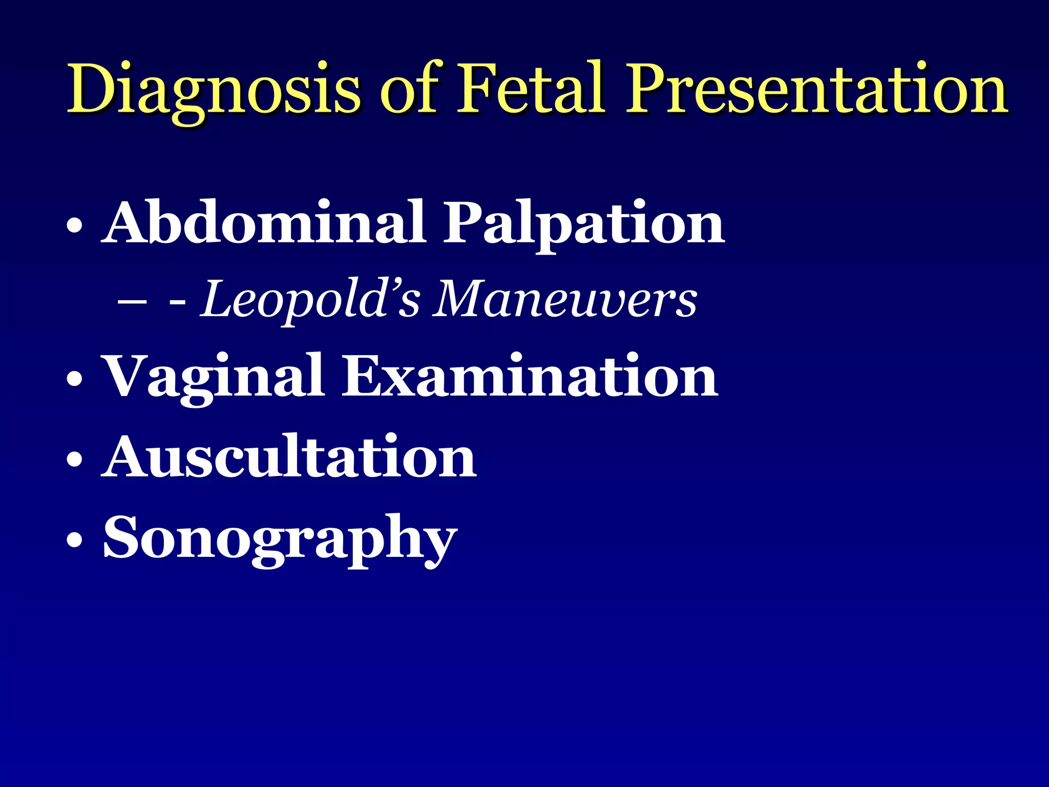
Intrapartum Care & Abnormal Labor Francis S. Nuthalapaty, MD Medical Student Lecture Series Department of Obstetrics & Gynecology Greenville Hospital System University Medical Center Greenville, South Carolina
Learning Objectives APGO ET-11: Intrapartum Care Understanding the process of normal labor and delivery allows optimal care and reassurance for the parturient and timely recognition of abnormal events. APGO Medical Student Educational Objectives 8 th  Ed. 2004
Learning Objectives ET-11 APGO Medical Student Educational Objectives 8 th  Ed. 2004 KH Describe the evaluation of common puerperal complications KH Describe the different methods of delivery with the indications and contraindications of each SH Describe the steps of a vaginal delivery KH Describe the three stages of labor and recognize common abnormalities KH List the signs and symptoms of labor Level of Competence Objective
Learning Objectives APGO Educational Topic 22: Abnormal Labor Labor is expected to progress in an orderly and predictable manner. Careful observation of the mother and fetus during labor will allow early detection of abnormalities so that management can be directed to optimize outcome. APGO Medical Student Educational Objectives 8 th  Ed. 2004
Learning Objectives ET-22 APGO Medical Student Educational Objectives 8 th  Ed. 2004 K List indications and contraindications for oxytocin administration K Discuss fetal and maternal complications of abnormal labor K Describe methods of fetal surveillance K List abnormal  labor patterns Level of Competence Objective
Learning Objectives ET-22 APGO Medical Student Educational Objectives 8 th  Ed. 2004 K Discuss strategies for emergency management  of breech, shoulder dystocia and cord prolapse K List indications for VBAC Level of Competence Objective
Terminology Gravidity #of  current  and  completed  pregnancies of any kind Parity # of  completed  pregnancies ≥ 20 weeks not delivered infants (e.g. twins)
Terminology Nullipara Primipara Multipara Grand Multipara
Terminology TPAL Nomenclature: T =  Term deliveries ≥ 37 wks P =  Preterm deliveries < 37 wks A =  Abortions (< 20 wks) L =  Living children
G3/P1-0-1-1: Terminology
G3/P1-0-1-1: 3rd Pregnancy 1 Term delivery 0 Preterm deliveries 1 Abortion 1 Living child Terminology
G5/P2-1-1-0: Terminology
G5/P2-1-1-0: 5th Pregnancy 2 Term deliveries 1 Preterm delivery 1 Abortion 0 Living children Terminology
G2/P0-2-0-3: Terminology
G2/P0203: 2nd Pregnancy 0 Term deliveries 2 Preterm deliveries  0 Abortions 3 Living children Terminology
Fetal Presentation Attitude Fetal Lie Fetal Position Fetal Station Terminology
Designates the fetal part over the pelvic inlet Fetal Presentation
Fetal Presentation Presentation %   Incidence Cephalic 96.8 --- Breech 2.7 1:36 Transverse 0.3 1:335 Compound 0.1 1:1000 Face 0.05 1:2000 Brow 0.01 1:10,000 Williams Obstetrics, 21 st  Ed. 452.
 
Cephalic Presenting Diameters FACE SINCIPUT MILITARY BROW VERTEX FLEXED Figure 9-2 Lateral view of the fetal skull showing the prominent landmarks and the anteroposterior diameters.
The degree of flexion a fetus assumes during labor or the relation of the fetal parts to each other Fetal Attitude
Variations in Fetal Attitude SINCIPUT, MILITARY BROW FACE VERTEX, FLEXED
Fetal Lie Refers to the relation of the long axis of fetus (back) to the long axis of the mother: ------ Transverse ------ -------------- Longitudinal ----------------
Diagnosis of Fetal Presentation Abdominal Palpation -  Leopold’s Maneuvers
 
Diagnosis of Fetal Presentation Abdominal Palpation -  Leopold’s Maneuvers Vaginal Examination Auscultation Sonography
Fetal Position Refers to the relation of an arbitrarily chosen portion of the fetal presenting part to the right or left side of the maternal birth canal Reference points (denominators) are:  Fetal occiput Fetal chin (mentum) Fetal sacrum
Fetal Head:  Landmarks Figure 9-1 Superior view of the fetal skull showing the sutures, fontanelles, and transverse diameters.
Occiput Presentation
Fetal Position  OP LOT OA ROT LOP ROP LOA ROA LOT:  40% ROT:  20% OP:  20%
Fetal Position  ?????
Fetal Position  Left Occiput Anterior
Fetal Position  ?????
Fetal Position  Right  Occiput Posterior
Fetal Position  ?????
Fetal Position  Left Occiput Transverse
The relationship of the fetal presenting part to the level of the ischial spines Fetal Station
Fetal Station World Health Organization:  Managing Complications in Pregnancy and Childbirth  www.who.int/reproductivehealth/impac/Clinical_Principles/Normal_labour_C57_C76.html
Clinical Correlation Correct identification of fetal position relative to the birth canal is critical! Document the following: Fetal Lie  Fetal Presentation   Fetal Position Fetal Station
Labor
Terminology False Labor (Braxton-Hicks ctx) May be present from first trimester Irregular, nonrhythmic True Labor Rhythmic contractions with cervical change
Essential Factors of Labor (The 3 P’s) Passage Powers Passenger
The 3 P’s of Labor Passage
Passage Bony Pelvis inlet midpelvis outlet Soft Tissue
Caldwell-Moloy Classification A P Gynecoid A P Android P A Platypelloid P A Anthropoid
Caldwell-Moloy Classification Gynecoid = 40 – 50% (10-15% AA) Android  = 30% Anthropoid  = 20% (40% in AA) Platypelloid  = 2- 5%
Gynecoid Pelvis Round at the inlet, with the widest transverse diameter only slightly greater than the anteroposterior diameter Side walls straight Ischial spines of average prominence Well-rounded sacrosciatic notch Well-curved sacrum Spacious subpubic arch, with an angle of approximately 90 degrees
Android Pelvis Triangular inlet with a flat posterior segment and the widest transverse diameter closer to the sacrum than in the gynecoid type Convergent side walls with prominent spines Shallow sacral curve Long and narrow sacrosciatic notch Narrow subpubic arch
Anthropoid Pelvis A much larger AP than transverse diameter, creating a long narrow oval at the inlet Side walls that do not converge Ischial spines that are not prominent but are close, owing to the overall shape Variable, but usually posterior, inclination of the sacrum Large sacrosciatic notch Narrow, outwardly shaped subpubic arch
Pelvic Landmarks - Inlet Sacral promontory Illiopectineal line Symphysis pubis
mywebpages.comcast.net/wnor/pelvis.htm  --Wesley Norman, PhD, DSc Georgetown University Pelvic Landmarks - Inlet
Pelvic Landmarks - Inlet
Vaginal Examination to Determine the Diagonal Conjugate
Pelvic Landmarks - Mid Ischial spines Sacrum Sacrosciatic notch
mywebpages.comcast.net/wnor/pelvis.htm  --Wesley Norman, PhD, DSc Georgetown University Pelvic Landmarks - Mid
Assessment of Mid-pelvis
Pelvic Landmarks - Outlet Pubic arch Ischial tuberosities Sacrococcygeal joint
Pelvic Landmarks - Outlet Figure 9-4 Pelvic outlet and its diameters.
Assessment of Pelvic Outlet
Minimum Pelvimetrics * Average measurement, no minimum defined 8 Bituberous Outlet 10.5* Bispinous Midplane 11.5 Diagonal conjugate   Inlet Length (cm) Diameter Pelvic Plane
The 3 P’s of Labor Powers
Powers 50 mm Hg or more Contractions occur q 2-3 minutes Upper uterus more active with pacemakers at cornual
The 3 P’s of Labor Passenger
Passenger Head is typically the largest structure Molding Smallest diameter of head: suboccipitobregmatic Abnormal lie or size or presentation can cause problems
Passenger: Fetal Head Considerations Bones in face fused but cranial vault has movable bones Molding  is when bones overlap under pressure Sutures   are membranous spaces between bones Fontanelles   or “soft spots” are the intersections between sutures
Molding
Passage and Passenger Relationship Engagement  –the fetal head is thru the pelvic inlet when fetal BPD reaches the ischial spines
Passage and Passenger Relationship Station  - descent of the fetal BPD, relative to the level of the ischial spines.
COURSE OF NORMAL LABOR
Labor Definition The physiologic process by which the gravid uterus evacuates its contents at or near term by a mechanism involving coordinated  sequence of periodic contractions of the myometrium effecting progressive cervical dilatation and fetal descent through the birth canal. -  Emanuel A. Friedmin
First Stage of Labor Onset of true labor until cervix fully dilated:   Latent Phase   Active Phase
First Stage of Labor
 
First Stage of Labor
Second Stage of Labor Complete dilatation to delivery of the infant
Cardinal Movements of Labor Engagement, descent, flexion Internal rotation Complete rotation with beginning of extension Complete extension
External Rotation (Restitution) External rotation with delivery of Anterior shoulder Expulsion with delivery of Posterior shoulder
Third Stage of Labor Delivery of the infant to delivery of the placenta
Third Stage of Labor Placental separation: Uterus becomes firmer Gush of blood Uterus rises in abdomen as placenta passes into lower segment Lengthening of umbilical cord
Power:  Forces of Labor First Stage of Labor Primary Forces = Uterine ctx to dilate cervix to 10 cm Second Stage of Labor Secondary Forces = Abdominal muscles to push fetus out after cervix is fully dilated Third Stage of Labor - Primary Forces = Uterine ctx to deliver placenta
INTRAPARTUM MANAGEMENT
Fetal Heart Rate Monitoring External:   Indirect - Doppler ultrasound Internal: Direct – Scalp electrode
Fetal Heart Rate Monitoring External: Assessment of: baseline, variability, accelerations, decelerations Limitations: Maternal Body Habitus Maternal/Fetal Movement Artifact
Fetal Heart Rate Monitoring Internal: Assessment of: baseline, variability, accelerations, decelerations Limitations: Membranes must be ruptured Minimally invasive Increase risks of Hep B/HIV if mother + Maternal HR detected if FHR absent
Contraction Monitoring External: Indirect:  Abdominal pressure electrode Internal: Direct: IUPC with pressure sensor
Contraction Monitoring External:   -   Can detect presence and interval of ctx,    but not strength -   Limitations:  Maternal Body Habitus Maternal/Fetal Movement Artifact Factitious contractions
Contraction Monitoring Internal:   -   Assessment of BOTH frequency and    intensity of contractions -  Limitations:  Membranes must be ruptured Minimally invasive
Intrapartum Monitors
Fetal Heart Rate Interpretation Assessment Quality of tracing Baseline fetal heart rate Describe overall variability Presence of accelerations? Presence of decelerations? Contraction frequency/intensity


Fetal Heart Rate Tracing
Fetal Monitoring Guidelines
Normal Labor Parameters Source: Modified from Friedman EA. Labor: Clinical Evaluation and Management, 2 nd  ed. New York. Appletion-Century-Cronz 1370.43 2.5 h 57 min Second Stage 1.2 cm/h 3.0 cm/h Maximum Slope 3.3 h 54 min Deceleration 11.5 h 4.9 h Active 20.6 h 8.6 h Latent Lower Limit (95%) Mean Nulliparous Labor
Normal Labor Parameters Source: Modified from Friedman EA. Labor: Clinical Evaluation and Management, 2 nd  ed. New York. Appletion-Century-Cronz 1370.43 50 min 14 min Second Stage 1.5 cm/h 5.7 cm / h Maximum Slope 53 min 14 min Deceleration 5.2 h 2.2 h Active 13.6 h 5.3 h Latent Lower Limit (95%) Mean Multiparous Labor
Abnormal Labor Assessment  Clinical Caveat Labor dystocia requires a close assessment of the 3-P’s to determine the etiology and implement appropriate management changes to address the problem identified.
Adequate Labor Defined as > 200 Montevideo units (MVU) as measured by IUPC MVU = Sum of contraction strength for each contraction occurring over 10 minutes
MVUs = ????
MVUs = 270 “Adequate Labor”
7 Labor Dysfunctions Prolonged Latent Phase Definition: > 20 hours nullipara > 14 hours multipara Treatment: “ Therapeutic rest” = sedatives 85% awaken in 6-10 hours and progress to active phase 10% have stopped contracting 5% continue to contract without progression, requiring uterine stimulation. Oxytocin
7 Labor Dysfunctions Protracted Active Phase Definition: Cervical dilation < 1.2 cm/h nullipara Cervical dilation < 1.4 cm/h multipara Treatment: Evaluate passenger, passageway, power IUPC to calculate MVU (goal > 200) Oxytocin augmentation
7 Labor Dysfunctions Protracted Deceleration Phase Definition: > 3 hours nullipara > 1 hour multipara Treatment: Same as for protracted active phase Evaluate passenger, passageway, power IUPC to calculate MVU (goal > 200) Oxytocin augmentation
7 Labor Dysfunctions Secondary Arrest of Dilatation in Active phase Definition: Absence of cervical change over 2 hours* MVU > 200 Treatment: Cesarean delivery * Extension to 4 hours results in higher rate of vaginal delivery (92%) and is also acceptable
7 Labor Dysfunctions Protracted Descent Definition: < 1 cm/h nullipara < 2 cm/h multipara Treatment: Same as for protracted active phase Evaluate passenger, passageway, power IUPC to calculate MVU (goal > 200) Oxytocin augmentation
7 Labor Dysfunctions Arrest of Descent in Second Stage Definition: No descent of presenting part in: > 2 hours (or > 3 hours with CLE) nullipara > 1 hour (or > 2 hours with CLE) multipara Treatment: Continued observation Operative vaginal delivery Cesarean delivery
7 Labor Dysfunctions Failure of Descent Definition: No descent in > 1 hour nullipara No descent in > 30 min multipara Treatment: Same as for protracted active phase Evaluate passenger, passageway, power IUPC to calculate MVU (goal > 200) Oxytocin augmentation
Labor Assessment  Case 1 32 yo G1P0 36 weeks presented with contractions.  Looks uncomfortable, and is contracting every 3 minutes but cervix is 2 cm and 50% effaced. Was seen the previous day with similar complaints and findings. Diagnosis: Management:
Labor Assessment  Case 1 32 yo G1P0 36 weeks presented with contractions.  Looks uncomfortable, and is contracting every 3 minutes but cervix is 2 cm and 50% effaced. Was seen the previous day with similar complaints and findings. Diagnosis: Prolonged latent phase Management: “ Therapeutic Rest”
Labor Assessment  Case 2 24 yo P1001 39 weeks presented in labor.  Contracting every 3 minutes but looks comfortable.  Progressed from 4 to 6 centimeters in 6 hours.  Membranes intact.  Estimated fetal weight – 3000 grams.  Pelvis adequate on examination.  Vertex presentation.    Diagnosis: Management:
Labor Assessment  Case 2 24 yo P1001 39 weeks presented in labor.  Contracting every 3 minutes but looks comfortable.  Progressed from 4 to 6 centimeters in 6 hours.  Membranes intact.  Estimated fetal weight – 3000 grams.  Pelvis adequate on examination.  Vertex presentation.    Diagnosis: Protracted active phase likely secondary to inadequate labor (insufficient power) Management: Amniotomy, Oxytocin augmentation +/- IUPC
Labor Assessment  Case 3 32 yo P0000 Class C diabetic at 40 weeks undergoing labor induction.  Contracting every 2-3 minutes.  7 cm dilation x 4 hours.  Confirmed adequate labor with intrauterine pressure catheter.  Membranes ruptured,  Estimated fetal weight – 4200 grams.  Pelvis adequate on examination. Vertex presentation.  Diagnosis:   Management:
Labor Assessment  Case 3 32 yo P0000 Class C diabetic at 40 weeks undergoing labor induction.  Contracting every 2-3 minutes.  7 cm dilation x 4 hours.  Confirmed adequate labor with intrauterine pressure catheter.  Membranes ruptured,  Estimated fetal weight – 4200 grams.  Pelvis adequate on examination. Vertex presentation.  Diagnosis: Arrest of dilatation likely secondary to cephalopelvic disproportion/fetal macrosomia (Passenger too big for pelvis) Management:   Cesarean Delivery
Labor Assessment  Case 4 28 yo P0101 at 42 weeks presented in labor.  History of previous MVA with pelvic fracture.  Contracting every 2-3 minutes.  6 cm dilation x 4 hours.  Confirmed adequate labor with intrauterine pressure catheter.  Membranes ruptured,  Estimated fetal weight – 3200 grams.  Constricted pelvic inlet with non-engaged fetal head. Vertex presentation.  Diagnosis:   Management:
Labor Assessment  Case 4 28 yo P0101 at 42 weeks presented in labor.  History of previous MVA with pelvic fracture.  Contracting every 2-3 minutes.  6 cm dilation x 4 hours.  Confirmed adequate labor with intrauterine pressure catheter.  Membranes ruptured,  Estimated fetal weight – 3200 grams.  Constricted pelvic inlet with non-engaged fetal head. Vertex presentation.  Diagnosis:   Arrest of dilatation likely secondary to cephalopelvic disproportion/abnormal pelvis (Pelvis too small for pelvis) Management:   Cesarean Delivery

Intrapartum Care and Abnormal Labor

  • 1.
    Intrapartum Care &Abnormal Labor Francis S. Nuthalapaty, MD Medical Student Lecture Series Department of Obstetrics & Gynecology Greenville Hospital System University Medical Center Greenville, South Carolina
  • 2.
    Learning Objectives APGOET-11: Intrapartum Care Understanding the process of normal labor and delivery allows optimal care and reassurance for the parturient and timely recognition of abnormal events. APGO Medical Student Educational Objectives 8 th Ed. 2004
  • 3.
    Learning Objectives ET-11APGO Medical Student Educational Objectives 8 th Ed. 2004 KH Describe the evaluation of common puerperal complications KH Describe the different methods of delivery with the indications and contraindications of each SH Describe the steps of a vaginal delivery KH Describe the three stages of labor and recognize common abnormalities KH List the signs and symptoms of labor Level of Competence Objective
  • 4.
    Learning Objectives APGOEducational Topic 22: Abnormal Labor Labor is expected to progress in an orderly and predictable manner. Careful observation of the mother and fetus during labor will allow early detection of abnormalities so that management can be directed to optimize outcome. APGO Medical Student Educational Objectives 8 th Ed. 2004
  • 5.
    Learning Objectives ET-22APGO Medical Student Educational Objectives 8 th Ed. 2004 K List indications and contraindications for oxytocin administration K Discuss fetal and maternal complications of abnormal labor K Describe methods of fetal surveillance K List abnormal labor patterns Level of Competence Objective
  • 6.
    Learning Objectives ET-22APGO Medical Student Educational Objectives 8 th Ed. 2004 K Discuss strategies for emergency management of breech, shoulder dystocia and cord prolapse K List indications for VBAC Level of Competence Objective
  • 7.
    Terminology Gravidity #of current and completed pregnancies of any kind Parity # of completed pregnancies ≥ 20 weeks not delivered infants (e.g. twins)
  • 8.
    Terminology Nullipara PrimiparaMultipara Grand Multipara
  • 9.
    Terminology TPAL Nomenclature:T = Term deliveries ≥ 37 wks P = Preterm deliveries < 37 wks A = Abortions (< 20 wks) L = Living children
  • 10.
  • 11.
    G3/P1-0-1-1: 3rd Pregnancy1 Term delivery 0 Preterm deliveries 1 Abortion 1 Living child Terminology
  • 12.
  • 13.
    G5/P2-1-1-0: 5th Pregnancy2 Term deliveries 1 Preterm delivery 1 Abortion 0 Living children Terminology
  • 14.
  • 15.
    G2/P0203: 2nd Pregnancy0 Term deliveries 2 Preterm deliveries 0 Abortions 3 Living children Terminology
  • 16.
    Fetal Presentation AttitudeFetal Lie Fetal Position Fetal Station Terminology
  • 17.
    Designates the fetalpart over the pelvic inlet Fetal Presentation
  • 18.
    Fetal Presentation Presentation% Incidence Cephalic 96.8 --- Breech 2.7 1:36 Transverse 0.3 1:335 Compound 0.1 1:1000 Face 0.05 1:2000 Brow 0.01 1:10,000 Williams Obstetrics, 21 st Ed. 452.
  • 19.
  • 20.
    Cephalic Presenting DiametersFACE SINCIPUT MILITARY BROW VERTEX FLEXED Figure 9-2 Lateral view of the fetal skull showing the prominent landmarks and the anteroposterior diameters.
  • 21.
    The degree offlexion a fetus assumes during labor or the relation of the fetal parts to each other Fetal Attitude
  • 22.
    Variations in FetalAttitude SINCIPUT, MILITARY BROW FACE VERTEX, FLEXED
  • 23.
    Fetal Lie Refersto the relation of the long axis of fetus (back) to the long axis of the mother: ------ Transverse ------ -------------- Longitudinal ----------------
  • 24.
    Diagnosis of FetalPresentation Abdominal Palpation - Leopold’s Maneuvers
  • 25.
  • 26.
    Diagnosis of FetalPresentation Abdominal Palpation - Leopold’s Maneuvers Vaginal Examination Auscultation Sonography
  • 27.
    Fetal Position Refersto the relation of an arbitrarily chosen portion of the fetal presenting part to the right or left side of the maternal birth canal Reference points (denominators) are: Fetal occiput Fetal chin (mentum) Fetal sacrum
  • 28.
    Fetal Head: Landmarks Figure 9-1 Superior view of the fetal skull showing the sutures, fontanelles, and transverse diameters.
  • 29.
  • 30.
    Fetal Position OP LOT OA ROT LOP ROP LOA ROA LOT: 40% ROT: 20% OP: 20%
  • 31.
  • 32.
    Fetal Position Left Occiput Anterior
  • 33.
  • 34.
    Fetal Position Right Occiput Posterior
  • 35.
  • 36.
    Fetal Position Left Occiput Transverse
  • 37.
    The relationship ofthe fetal presenting part to the level of the ischial spines Fetal Station
  • 38.
    Fetal Station WorldHealth Organization: Managing Complications in Pregnancy and Childbirth www.who.int/reproductivehealth/impac/Clinical_Principles/Normal_labour_C57_C76.html
  • 39.
    Clinical Correlation Correctidentification of fetal position relative to the birth canal is critical! Document the following: Fetal Lie Fetal Presentation Fetal Position Fetal Station
  • 40.
  • 41.
    Terminology False Labor(Braxton-Hicks ctx) May be present from first trimester Irregular, nonrhythmic True Labor Rhythmic contractions with cervical change
  • 42.
    Essential Factors ofLabor (The 3 P’s) Passage Powers Passenger
  • 43.
    The 3 P’sof Labor Passage
  • 44.
    Passage Bony Pelvisinlet midpelvis outlet Soft Tissue
  • 45.
    Caldwell-Moloy Classification AP Gynecoid A P Android P A Platypelloid P A Anthropoid
  • 46.
    Caldwell-Moloy Classification Gynecoid= 40 – 50% (10-15% AA) Android = 30% Anthropoid = 20% (40% in AA) Platypelloid = 2- 5%
  • 47.
    Gynecoid Pelvis Roundat the inlet, with the widest transverse diameter only slightly greater than the anteroposterior diameter Side walls straight Ischial spines of average prominence Well-rounded sacrosciatic notch Well-curved sacrum Spacious subpubic arch, with an angle of approximately 90 degrees
  • 48.
    Android Pelvis Triangularinlet with a flat posterior segment and the widest transverse diameter closer to the sacrum than in the gynecoid type Convergent side walls with prominent spines Shallow sacral curve Long and narrow sacrosciatic notch Narrow subpubic arch
  • 49.
    Anthropoid Pelvis Amuch larger AP than transverse diameter, creating a long narrow oval at the inlet Side walls that do not converge Ischial spines that are not prominent but are close, owing to the overall shape Variable, but usually posterior, inclination of the sacrum Large sacrosciatic notch Narrow, outwardly shaped subpubic arch
  • 50.
    Pelvic Landmarks -Inlet Sacral promontory Illiopectineal line Symphysis pubis
  • 51.
    mywebpages.comcast.net/wnor/pelvis.htm --WesleyNorman, PhD, DSc Georgetown University Pelvic Landmarks - Inlet
  • 52.
  • 53.
    Vaginal Examination toDetermine the Diagonal Conjugate
  • 54.
    Pelvic Landmarks -Mid Ischial spines Sacrum Sacrosciatic notch
  • 55.
    mywebpages.comcast.net/wnor/pelvis.htm --WesleyNorman, PhD, DSc Georgetown University Pelvic Landmarks - Mid
  • 56.
  • 57.
    Pelvic Landmarks -Outlet Pubic arch Ischial tuberosities Sacrococcygeal joint
  • 58.
    Pelvic Landmarks -Outlet Figure 9-4 Pelvic outlet and its diameters.
  • 59.
  • 60.
    Minimum Pelvimetrics *Average measurement, no minimum defined 8 Bituberous Outlet 10.5* Bispinous Midplane 11.5 Diagonal conjugate   Inlet Length (cm) Diameter Pelvic Plane
  • 61.
    The 3 P’sof Labor Powers
  • 62.
    Powers 50 mmHg or more Contractions occur q 2-3 minutes Upper uterus more active with pacemakers at cornual
  • 63.
    The 3 P’sof Labor Passenger
  • 64.
    Passenger Head istypically the largest structure Molding Smallest diameter of head: suboccipitobregmatic Abnormal lie or size or presentation can cause problems
  • 65.
    Passenger: Fetal HeadConsiderations Bones in face fused but cranial vault has movable bones Molding is when bones overlap under pressure Sutures are membranous spaces between bones Fontanelles or “soft spots” are the intersections between sutures
  • 66.
  • 67.
    Passage and PassengerRelationship Engagement –the fetal head is thru the pelvic inlet when fetal BPD reaches the ischial spines
  • 68.
    Passage and PassengerRelationship Station - descent of the fetal BPD, relative to the level of the ischial spines.
  • 69.
  • 70.
    Labor Definition Thephysiologic process by which the gravid uterus evacuates its contents at or near term by a mechanism involving coordinated sequence of periodic contractions of the myometrium effecting progressive cervical dilatation and fetal descent through the birth canal. - Emanuel A. Friedmin
  • 71.
    First Stage ofLabor Onset of true labor until cervix fully dilated: Latent Phase Active Phase
  • 72.
  • 73.
  • 74.
  • 75.
    Second Stage ofLabor Complete dilatation to delivery of the infant
  • 76.
    Cardinal Movements ofLabor Engagement, descent, flexion Internal rotation Complete rotation with beginning of extension Complete extension
  • 77.
    External Rotation (Restitution)External rotation with delivery of Anterior shoulder Expulsion with delivery of Posterior shoulder
  • 78.
    Third Stage ofLabor Delivery of the infant to delivery of the placenta
  • 79.
    Third Stage ofLabor Placental separation: Uterus becomes firmer Gush of blood Uterus rises in abdomen as placenta passes into lower segment Lengthening of umbilical cord
  • 80.
    Power: Forcesof Labor First Stage of Labor Primary Forces = Uterine ctx to dilate cervix to 10 cm Second Stage of Labor Secondary Forces = Abdominal muscles to push fetus out after cervix is fully dilated Third Stage of Labor - Primary Forces = Uterine ctx to deliver placenta
  • 81.
  • 82.
    Fetal Heart RateMonitoring External: Indirect - Doppler ultrasound Internal: Direct – Scalp electrode
  • 83.
    Fetal Heart RateMonitoring External: Assessment of: baseline, variability, accelerations, decelerations Limitations: Maternal Body Habitus Maternal/Fetal Movement Artifact
  • 84.
    Fetal Heart RateMonitoring Internal: Assessment of: baseline, variability, accelerations, decelerations Limitations: Membranes must be ruptured Minimally invasive Increase risks of Hep B/HIV if mother + Maternal HR detected if FHR absent
  • 85.
    Contraction Monitoring External:Indirect: Abdominal pressure electrode Internal: Direct: IUPC with pressure sensor
  • 86.
    Contraction Monitoring External: - Can detect presence and interval of ctx, but not strength - Limitations: Maternal Body Habitus Maternal/Fetal Movement Artifact Factitious contractions
  • 87.
    Contraction Monitoring Internal: - Assessment of BOTH frequency and intensity of contractions - Limitations: Membranes must be ruptured Minimally invasive
  • 88.
  • 89.
    Fetal Heart RateInterpretation Assessment Quality of tracing Baseline fetal heart rate Describe overall variability Presence of accelerations? Presence of decelerations? Contraction frequency/intensity
  • 90.
  • 91.
  • 92.
  • 93.
  • 94.
  • 95.
  • 96.
  • 97.
  • 98.
  • 99.
  • 100.
  • 101.
  • 102.
  • 103.
  • 104.
    Normal Labor ParametersSource: Modified from Friedman EA. Labor: Clinical Evaluation and Management, 2 nd ed. New York. Appletion-Century-Cronz 1370.43 2.5 h 57 min Second Stage 1.2 cm/h 3.0 cm/h Maximum Slope 3.3 h 54 min Deceleration 11.5 h 4.9 h Active 20.6 h 8.6 h Latent Lower Limit (95%) Mean Nulliparous Labor
  • 105.
    Normal Labor ParametersSource: Modified from Friedman EA. Labor: Clinical Evaluation and Management, 2 nd ed. New York. Appletion-Century-Cronz 1370.43 50 min 14 min Second Stage 1.5 cm/h 5.7 cm / h Maximum Slope 53 min 14 min Deceleration 5.2 h 2.2 h Active 13.6 h 5.3 h Latent Lower Limit (95%) Mean Multiparous Labor
  • 106.
    Abnormal Labor Assessment Clinical Caveat Labor dystocia requires a close assessment of the 3-P’s to determine the etiology and implement appropriate management changes to address the problem identified.
  • 107.
    Adequate Labor Definedas > 200 Montevideo units (MVU) as measured by IUPC MVU = Sum of contraction strength for each contraction occurring over 10 minutes
  • 108.
  • 109.
    MVUs = 270“Adequate Labor”
  • 110.
    7 Labor DysfunctionsProlonged Latent Phase Definition: > 20 hours nullipara > 14 hours multipara Treatment: “ Therapeutic rest” = sedatives 85% awaken in 6-10 hours and progress to active phase 10% have stopped contracting 5% continue to contract without progression, requiring uterine stimulation. Oxytocin
  • 111.
    7 Labor DysfunctionsProtracted Active Phase Definition: Cervical dilation < 1.2 cm/h nullipara Cervical dilation < 1.4 cm/h multipara Treatment: Evaluate passenger, passageway, power IUPC to calculate MVU (goal > 200) Oxytocin augmentation
  • 112.
    7 Labor DysfunctionsProtracted Deceleration Phase Definition: > 3 hours nullipara > 1 hour multipara Treatment: Same as for protracted active phase Evaluate passenger, passageway, power IUPC to calculate MVU (goal > 200) Oxytocin augmentation
  • 113.
    7 Labor DysfunctionsSecondary Arrest of Dilatation in Active phase Definition: Absence of cervical change over 2 hours* MVU > 200 Treatment: Cesarean delivery * Extension to 4 hours results in higher rate of vaginal delivery (92%) and is also acceptable
  • 114.
    7 Labor DysfunctionsProtracted Descent Definition: < 1 cm/h nullipara < 2 cm/h multipara Treatment: Same as for protracted active phase Evaluate passenger, passageway, power IUPC to calculate MVU (goal > 200) Oxytocin augmentation
  • 115.
    7 Labor DysfunctionsArrest of Descent in Second Stage Definition: No descent of presenting part in: > 2 hours (or > 3 hours with CLE) nullipara > 1 hour (or > 2 hours with CLE) multipara Treatment: Continued observation Operative vaginal delivery Cesarean delivery
  • 116.
    7 Labor DysfunctionsFailure of Descent Definition: No descent in > 1 hour nullipara No descent in > 30 min multipara Treatment: Same as for protracted active phase Evaluate passenger, passageway, power IUPC to calculate MVU (goal > 200) Oxytocin augmentation
  • 117.
    Labor Assessment Case 1 32 yo G1P0 36 weeks presented with contractions. Looks uncomfortable, and is contracting every 3 minutes but cervix is 2 cm and 50% effaced. Was seen the previous day with similar complaints and findings. Diagnosis: Management:
  • 118.
    Labor Assessment Case 1 32 yo G1P0 36 weeks presented with contractions. Looks uncomfortable, and is contracting every 3 minutes but cervix is 2 cm and 50% effaced. Was seen the previous day with similar complaints and findings. Diagnosis: Prolonged latent phase Management: “ Therapeutic Rest”
  • 119.
    Labor Assessment Case 2 24 yo P1001 39 weeks presented in labor. Contracting every 3 minutes but looks comfortable. Progressed from 4 to 6 centimeters in 6 hours. Membranes intact. Estimated fetal weight – 3000 grams. Pelvis adequate on examination. Vertex presentation. Diagnosis: Management:
  • 120.
    Labor Assessment Case 2 24 yo P1001 39 weeks presented in labor. Contracting every 3 minutes but looks comfortable. Progressed from 4 to 6 centimeters in 6 hours. Membranes intact. Estimated fetal weight – 3000 grams. Pelvis adequate on examination. Vertex presentation. Diagnosis: Protracted active phase likely secondary to inadequate labor (insufficient power) Management: Amniotomy, Oxytocin augmentation +/- IUPC
  • 121.
    Labor Assessment Case 3 32 yo P0000 Class C diabetic at 40 weeks undergoing labor induction. Contracting every 2-3 minutes. 7 cm dilation x 4 hours. Confirmed adequate labor with intrauterine pressure catheter. Membranes ruptured, Estimated fetal weight – 4200 grams. Pelvis adequate on examination. Vertex presentation. Diagnosis: Management:
  • 122.
    Labor Assessment Case 3 32 yo P0000 Class C diabetic at 40 weeks undergoing labor induction. Contracting every 2-3 minutes. 7 cm dilation x 4 hours. Confirmed adequate labor with intrauterine pressure catheter. Membranes ruptured, Estimated fetal weight – 4200 grams. Pelvis adequate on examination. Vertex presentation. Diagnosis: Arrest of dilatation likely secondary to cephalopelvic disproportion/fetal macrosomia (Passenger too big for pelvis) Management: Cesarean Delivery
  • 123.
    Labor Assessment Case 4 28 yo P0101 at 42 weeks presented in labor. History of previous MVA with pelvic fracture. Contracting every 2-3 minutes. 6 cm dilation x 4 hours. Confirmed adequate labor with intrauterine pressure catheter. Membranes ruptured, Estimated fetal weight – 3200 grams. Constricted pelvic inlet with non-engaged fetal head. Vertex presentation. Diagnosis: Management:
  • 124.
    Labor Assessment Case 4 28 yo P0101 at 42 weeks presented in labor. History of previous MVA with pelvic fracture. Contracting every 2-3 minutes. 6 cm dilation x 4 hours. Confirmed adequate labor with intrauterine pressure catheter. Membranes ruptured, Estimated fetal weight – 3200 grams. Constricted pelvic inlet with non-engaged fetal head. Vertex presentation. Diagnosis: Arrest of dilatation likely secondary to cephalopelvic disproportion/abnormal pelvis (Pelvis too small for pelvis) Management: Cesarean Delivery