
Recommended
Recommended
More Related Content
Similar to See discussions, stats, and author profiles for this publicati.docx
Similar to See discussions, stats, and author profiles for this publicati.docx (20)
More from edgar6wallace88877
More from edgar6wallace88877 (20)
Recently uploaded
Recently uploaded (20)
See discussions, stats, and author profiles for this publicati.docx
- 1. See discussions, stats, and author profiles for this publication at: https://www.researchgate.net/publication/231740008 Cruciate ligament loading during common knee rehabilitation exercises Article in Proceedings of the Institution of Mechanical Engineers Part H Journal of Engineering in Medicine · September 2012 DOI: 10.1177/0954411912451839 · Source: PubMed CITATIONS 11 READS 2,866 5 authors, including: Some of the authors of this publication are also working on these related projects: 1) Pitching Biomechanics View project Rafael F Escamilla California State University, Sacramento 99 PUBLICATIONS 5,543 CITATIONS
- 2. SEE PROFILE Toran D MacLeod California State University, Sacramento 61 PUBLICATIONS 477 CITATIONS SEE PROFILE James R Andrews American Sports Medicine Institute 188 PUBLICATIONS 9,583 CITATIONS SEE PROFILE All content following this page was uploaded by Toran D MacLeod on 05 April 2015. The user has requested enhancement of the downloaded file. https://www.researchgate.net/publication/231740008_Cruciate_l igament_loading_during_common_knee_rehabilitation_exercise s?enrichId=rgreq-8852a600dafd042a7f3518b6537ffee9- XXX&enrichSource=Y292ZXJQYWdlOzIzMTc0MDAwODtBUz oyMTQ2OTAzMzM0OTkzOTJAMTQyODE5NzU3Mjg2Mg%3D %3D&el=1_x_2&_esc=publicationCoverPdf https://www.researchgate.net/publication/231740008_Cruciate_l igament_loading_during_common_knee_rehabilitation_exercise s?enrichId=rgreq-8852a600dafd042a7f3518b6537ffee9- XXX&enrichSource=Y292ZXJQYWdlOzIzMTc0MDAwODtBUz oyMTQ2OTAzMzM0OTkzOTJAMTQyODE5NzU3Mjg2Mg%3D %3D&el=1_x_3&_esc=publicationCoverPdf
- 5. oyMTQ2OTAzMzM0OTkzOTJAMTQyODE5NzU3Mjg2Mg%3D %3D&el=1_x_7&_esc=publicationCoverPdf https://www.researchgate.net/profile/Toran_MacLeod?enrichId= rgreq-8852a600dafd042a7f3518b6537ffee9- XXX&enrichSource=Y292ZXJQYWdlOzIzMTc0MDAwODtBUz oyMTQ2OTAzMzM0OTkzOTJAMTQyODE5NzU3Mjg2Mg%3D %3D&el=1_x_10&_esc=publicationCoverPdf Special Issue Article Proc IMechE Part H: J Engineering in Medicine 226(9) 670–680 � IMechE 2012 Reprints and permissions: sagepub.co.uk/journalsPermissions.nav DOI: 10.1177/0954411912451839 pih.sagepub.com Cruciate ligament loading during common knee rehabilitation exercises Rafael F Escamilla1, Toran D MacLeod2, Kevin E Wilk3, Lonnie Paulos4 and James R Andrews5,6 Abstract Cruciate ligament injuries are common and may lead to dysfunction if not rehabilitated. Understanding how to progress anterior cruciate ligament and posterior cruciate ligament loading, early after injury or reconstruction, helps clinicians prescribe rehabilitation exercises in a safe manner to enhance recovery. Commonly prescribed therapeutic exercises include both weight-bearing exercise and non-weight-bearing exercise. This review was written to summarize and pro- vide an update on the available literature on cruciate ligament
- 6. loading during commonly used therapeutic exercises. In general, weight-bearing exercise produces smaller loads on the anterior cruciate ligament and posterior cruciate liga- ment compared with non-weight-bearing exercise. The anterior cruciate ligament is loaded less at higher knee angles (i.e. 50–100�). Squatting and lunging with a more forward trunk tilt and moving the resistance pad proximally on the leg during the seated knee extension unloads the anterior cruciate ligament. The posterior cruciate ligament is less loaded at lower knee angles (i.e. 0–50�), and may be progressed from level ground walking to a one-leg squat, lunges, wall squat, leg press, and the two-leg squat (from smallest to greatest). Exercise type and technique variation affect cruciate ligament loading, such that the clinician may prescribe therapeutic exercises to progress ligament loading safely, while ensuring optimal recovery of the musculoskeletal system. Keywords Anterior cruciate ligament, anterior shear force, exercise therapy, reconstruction, strain Date received: 3 May 2011; accepted: 23 March 2012 Introduction Cruciate ligament injuries are common. After sustaining an injury to the cruciate ligaments, or after anterior cruci- ate ligament (ACL) or posterior cruciate ligament (PCL) reconstruction, it is important to properly rehabilitate the tibiofemoral joint to ensure optimal recovery of the heal- ing tissues, keep the joint healthy, and to prevent lower extremity muscle atrophy. Understanding cruciate liga- ment loading during commonly prescribed rehabilitation exercises helps the clinician maximize treatment efficacy and minimize the likelihood of injury.
- 7. The scientific literature on cruciate ligament loading has not been recently reviewed and summarized to give the clinician a current understanding of how ligament loading is quantified, or to give an updated understand- ing of loading across a wider range of exercises than pre- viously described. 1–13 Rehabilitation exercises commonly used include both weight-bearing exercises (WBE), also referred to as closed kinetic chain exercises, and non- weight-bearing exercises (NWBE), also referred to as open kinetic chain exercises. The specific exercises included in this review include squatting, lunging, step- ping (e.g. stepping up and down stairs), leg press, seated knee extension and knee flexion, stair climbing, station- ary bicycling, drop landing, and walking (samples of squatting and lunging exercises are shown in Figures 1 and 2). These exercises were chosen because using these exercises as part of a cruciate ligament rehabilitation 1 Department of Physical Therapy, California State University Sacramento, USA 2 Department of Radiology and Biomedical Imaging, University of California San Francisco, USA 3 Champion Sports Medicine, USA 4
- 8. Paulos Sports Injury and Joint Preservation Clinic, USA 5American Sports Medicine Institute, USA 6 Andrews Institute, USA This paper was submitted as part of the Lower Limb Musculoskeletal Modelling Special Issue. Corresponding author: Rafael F Escamilla, Department of Physical Therapy, California State University Sacramento, 6000 J Street, Sacramento, CA 95819- 6020, USA. Email: [email protected] program after cruciate ligament injury or reconstruction has been shown to significantly improve short- and long- term knee function and enhance a successful return to sport or activity. 14–16 Cruciate ligament loading will be investigated during these exercises with varying resistance, speeds of movement, and techniques. Understanding how the cruciate ligaments are loaded during WBE and NWBE rehabilitation can help clinicians better prescribe train-
- 9. ing and rehabilitation regimens in a safe manner, to enhance recovery and the rehabilitation process. Techniques commonly used to measure cruciate ligament biomechanics Both in-vivo 1–7 and experimental 8–13 biomechanical models have been developed to evaluate ACL strain or tensile force during WBE and NWBE, and both these approaches have advantages and limitations. The obvi- ous advantage of in vivo studies is that they calculate ACL strain directly by using strain sensors within the ACL. The subjects in these in vivo studies were patients that had strain sensors implanted within the anterome- dial bundle of their healthy ACL during arthroscopic surgery to repair damaged knee structures (partial meniscectomy; capsule, and patellofemoral joint debri- dement). Immediately after surgery, these patients were asked to perform a variety of NWBE and WBE, includ- ing lunging, squatting, leg press, step-up and step-down, stair climbing, bicycling, and seated knee extension and knee flexion. The strain within the anteromedial bundle of the ACL was measured and referenced to an instru- mented Lachman Test with 150 N of resistance. There are several limitations to measuring ACL strain in vivo, such as, the procedure is invasive, time consum- ing, costly, performed in a patient population under sur- gical conditions, and that the types of activities are limited. Moreover, the exercise technique employed
- 10. while these patients performed selected WBE was gener- ally not controlled. For example, there are many ways to perform a squat that could affect muscle forces and cruciate ligament loading, such as, using narrow stance or a wide stance, turning the feet in or out, having a near vertical trunk position or tilting the trunk forward 30�– 45� relative to vertical, and during the squat descent moving the knees forward beyond the toes or keeping the knees from moving forward beyond the toes. Another limitation to in vivo studies is that both athletes and non-athletes are employed in performing WBE, gen- erally only body weight or light external resistance is employed during the exercises, and usually only selected knee flexion angles are chosen for ACL strain data col- lection. Therefore, the ability to generalize the results of ACL strain in vivo from studies during WBE to the active athletic population, which comprise the majority of ACL injuries and who often trains with moderate to heavy external resistance over a large knee range of motion, is limited and should be interpreted cautiously. Experimental biomechanical knee models, which also have advantages and limitations, have been previ- ously developed and described. 8,9,11–13,17–21 The advan- tage of using experimental models is that the estimated loads are better generalized to the active athletic popu- lation because variables are often better controlled. For Figure 2. One leg squat. Figure 1. Forward lunge.
- 11. Escamilla et al. 671 example, moderate to heavy resistance could be used during exercise using the experimental model, such is more consistent to how athletes train, but only body weight or light resistance could be used with the in vivo model, because the subjects are all patients that are just coming out of knee surgery. The obvious limitation of experimental biomechanical knee models is that they do not measure ACL loading directly, but only esti- mate its value. However, if the same experimental model is used for all the exercises, it still provides a good relative comparison (assuming that the models are physiologically realistic). Another limitation in using experimental biomechanical knee models is that these models were primarily limited to sagittal plane motion because squatting, lunging, and similar exer- cises are performed primarily in the sagittal plane with only minimal transverse plane rotary motions and fron- tal plane valgus/varus motions. However, performing these types of exercises with excessive transverse plane rotary motions and frontal plane valgus/varus motions could affect cruciate ligament loading, and this should be the focus of future studies. Further, most experimen- tal models are constructed with the assumption that the cruciate ligaments are the only restraints to tibiofe- moral shear forces, and do not account for other soft or hard tissues (meniscus, tibial slope, etc.) that likely play a role. Both in vivo and experimental models have draw- backs, but there is evidence to suggest their validity because several in vivo experiments found similar results to studies using experimental models examining ACL
- 12. loading. Studies using in vivo modeling 1–7 reported peak ACL loading for squatting and lunging of approxi- mately 2.8% to 4% (about 100–150 N) at knee flexion angles between 0� and 30�, corresponding to the peak ACL forces calculated from experimental models 8,9,11– 13,17–21 for the same exercises. This example demon- strates that the magnitude of predicted forces from experimental models are in general agreement with in vivo direct measurement, thus providing some validity to the measurements and suggesting that the two mea- sures may be very cautiously compared between and within techniques. There is another drawback related to the two models, in that the ultimate tensile force is not readily determined using live subjects, representing a potential disagreement between at maximal force lev- els between in vivo and experimental modeling studies. Finally, because in vivo studies only include subjects that have otherwise undergone surgery, the majority of studies use experimental models and, therefore, the majority of the work presented within this article is based upon experimental models. Commonly used graft mechanical properties Both autograft, which is tissue harvested from the patient undergoing surgery, and allograft, which is
- 13. tissue harvested from a cadaver, are commonly used to reconstruct the cruciate ligaments in the United States. 22,23 In healthy adults, the ultimate strength of the native ACL is approximately 2000 N, 24 and the reconstructed ACL has similar ultimate strengths com- pared with the healthy ACL, although these values can change considerably depending on graft type, donor’s age, and donor characteristics (e.g, autograft versus allograft, patellar tendon versus hamstrings graft, etc.). 25 However, the healing graft and graft site may be injured with considerably less force compared with the ultimate strength of the graft, especially when it involves soft tissue to bone fixation, such as the ham- strings graft. The graft must mature, and as the maturation process continues, the ultimate tensile prop- erties of the graft increase in strength. Unfortunately it is not known how much force to the graft site is too much and how soon force can be applied to the healing tissues after reconstruction. However, concerns regard- ing the loading properties of the cruciate ligaments highlight the importance of biomechanical testing of the cruciate ligaments. ACL loading during selected rehabilitation
- 14. exercises Both WBE and NWBE have been employed and shown to be effective in enhancing ACL rehabilitation and return to sport. 26 However, it is believed by some clini- cians that, compared with NWBE, individuals that per- form predominately WBE in their rehabilitation tend to have less knee pain, more stable knees, are generally more satisfied with the end result, and return to their sport sooner than expected. Tables 1–3 present ACL strain, ACL tensile force, and anterior shear force (the force on the tibia in the anterior direction that loads the ACL) data from selected articles in scientific litera- ture, and each of these will now be summarized. ACL strain The ACL strains reported in Table 1 are from several in vivo studies 1–6 performed during a variety of NWBE and WBE. Key points from Table 1 are as follows. 1. It should be emphasized that peak ACL strain occurs at knee angles of less than 30�. Therefore, if the rehabilitation goal is to minimize ACL loading, such as during the early phases after ACL recon- struction surgery, training both NWBE and WBE at higher knee angles (i.e. 50�–100�) is recom- mended, compared with training these exercises at
- 15. lower knee angles (i.e. 0�–50�). In addition, it should be emphasized that ACL loading from both NWBE and WBE at knee angles less than 60� are of relatively small strain magnitudes (typically less than 3.7% from Table 1), which is similar to the ACL strain observed with a 150 N Lachman test, 672 Proc IMechE Part H: J Engineering in Medicine 226(9) which produced 3.7% strain at a 30� knee flexion angle. 2. Peak ACL strain is typically greatest at around 10�–15� knee flexion, and gradually decreases between 15�–50� knee angle, and between approxi- mately 50�–90� knee angles there is minimal or no ACL strain. For example, ACL strains during the isometric seated leg extension using a 30 Nm tor- que as resistance were 4.4% at 15� knee angle, 2% at 30� knee angle, and no ACL strain at 60� and 90� knee angles.6 Moreover, when tested at 30�, 50�, and 70� knee angles, squatting, lunging and step-up and step-down exercises had the greatest ACL strain at 30� knee angle.6 ACL strain at full knee extension (0�) has not been reported during exercise, but is assumed to be minimal owing to the knee being in a very stable closed pack position. 3. Peak ACL strain was not significantly different between squatting with or without 136 N of exter- nal resistance, or between stair climbing at slower versus faster rates. 1,4,6,30
- 16. It can be concluded from these WBE data that increasing resistance during the squat, or increasing the rate of stepping during stair climbing, may not increase ACL strain. This may have occurred because adding resistance or stepping faster may affect muscle recruitment pat- terns, such as recruiting the hamstrings to a greater extent (perhaps owing to changes in technique, such as a greater forward trunk tilt). Muscle force from the hamstrings helps unload the ACL owing to their posterior directed force on the leg. This finding, during the WBE (such as squatting and lunging), is different compared with the NWBE seated knee extension, in which ACL strain increased from 2.8% without external resistance to 3.8% with adding only 45 N (10 lbs) of external resistance 5 One possible explanation for this is that technique variations typically do not occur during the seated knee extension exercise (and the ham- strings are not recruited to unload the ACL), but techniques variations do occur during WBE, such as the squat and lunge. 4. Peak ACL strain was generally greater in the NWBE seated knee extension compared with most WBE. 5 For example, performing a leg press type
- 17. exercise with 40% bodyweight resistance, stair climbing, and forward lunging, all produced less ACL strain compared with performing a seated knee extension with no external resistance. 5 Interestingly, performing a NWBE seated knee extension with no external resistance (quadriceps activation only), produced the same amount of ACL strain compared with performing a WBE one leg sit-to-stand or stair climbing, with the WBE being much more challenging in recruiting impor- tant hip and thigh musculature (e.g. quadriceps, hamstrings, and gluteals) that help stabilize the knee and protect the ACL. 6 Therefore, WBEs minimize ACL strain to a greater extent compared with the NWBE seated knee extension, and WBEs Table 1. Peak ACL strain and knee angle for commonly performed rehabilitation exercises. Author Rehabilitation exercise Peak ACL strain (%) Knee flexion angle (�) Beynnon et al. (1997)1 Squatting with or without 136 N (30 lb) resistance 3.6-4.0 10 Beynnon et al. (1995)
- 18. 4 Dynamic seated leg extension using with a 45 N (10 lb) force as resistance 3.8 10 Dynamic seated leg extension without external resistance 2.8 10 Isometric seated leg extension using a 30 Nm torque as resistance 4.4 15 Isometric seated leg extension using a 30 Nm torque as resistance 2.0 30 Isometric seated leg extension using a 30 Nm torque as resistance 0 60 Isometric seated leg extension using a 30 Nm torque as resistance 0 90 Beynnon et al. (1992) 3 Isometric seated leg extension using a 27 Nm torque as resistance 3.2 30 Isometric seated leg extension using a 27 Nm torque as resistance 0 90 150 N (33 lbs) Lachman test 3.7 30 Anterior drawer test 150 N (33 lbs) 1.8 90 Heijne et al. (2004)6 One-legged sit to stand (without external resistance) - tested at 30�, 50�, and 70� knee angle 2.8 30 Step-up (without external resistance) – tested at 30�, 50�, and 70� knee angle 2.5 30 Step-down (without external resistance) – tested at 30�, 50�, and 70� knee angle 2.5–2.6 30
- 19. Leg press using 40% bodyweight resistance 2.1 20 Forward lunge (without external resistance) – tested at 30�, 50�, and 70� knee angle 1.8–2.0 30 Stationary bicycling 1.7 30 Fleming et al. (1999) 5 Stair climbing (112 steps per minute) (without external resistance) 2.8 20 Stair climbing (80 steps per minute) (without external resistance) 2.7 11 Fleming et al. (1998) 7 Stationary bicycling (175 W, 60 r/min) 0 Near full extension 150 N (33 lbs) Lachman test 3.0 30 ACL: anterior cruciate ligament. Escamilla et al. 673 Table 2. Peak ACL tensile forces and knee angles for commonly performed rehabilitation exercises. Author Rehabilitation exercise ACL
- 20. Peak force (N) Knee flexion angle (�) Escamilla et al. (1998)9 Barbell squat using 12 repetition maximum resistance** 0 Leg press using 12 repetition maximum resistance** 0 Dynamic seated knee extension using 12 repetition maximum resistance** 158 15 Escamilla et al. (2001)27 Barbell squat with narrow stance using 12 repetition maximum resistance** 0 Barbell squat with wide stance using 12 repetition maximum resistance** 0 Leg press with narrow stance with high foot placement using 12 repetition maximum resistance** 0 Leg press with wide stance with high foot placement using 12 repetition maximum resistance** 0 Leg press with narrow stance with low foot placement using 12 repetition maximum resistance** 0 Leg press with wide stance with low foot placement using 12 repetition
- 21. maximum resistance** 0 Escamilla et al. (2009)8 Wall squat with heels position far from wall using 12 repetition maximum dumbbell resistance** 0 Wall squat with heels positioned close to wall using 12 repetition maximum dumbbell resistance** 0 One-leg squat using 12 repetition maximum dumbbell resistance** 59 30 Escamilla et al. (2010)11 Forward lunge while taking a long step forward using 12 repetition maximum dumbbell resistance** 0 Forward lunge while taking a short step forward using 12 repetition maximum dumbbell resistance** 0 Escamilla et al. (2010) 11 Forward lunge while taking a normal length step forward using 12 repetition maximum dumbbell resistance**
- 22. 0 Side lunge while taking a normal length step sideways using 12 repetition maximum dumbbell resistance** 0 Lunging forward and sideways while taking a normal length step using 12 repetition maximum dumbbell resistance** 0 Lunging forward and sideways while keeping both feet stationary using 12 repetition maximum dumbbell resistance** 0 Toutoungi et al. (2000) 18 Isokinetic seated knee extension at 60�/s 349 35–40 Isokinetic seated knee extension at 120�/s 325 35–40 Isokinetic seated knee extension at 180�/s 254 35–40 Isokinetic seated knee flexion at 60�/s 0 Isokinetic seated knee flexion at 120�/s 0 Isokinetic seated knee flexion at 180�/s 0 Isometric seated knee extension 396 35–40 Isometric seated knee flexion 0 Squat with heel-off-ground without external resistance 95 50 Squat with heel-on-ground without external resistance 28 50 Squat one-legged without external resistance 142 50
- 23. Shelburne et al. (2005)19 Level ground walking 303 15–20 Shelburne et al. (2002) 20 Dynamic squat-to-stand 20 25 Pflum et al. (2004) 13 Two-feet drop landing stepping off of 60 cm height platform 253 33–48 Shin et al. (2007)21 Single leg landing from running to a stop 1294 25–30� **Used the heaviest resistance possible that allowed the performance of 12 consecutive repetitions with proper form and technique. ACL: anterior cruciate ligament. Table 3. Peak anterior shear force (N) (ACL loading) and knee angle (�) for commonly performed rehabilitation exercises. Author Rehabilitation exercise Anterior shear force (N) Knee flexion angle (�) Wilk et al. (1996)28 Barbell squat using 12 repetition maximum resistance** 0 Leg press using 12 repetition maximum resistance** 0 Dynamic seated knee extension using 12 repetition maximum resistance** 248 14
- 24. Nagura et al. (2006) 29 Full squat using no external resistance 66 10.9 Rising from kneeling 111 40.9 Level ground walking 355 16.8 Stair climbing 146 50.8 Pflum et al. (2004) 13 Drop landing 220 33–48 **Used the heaviest resistance possible that allowed the performance of 12 consecutive repetitions with proper form and technique. 674 Proc IMechE Part H: J Engineering in Medicine 226(9) are more functional multi-joint, multi-muscle exer- cises that are effective in developing important hip and thigh musculature, such as the gluteals, ham- strings, quadriceps, and adductors and abductors. Peak ACL tensile force ACL tensile force levels that are injurious to the recon- structed ACL are unknown, although it likely depends, in part, on the number of weeks post reconstruction. ACL tensile forces are generally lower during WBE compared with NWBE, 6,9,28 and are typically absent in
- 25. both WBE and NWBE between 50�–100� knee angles. 6,12,28 Therefore, employing higher knee angles of between 50�–100� during WBE and NWBE mini- mizes the risk of injury to the healing graft site. Table 2 presents ACL tensile force during a variety of NWBE and WBE. Key points of emphasis from Table 2 are as follows. First, like the ACL strain data from Table 1, peak ACL tensile force is of relatively low magnitude (typi- cally under 150 N for WBE, between approximately 150–350 N for the NWBE seated knee extension) com- pared with loading of the posterior cruciate ligament, 9 and occurred at lower knee angles, typically between 15�–35�. The highest ACL tensile forces between NWBE and WBE occurred during maximal-effort iso- kinetic seated knee extension exercises, in which ACL tensile force was approximately 40% greater at a slower 60�/s speed, compared with faster 180�/s speed. Rapid deceleration activities, such as one-leg landing from a jump, or running and cutting movements, have been shown to generate very high ACL loading and are often implicated in ACL injuries. 31 For example, dur-
- 26. ing a running plyometric-type exercise involving a single-leg landing and rapidly coming to a stop, high deceleration forces are produced that result in approxi- mately 1300 N of ACL tensile force. 31 This high ACL loading demonstrates that high explosive deceleration- type plyometric exercises should not be performed until the later stages of ACL rehabilitation, after the ACL graft has healed, revascularized, and strengthened ade- quately. In contrast, a two-leg drop jump from a 60 cm platform only resulted in approximately 250 N of ACL tensile force, 31 which is similar to the ACL loading that occurred during the NWBE seated knee extension. Therefore, lower-intensity plyometric exercises, such as the two-leg drop jump, should precede higher intensity plyometric exercises, such as the single-leg drop jump. The rate of deceleration should also be considered when performing plyometric exercises, as a higher rate of deceleration will result in greater ACL loading. Second, squatting typically resulted in minimal or no ACL tensile force, and one-leg squatting producing slightly greater ACL loading compared with two-leg squatting. The minimal or absence of ACL loading dur- ing the squat is, in part, owing to the increased ham- strings activity and force generated during squatting. For example, peak hamstring activity during the barbell
- 27. squat was approximately 50% of a maximum voluntary isometric contraction, which helps unload the ACL. 9 Moreover, peak hamstring force reported during the one-leg squat has been reported to be approximately 200 N. 8 The increased hamstring activity and force from one- and two-leg squatting was, in part, owing to a forward trunk tilt of approximately 30�–40� at maxi- mum knee flexion. 8,9 Progressively increasing the for- ward trunk tilt during the squat tends to increase hamstring activity and decrease quadricep activity, both which result in ACL unloading at knee angles of less than 60�.32 Also, squatting with the heels off the ground, which typically results in increased forward knee movement beyond the toes, resulted in over three times the ACL loading compared with squatting with the heels on the ground. Like the squat, the absence of ACL loading during the forward and side lunge is, in part, owing to the relatively high hamstring force, peak- ing at approximately 150 N at knee angles of less than 30�.11,12 Wall squat exercises may be a better choice com- pared with the one-leg squat early after ACL recon- struction because of greater ACL forces generated during the one-leg squat compared with the wall squat.
- 28. However, because peak ACL force during the one leg squat was only approximately 60 N, it is not likely that the one leg squat will produce forces that would be injurious to the healing ACL graft, and mild strain to the graft may enhance the healing process. 33 During both the wall squat and one-leg squat, as well as other WBE such as the leg press and lunge, employing larger knee angles (i.e. 50�–100�) before progressing to smaller knee angles (i.e. 0�–50�) may be desirable during the early stages of ACL rehabilitation because ACL forces primarily occur at smaller knee angles of less than 50�. The knees moving forward beyond the toes during squatting and lunging may also increase ACL loading, especially if excessive (approximately 8–10 cm or more). 8,11,12 ACL loading was significantly greater in the one-leg squat, in which the knees moved forward beyond the toes 10 6 2 cm, and in the lunge using a short step, in which the knees moved forward beyond the toes 9 6 2 cm, compared with a lunge with a long step and a wall squat exercise in which the knees did not move forward beyond the toes. 8,11,12 Moreover, squatting with a more erect trunk position at the lowest position of the squat compared with squatting with a
- 29. 30�–40� forward trunk tilt position tends to cause more forward movement of the knees beyond the toes, as well as greater quadricep activation (which increases ACL tensile force at lower knee angles) and less ham- string activation (which results in less unloading of the ACL). 8,9 Furthermore, as the knee goes forward beyond the toes, the tibia plateau slopes anteriorly, resulting in an increase in ACL loading. 29,34 Forward trunk tilt may also affect ACL loading dur- ing the squat and forward lunge exercises. Squatting and lunging with increased forward trunk tilt, com- pared with a more erect trunk position, has been shown Escamilla et al. 675 to increase hamstrings, which may decrease ACL load- ing. 9,28,32,35 For example, Ohkoshi et al. 32 reported no ACL loading at all knee angles tested (15�, 30�, 60�, and 90�) while maintaining a squat position with trunk tilted forward from 0�–90�, with 30� or more forward
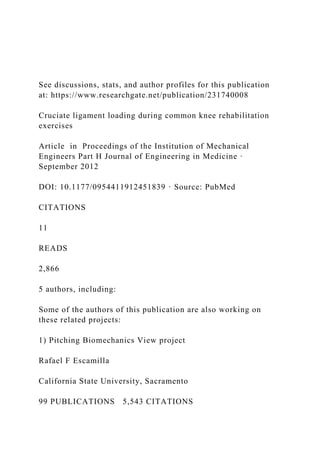
- 30. trunk tilt optimal for recruiting relatively high ham- strings activity and minimizing ACL loading. Technique variations during the NWBE seated knee extension also can affect ACL tensile force. For exam- ple, given a constant external knee torque applied to the leg, ACL force decreases when the resistance pad is moved up the leg more proximal to the knee compared with being more distal to the knee closer to the ankle (Figure 3). 17 From Figure 3, when a constant external knee torque is applied to the leg at 30� knee angle, the ACL tensile force is approximately twice as great when the resistance pad is positioned near the ankle (approxi- mately 400 N) compared with when it is placed near the middle of the leg (approximately 200 N). Also, Figure 3 demonstrates that ACL loading decreases progressively from 15� knee angle (approximately 500 N when the resistance pad is near the ankle and approximately 325 N when the resistance pad is placed near the middle of the leg) to 60� knee angle (approximately 100 N when the resistance pad is near the ankle and approximately 0 N when the resistance pad is positioned near the mid- dle of the leg), with no ACL loading at knee angles greater than 60�. Nisell et al.34 reported a similar find- ing of less ACL loading with a more proximally posi- tion resistance pad on the leg during isokinetic seated knee extension at 30�/s and 180�/s. It can be concluded from these data that, when the goal is to minimize ACL loading while using the seated knee extension exercise, this exercise should be performed at higher knee angles (50�–100�) and with the resistance pad position more
- 31. proximal on the leg compared with a more distal posi- tion. Moreover, it should be emphasized that if the ACL is torn, there is no ligament to restrain anterior tibial translation on the femur. Therefore, performing exercises that would normally load the ACL may cause anterior tibial translation, which may result in altered and possibly injurious tibiofemoral joint loading. 36,37 Wilk and Andrews 38 have reported that ACL-deficient knees during isokinetic exercises tibial translation can be reduced by utilizing a proximal pad and performing higher angular velocities (e.g. 180 �/s and 300 �/s) com- pared with slower speeds (e.g. 60 �/s). Peak anterior shear force Peak anterior shear force during WBE and NWBE are shown in Table 3. From Table 3, it is interesting that level ground walking resulted in greater anterior shear force (ACL loading) compared with both NWBE and WBE. Compared with WBE, anterior shear forces were greater in the NWBE seated knee extension. Peak ante- rior shear forces occurred at knee angles of 50� or less. Moreover, from Table 2 peak ACL tensile force during level walking was approximately 300 N and occurred near opposite foot toe-off (approximately 15�–20� knee flexion). Therefore, peak ACL loading during level walking is similar to peak ACL loading during NWBE seated isokinetic and isometric knee extension exercises, and these ACL loading magnitudes are several times greater than the ACL tensile forces reported for the
- 32. WBE. Figure 3. Changes in ACL loading during the seated knee extension exercise with proximal or distal resistance applied on the leg. The location of the restraining force is given relative to the distance from the knee joint. Given a constant external knee torque applied to the leg, moving the restraining force closer to the knee joint axis decreases ACL force. Adapted from Pandy and Shelburne17 with kind permission of Elsevier. ACL: anterior cruciate ligament. 676 Proc IMechE Part H: J Engineering in Medicine 226(9) PCL loading during selected rehabilitation exercises Table 4 presents PCL tensile force data from selected articles in the scientific literature during a variety of NWBE and WBE commonly used in cruciate ligament rehabilitation. Key points of emphasis from Table 4 are as follows. First, in contrast to the peak, ACL loading at lower knee angles between approximately 0�–50� shown in Tables 1 and 2, peak PCL loading occurs at higher knee angles between approximately 50�–100� (typically around 80�–90� knee angles). Thus, to control PCL loading, the rehabilitation process is the complete opposite of that of the ACL program. Therefore, if the rehabilitation goal is to minimize PCL loading, such as during the early phases after PCL reconstruction sur-
- 33. gery, training both NWBE and WBE at lower knee angles (e.g. 0�–50�) would be recommended compared with training these exercises as higher knee angles (e.g. 50�–100�). Second, peak PCL tensile force was generally greater in the NWBE seated knee flexion compared with WBE. For example, performing the NWBE seated isometric knee flexion at 90� knee angle produced greater PCL Table 4. Peak PCL tensile force and knee angle for commonly performed rehabilitation exercises. Author Rehabilitation exercise Peak PCL force (N) Knee flexion angle (�) Escamilla et al. (2009)8 Wall squat with heels position far from wall using 12 repetition maximum dumbbell resistance** 757 80 Wall squat with heels positioned close to wall using 12 repetition maximum dumbbell resistance** 786 90 One-leg squat using 12 repetition maximum dumbbell resistance** 414 90
- 34. Escamilla et al. (2010) 11 Forward lunge while taking a long step forward using 12 repetition maximum dumbbell resistance** 765 70 Forward lunge while taking a short step forward using 12 repetition maximum dumbbell resistance** 612 90 Escamilla et al. (2010)11 Forward lunge while taking a normal length step forward using 12 repetition maximum dumbbell resistance** 765 70 Side lunge while taking a normal length step sideways using 12 repetition maximum dumbbell resistance** 641 50 Lunging forward and sideways while taking a normal length step using 12 repetition maximum dumbbell resistance** 733 60 Lunging forward and sideways while keeping both feet stationary using 12 repetition maximum dumbbell resistance** 652 80
- 35. Escamilla et al. (1998)9 Barbell squat using 12 repetition maximum resistance** 1868 63 Leg press using 12 repetition maximum resistance** 1866 95 Dynamic seated knee extension using 12 repetition maximum resistance** 959 79 Escamilla et al. (2001) 39 Barbell squat with narrow stance using 12 repetition maximum resistance** 2066 77 Barbell squat with wide stance using 12 repetition maximum resistance** 2212 76 Leg press with narrow stance with high foot placement using 12 repetition maximum resistance** 1703 94 Leg press with wide stance with high foot placement using 12 repetition maximum resistance** 1726 88 Leg press with narrow stance with low foot placement using 12 repetition maximum resistance** 1690 95
- 36. Leg press with wide stance with low foot placement using 12 repetition maximum resistance** 1726 95 Toutoungi et al. (2000)18 Isokinetic seated knee extension at 60 �/s 74 90 Isokinetic seated knee extension at 120 �/s 59 90 Isokinetic seated knee extension at 180 �/s 55 90 Isokinetic seated knee flexion at 60 �/s 2701 90 Isokinetic seated knee flexion at 120 �/s 2394 90 Isokinetic seated knee flexion at 180 �/s 1952 90 Isometric seated knee extension 0 Isometric seated knee flexion 3330 90 Squat with heel-off-ground without external resistance 2222 90– 100 Squat with heel-on-ground without external resistance 2704 90– 100 Shelburne et al. (2005)19 Level ground walking Approximately 160 15–20 **Used the heaviest resistance possible that allowed the performance of 12 consecutive repetitions with proper form and technique. PCL: posterior cruciate ligament. Escamilla et al. 677 tensile force (3330 N) compared with all WBE. The NWBE seated isokinetic knee flexion at 60 �/s, 120 �/s, and 180 �/s, and the two-leg squat, produced the next highest PCL tensile force, ranging between approxi-
- 37. mately 1900�2700 N. PCL tensile forces were approxi- mately 1700�1900 N during the leg press, approximately 750�800 N during the wall squat, approximately 650�750 N during the forward and side lunges, approxi- mately 400 N during the one-leg squat, and approxi- mately 160 N during level ground walking. Comparing technique variations within exercises, performing the forward lunge while taking a long step forward, produces a significantly greater PCL tensile force compared with performing the forward lunge while taking a short step forward (the short step lunge causes the knees to move forward over the toes approx- imately 8 cm). Moreover, performing the forward lunge produced a significantly greater PCL tensile force com- pared with performing the side lunge, and performing the forward and side lunge while taking a normal length step (and then pushing back to the upright start- ing position) produced a significantly greater PCL ten- sile force compared with performing the forward and side lunge while keeping both feet stationary (and sim- ply lunging up and down). Therefore, there are differ- ent progressions both within an exercise and between exercises that can be employed during PCL rehabilita- tion, and as a general rule NWBE and WBE should first begin with no external resistance and progress to increasing amounts of external resistance. Given the PCL loading shown in Table 4, one example of exercise progression for lower to higher PCL loading may include performing NWBE and WBE initially between 0�–50� knee flexion and progressing to 50�–100� knee flexion. Exercises that may be appropriate early in rehabilitation might include seated knee extensions between 0�–50� knee flexion, level ground walking, one-leg squats with no resistance, and forward and side lunges with no resistance. Resistance can slowly be
- 38. added to these exercises, and the leg press, squat, and seated knee flexion exercises can be added later, ini- tially without resistance and progressing to resistance. It is not well understood what PCL force magnitudes become injurious to the healthy or reconstructed PCL. In healthy adults, the ultimate strength of the PCL is approximately 4000 N, 40 although these values depend on age and anatomical factors. Therefore, the PCL loads generated during both NWBE and WBE appear to be well within a safe limit for the healthy PCL. The reconstructed PCL has similar ultimate strengths com- pared with the healthy PCL. However, the healing graft site may be injured with considerably less force com- pared with the ultimate strength of the graft, although it is not well understood how much force to the healing graft site is too much and how soon force can be applied after reconstruction. Therefore, the peak PCL forces that occur during NWBE and WBE may be pro- blematic early after PCL reconstruction when the graft site is still healing, especially between 50�–100� of knee angles. It may be prudent to employ smaller knee angles (e.g. 0�–50�) before progressing to larger knee angles (e.g. 50�–100�) during NWBE and WBE, because PCL forces increase as knee angle increases. Summary This review allows the clinician to select specific thera- peutic exercises, broken down by weight bearing status, knee range of motion, and technique variations, to
- 39. progress cruciate ligament loading over the course of rehabilitation safely while ensuring optimal recovery of the musculoskeletal system. In general, WBE produces smaller loads on the ACL and PCL compared with NWBE. For the ACL, performing seated knee exten- sions with resistance produces significantly greater ACL loading compared with most WBE. Further, peak ACL loading during level walking is similar to peak ACL loading during NWBE seated isokinetic and iso- metric knee extension exercises, and these ACL loading magnitudes are several times greater than the ACL ten- sile forces reported for the WBE. For the PCL, the highest loading occurred in the two-leg squat, followed by the leg press, wall squat, forward and side lunges, one-leg squat, and level ground walking. WBE has the benefit of being much more functional and challenging in terms of hip and thigh muscle recruitment compared with NWBE. Therefore, early after injury or recon- struction of the cruciate ligament the clinician should prescribe WBE rather than NWBE, and progress to NWBE as tolerated and to facilitate isolated muscle functional groups – such as the quadriceps. Peak ACL loading occurs at knee angles of between 10�–15�, and progressively decreases between 15�–60� knee angles. Beyond 50�–60� knee angles there is mini- mal or no ACL loading. In contrast, PCL loading occurs at higher knee angles (i.e. 50�–100�), with peak PCL loading typically occurring around 80�–90� knee angles. These arc ranges of motion should serve as guidelines to limit the therapeutic exercise technique early on in the rehabilitation protocol. Exercise technique variation should also be considered when prescribing a rehabilitation protocol. For ACL rehabilitation, anterior knee movement of 8 cm or more
- 40. beyond the toes may also increase ACL loading during squatting, lunging, leg press, and other WBE. Moreover, squatting with the heels off the ground, which typically results in increased anterior knee movement beyond the toes, resulted in over three times the ACL loading com- pared with squatting with the heels on the ground. Squatting and lunging with a more forward trunk tilt tends to unload the ACL to a greater extent compared with squatting and lunging with a more erect trunk posi- tion. Moving the resistance pad up the leg proximally towards the knee when performing the seated knee extension, rather than being positioned closer to the ankle, decreases ACL loading. Rapid deceleration activi- ties, such as one-leg landing from a jump, or running 678 Proc IMechE Part H: J Engineering in Medicine 226(9) and cutting movements, have been shown to generate very high ACL loading and are often implicated in ACL injuries, so cautious progression is required. These higher intensity plyometric type exercises should be performed only during the later stages of ACL rehabilitation. When prescribing therapeutic exercise for the PCL, the forward lunge with a long step forward produced greater PCL loading compared with forward lunge with a short step forward. The forward lunge produced greater PCL load- ing compared with the side lunge, and lunging by taking a forward or sideways step and pushing back to the starting position produced greater PCL loading com- pared with lunging up and down with both feet station- ary. The exercise guidelines provided in this review may be used by the clinician to progress a cruciate ligament injured individual to maximize the potential benefits and minimize the chance for injury.
- 41. Funding This research received no specific grant from any fund- ing agency in the public, commercial, or not-for-profit sectors. This article was submitted as part of the Lower Limb Musculoskeletal Modelling Special Issue. References 1. Beynnon BD, Johnson RJ, Fleming BC, et al. The strain behavior of the anterior cruciate ligament during squat- ting and active flexion-extension. A comparison of an open and a closed kinetic chain exercise. Am J Sports Med. 1997; 25: 823–829 2. Beynnon BD and Fleming BC. Anterior cruciate liga- ment strain in-vivo: a review of previous work. J Bio- mech 1998; 31(6): 519–525. 3. Beynnon B, Howe JG, Pope MH, et al. The measure- ment of anterior cruciate ligament strain in vivo. Int Orthop 1992; 16(1): 1–12. 4. Beynnon BD, Fleming BC, Johnson RJ, et al. Anterior cruciate ligament strain behavior during rehabilitation exercises in vivo. Am J Sports Med 1995; 23(1): 24–34.
- 42. 5. Fleming BC, Beynnon BD, Renstrom PA, et al. The strain behavior of the anterior cruciate ligament during stair climbing: an in vivo study. Arthroscopy 1999; 15(2): 185–191. 6. Heijne A, Fleming BC, Renstrom PA, et al. Strain on the anterior cruciate ligament during closed kinetic chain exercises. Med Sci Sports Exerc 2004; 36(6): 935–941. 7. Fleming BC, Beynnon BD, Renstrom PA, et al. The strain behavior of the anterior cruciate ligament during bicycling. An in vivo study. Am J Sports Med 1998; 26(1): 109–118. 8. Escamilla RF, Zheng N, Imamura R, et al. Cruciate liga- ment force during the wall squat and the one-leg squat. Med Sci Sports Exerc 2009; 41(2): 408–417. 9. Escamilla RF, Fleisig GS, Zheng N, et al. Biomechanics of the knee during closed kinetic chain and open kinetic chain exercises. Med Sci Sports Exerc 1998; 30(4): 556–569. 10. Zheng N, Fleisig GS, Escamilla RF, et al. An analytical model of the knee for estimation of internal forces during
- 43. exercise. J Biomech 1998; 31(10): 963–967. 11. Escamilla RF, Zheng N, Macleod TD, et al. Cruciate ligament forces between a short and long step forward lunge. Med Sci Sports Exerc 2010; 42(10): 1932–1942. 12. Escamilla RF, Zheng N, MacLeod TD, et al. Cruciate ligament tensile forces during the forward and side lunge. Clinical Biomech 2010; 25(3): 213–221. 13. Pflum MA, Shelburne KB, Torry MR, et al. Model pre- diction of anterior cruciate ligament force during drop- landings. Med Sci Sports Exerc 2004; 36(11): 1949–1958. 14. Eitzen I, Moksnes H, Snyder-Mackler L, et al. A progres- sive 5-week exercise therapy program leads to significant improvement in knee function early after anterior cruciate ligament injury. J Orthop Sports Phys Ther 2010; 40(11): 705–721. 15. Wilk KE, Reinold MM and Hooks TR. Recent advances in the rehabilitation of isolated and combined anterior cruciate ligament injuries. Orthop Clin North Am 2003; 34(1): 107–137. 16. Risberg MA and Holm I. The long-term effect of 2 post- operative rehabilitation programs after anterior cruciate
- 44. ligament reconstruction: a randomized controlled clinical trial with 2 years of follow-up. Am J Sports Med 2009; 37(10): 1958–1966. 17. Pandy MG and Shelburne KB. Dependence of cruciate- ligament loading on muscle forces and external load. J Biomech 1997; 30(10): 1015–1024. 18. Toutoungi DE, Lu TW, Leardini A, et al. Cruciate liga- ment forces in the human knee during rehabilitation exer- cises. Clin Biomech (Bristol, Avon) 2000; 15(3): 176–187. 19. Shelburne KB, Torry MR and Pandy MG. Muscle, liga- ment, and joint-contact forces at the knee during walking. Med Sci Sports Exerc 2005; 37(11): 1948–1956. 20. Shelburne KB and Pandy MG. A dynamic model of the knee and lower limb for simulating rising movements. Comput Methods Biomech Biomed Engng 2002; 5(2): 149–159. 21. Shin CS, Chaudhari AM and Andriacchi TP. The influ- ence of deceleration forces on ACL strain during single- leg landing: a simulation study. J Biomech 2007; 40(5): 1145–1152. 22. Delay BS, Smolinski RJ, Wind WM, et al. Current prac-
- 45. tices and opinions in ACL reconstruction and rehabilita- tion: results of a survey of the American Orthopaedic Society for Sports Medicine. Am J Knee Surg 2001; 14(2): 85–91. 23. Francis A, Thomas RD and McGregor A. Anterior cruci- ate ligament rupture: reconstruction surgery and rehabili- tation. A nation-wide survey of current practice. Knee 2001; 8(1): 13–18. 24. Woo SL, Hollis JM, Adams DJ, et al. Tensile properties of the human femur-anterior cruciate ligament-tibia com- plex. The effects of specimen age and orientation. Am J Sports Med 1991; 19(3): 217–225. 25. Brown Jr CH, Steiner ME and Carson EW. The use of hamstring tendons for anterior cruciate ligament recon- struction. Technique and results. Clin Sports Med 1993; 12(4): 723–756. 26. Shelbourne KD and Nitz P. Accelerated rehabilitation after anterior cruciate ligament reconstruction. Am J Sports Med 1990; 18(3): 292–299. Escamilla et al. 679
- 46. 27. Escamilla RF, Fleisig GS, Zheng N, et al. Effects of tech- nique variations on knee biomechanics during the squat and leg press. Med Sci Sports Exerc 2001; 33(9): 1552– 1566. 28. Wilk KE, Escamilla RF, Fleisig GS, et al. A comparison of tibiofemoral joint forces and electromyographic activ- ity during open and closed kinetic chain exercises. Am J Sports Med 1996; 24(4): 518–527. 29. Nagura T, Matsumoto H, Kiriyama Y, et al. Tibiofe- moral joint contact force in deep knee flexion and its con- sideration in knee osteoarthritis and joint replacement. J Appl Biomech 2006; 22(4): 305–313. 30. Beynnon BD and Amis AA. In vitro testing protocols for the cruciate ligaments and ligament reconstructions. Knee Surg Sports Traumatol Arthrosc 1998; 6(Suppl 1): S70–76. 31. Shimokochi Y and Shultz SJ. Mechanisms of noncontact anterior cruciate ligament injury. J Athl Train 2008; 43(4): 396–408. 32. Ohkoshi Y, Yasuda K, Kaneda K, et al. Biomechanical analysis of rehabilitation in the standing position. Am J Sports Med 1991; 19(6): 605–611. 33. Shelbourne KD, Klootwyk TE, Wilckens JH, et al. Liga- ment stability two to six years after anterior cruciate liga- ment reconstruction with autogenous patellar tendon graft and participation in accelerated rehabilitation pro- gram. Am J Sports Med 1995; 23(5): 575–579.
- 47. 34. Nisell R, Ericson MO, Nemeth G, et al. Tibiofemoral joint forces during isokinetic knee extension. Am J Sports Med 1989; 17(1): 49–54. 35. Farrokhi S, Pollard CD, Souza RB, et al. Trunk position influences the kinematics, kinetics, and muscle activity of the lead lower extremity during the forward lunge exer- cise. J Orthop Sports Phys Ther 2008; 38(7): 403–409. 36. Jacobsen K. Osteoarthrosis following insufficiency of the cruciate ligaments in man. A clinical study. Acta Orthop Scand 1977; 48(5): 520–526. 37. Vilensky JA, O’Connor BL, Brandt KD, et al. Serial kinematic analysis of the unstable knee after transection of the anterior cruciate ligament: temporal and angular changes in a canine model of osteoarthritis. J Orthop Res 1994; 12(2): 229–237. 38. Wilk KE and Andrews JR. The effects of pad placement and angular velocity on tibial displacement during isoki- netic exercise. J Orthop Sports Phys Ther 1993; 17(1): 24–30. 39. Escamilla RF. Knee biomechanics of the dynamic squat
- 48. exercise. Med Sci Sports Exerc 2001; 33(1): 127–141. 40. Race A and Amis AA. The mechanical properties of the two bundles of the human posterior cruciate ligament. J Biomech 1994; 27(1): 13–24. 680 Proc IMechE Part H: J Engineering in Medicine 226(9) View publication statsView publication stats https://www.researchgate.net/publication/231740008 << /ASCII85EncodePages false /AllowTransparency false /AutoPositionEPSFiles true /AutoRotatePages /None /Binding /Left /CalGrayProfile () /CalRGBProfile (Adobe RGB 0501998051) /CalCMYKProfile () /sRGBProfile (sRGB IEC61966-2.1) /CannotEmbedFontPolicy /Error /CompatibilityLevel 1.3 /CompressObjects /Off /CompressPages true /ConvertImagesToIndexed true /PassThroughJPEGImages true /CreateJobTicket false /DefaultRenderingIntent /Default /DetectBlends true /DetectCurves 0.1000 /ColorConversionStrategy /LeaveColorUnchanged /DoThumbnails false /EmbedAllFonts true /EmbedOpenType false
- 49. /ParseICCProfilesInComments true /EmbedJobOptions true /DSCReportingLevel 0 /EmitDSCWarnings false /EndPage -1 /ImageMemory 1048576 /LockDistillerParams true /MaxSubsetPct 100 /Optimize false /OPM 1 /ParseDSCComments true /ParseDSCCommentsForDocInfo true /PreserveCopyPage false /PreserveDICMYKValues true /PreserveEPSInfo true /PreserveFlatness true /PreserveHalftoneInfo false /PreserveOPIComments false /PreserveOverprintSettings true /StartPage 1 /SubsetFonts true /TransferFunctionInfo /Apply /UCRandBGInfo /Remove /UsePrologue false /ColorSettingsFile () /AlwaysEmbed [ true ] /NeverEmbed [ true ] /AntiAliasColorImages false /CropColorImages true /ColorImageMinResolution 266 /ColorImageMinResolutionPolicy /Warning /DownsampleColorImages false /ColorImageDownsampleType /Bicubic /ColorImageResolution 266
- 50. /ColorImageDepth 8 /ColorImageMinDownsampleDepth 1 /ColorImageDownsampleThreshold 1.50000 /EncodeColorImages true /ColorImageFilter /FlateEncode /AutoFilterColorImages false /ColorImageAutoFilterStrategy /JPEG /ColorACSImageDict << /QFactor 0.15 /HSamples [1 1 1 1] /VSamples [1 1 1 1] >> /ColorImageDict << /QFactor 0.15 /HSamples [1 1 1 1] /VSamples [1 1 1 1] >> /JPEG2000ColorACSImageDict << /TileWidth 256 /TileHeight 256 /Quality 30 >> /JPEG2000ColorImageDict << /TileWidth 256 /TileHeight 256 /Quality 30 >> /AntiAliasGrayImages false /CropGrayImages true /GrayImageMinResolution 266 /GrayImageMinResolutionPolicy /Warning /DownsampleGrayImages false /GrayImageDownsampleType /Bicubic /GrayImageResolution 266 /GrayImageDepth 8 /GrayImageMinDownsampleDepth 2 /GrayImageDownsampleThreshold 1.50000 /EncodeGrayImages true
- 51. /GrayImageFilter /FlateEncode /AutoFilterGrayImages false /GrayImageAutoFilterStrategy /JPEG /GrayACSImageDict << /QFactor 0.15 /HSamples [1 1 1 1] /VSamples [1 1 1 1] >> /GrayImageDict << /QFactor 0.15 /HSamples [1 1 1 1] /VSamples [1 1 1 1] >> /JPEG2000GrayACSImageDict << /TileWidth 256 /TileHeight 256 /Quality 30 >> /JPEG2000GrayImageDict << /TileWidth 256 /TileHeight 256 /Quality 30 >> /AntiAliasMonoImages false /CropMonoImages true /MonoImageMinResolution 900 /MonoImageMinResolutionPolicy /Warning /DownsampleMonoImages false /MonoImageDownsampleType /Bicubic /MonoImageResolution 900 /MonoImageDepth -1 /MonoImageDownsampleThreshold 1.33333 /EncodeMonoImages true /MonoImageFilter /CCITTFaxEncode /MonoImageDict << /K -1 >> /AllowPSXObjects false
- 52. /CheckCompliance [ /PDFX1a:2001 ] /PDFX1aCheck true /PDFX3Check false /PDFXCompliantPDFOnly true /PDFXNoTrimBoxError false /PDFXTrimBoxToMediaBoxOffset [ 0.00000 0.00000 0.00000 0.00000 ] /PDFXSetBleedBoxToMediaBox true /PDFXBleedBoxToTrimBoxOffset [ 0.00000 0.00000 0.00000 0.00000 ] /PDFXOutputIntentProfile (ISO Coated v2 300% 050ECI051) /PDFXOutputConditionIdentifier () /PDFXOutputCondition () /PDFXRegistryName () /PDFXTrapped /False /CreateJDFFile false /Description << /ARA <FEFF06270633062A062E062F06450020064706300647002006 27064406250639062F0627062F0627062A002006440625064606 340627062100200648062B062706260642002000410064006F00 620065002000500044004600200645062A064806270641064206 290020062A0645062706450627064B0020064506390020064506 420627064A064A06330020005000440046002F0058002D00310 061003A003200300030003100200630064800200627064406450
- 64. PDF/X-1a:2001, uno standard ISO per lo scambio di contenuto grafico. Per ulteriori informazioni sulla creazione di documenti PDF compatibili con PDF/X-1a, consultare la Guida dell'utente di Acrobat. I documenti PDF creati possono essere aperti con Acrobat e Adobe Reader 4.0 e versioni successive.) /JPN <FEFF30b030e930d530a330c330af30b330f330c630f330c4306e5 90963db306b5bfe3059308b002000490053004f00206a196e9689 8f683c306e0020005000440046002f0058002d00310061003a003 20030003000310020306b6e9662e03057305f002000410064006f 0062006500200050004400460020658766f830924f5c621030593 08b305f3081306b4f7f75283057307e30593002005000440046002 f0058002d0031006100206e9662e0306e00200050004400460020 658766f84f5c6210306b306430443066306f300100410063007200 6f006200610074002030e630fc30b630ac30a430c9309253c27167 30573066304f30603055304430023053306e8a2d5b9a30674f5c62 103055308c305f0020005000440046002030d530a130a430eb306f 3001004100630072006f0062006100740020304a3088307300200 0410064006f006200650020005200650061006400650072002000 34002e003000204ee5964d3067958b304f30533068304c3067304 d307e30593002> /KOR <FEFFc7740020c124c815c7440020c0acc6a9d558c5ec0020c791 c131d558b294002000410064006f0062006500200050004400460 020bb38c11cb2940020d655c778c7740020d544c694d558ba7000 20adf8b798d53d0020cee8d150d2b8b97c0020ad50d658d558b29 40020bc29bc95c5d00020b300d55c002000490053004f0020d45c c900c7780020005000440046002f0058002d00310061003a00320 03000300031c7580020addcaca9c5d00020b9dec544c57c0020d56 9b2c8b2e4002e0020005000440046002f0058002d003100610020 d638d65800200050004400460020bb38c11c0020c791c131c5d00 020b300d55c0020c790c138d55c0020c815bcf4b2940020004100 630072006f0062006100740020c0acc6a90020c124ba85c11cb97c 0020cc38c870d558c2edc2dcc624002e0020c774b807ac8c0020c7 91c131b41c00200050004400460020bb38c11cb29400200041006 30072006f0062006100740020bc0f002000410064006f006200650
- 66. 6f00740075002000410064006f006200650020005000440046002 00064006f006b0075006d0065006e007400750073002c0020006b 0075007200690020006900720020006a010100700101007200620 06100750064006100200076006100690020006b00750072006900 65006d0020006900720020006a01010061007400620069006c007 300740020005000440046002f0058002d00310061003a00320030 00300031002c002000490053004f0020007300740061006e00640 061007200740061006d002000610070006d006100690146006100 690020006100720020006700720061006600690073006b006f002 0007300610074007500720075002e00200050006c006101610101 006b007500200069006e0066006f0072006d010100630069006a0 07500200070006100720020005000440046002f0058002d003100 61002000730061006400650072012b00670075002000500044004 600200064006f006b0075006d0065006e0074007500200069007a 00760065006900640069002c0020006c016b0064007a0075002c0 0200073006b006100740069006500740020004100630072006f00 62006100740020006c006900650074006f00740101006a0061002 00072006f006b00610073006700720101006d006100740101002e 00200049007a0076006500690064006f006a00690065007400200 0500044004600200064006f006b0075006d0065006e0074007500 73002c0020006b006f00200076006100720020006100740076011 3007200740020006100720020004100630072006f006200610074 00200075006e002000410064006f0062006500200052006500610 0640065007200200034002e0030002c0020006b01010020006100 72012b00200074006f0020006a00610075006e0101006b0101006 d002000760065007200730069006a0101006d002e> /NLD (Gebruik deze instellingen om Adobe PDF-documenten te maken die moeten worden gecontroleerd of moeten voldoen aan PDF/X-1a:2001, een ISO-standaard voor het uitwisselen van grafische gegevens. Raadpleeg de gebruikershandleiding van Acrobat voor meer informatie over het maken van PDF- documenten die compatibel zijn met PDF/X-1a. De gemaakte PDF-documenten kunnen worden geopend met Acrobat en Adobe Reader 4.0 en hoger.) /NOR <FEFF004200720075006b0020006400690073007300650020006