Call Girls Horamavu WhatsApp Number 7001035870 Meeting With Bangalore Escorts
Needle in Foot
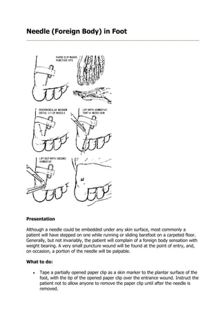
1. Needle (Foreign Body) in Foot
Presentation
Although a needle could be embedded under any skin surface, most commonly a
patient will have stepped on one while running or sliding barefoot on a carpeted floor.
Generally, but not invariably, the patient will complain of a foreign body sensation with
weight bearing. A very small puncture wound will be found at the point of entry, and,
on occasion, a portion of the needle will be palpable.
What to do:
• Tape a partially opened paper clip as a skin marker to the plantar surface of the
foot, with the tip of the opened paper clip over the entrance wound. Instruct the
patient not to allow anyone to remove the paper clip until after the needle is
removed.
2. • Send the patient for PA and lateral radiographs of the foot with the skin marker
in place.
• Evaluate the x rays. If the needle appears to be very deep you may choose to
call in a consultant who can remove the needle under fluoroscopy. If the needle
is relatively superficial, inform the patient that removing a needle is not as easy
as it appears. Let him know that you are going to use a simple technique for
locating and removing the needle, but that sometimes the needle is hidden
within the tissue of the foot ("like a needle in a haystack"). If you cannot locate
the needle within 10-15 minutes, because you do not want to further damage his
foot, you will call in a consultant or arrange for fluoroscopy.
• Establish a bloodless field by elevating the leg above the level of the heart,
tightly wrapping an ACE bandage around the foot and lower leg, and then
inflating and clamping off a thigh cuff at approximately 200mmHg. This will
become uncomfortable within l0-15 minutes and thereby serve as an automatic
timer for your procedure.
• Remove the ACE wrap, clean and then paint the area with Betadine solution, and
locally infiltrate the appropriate area with plain 1% Xylocaine. (It will be
somewhat more comfortable if the needle stick is accomplished from the medial
or lateral aspect of the foot rather than directly into the plantar surface.)
• The x rays should give you an idea of the location of the needle relative to the
paper clip skin marker.
• With the patient lying prone and the plantar surface of his foot facing upward,
make an incision that crosses perpendicular to the needle's apparent position at
its midpoint or 1/3 of the way toward the most superficial end of the needle. Do
not cut deep to the plantar fascia. With any deep entry into the foot, use iris
scissors with the blades open to advance a few millimeters at at time before
closing the scissor blades. Continue repeating this process until the needle
prevents closure of the scissors. If you are using a scalpel blade, as you cut
across the needle, there will be an audible clicking sound. Spread the incision
apart, visualize the needle and grasp it firmly with a hemostat or small Kelly
clamp.
• Now, push the needle out in the direction from which it entered. Even the eye or
back end of a broken needle is sharp enough to be pushed to the skin surface. If
the needle tents up the skin and will not push through, nick the overlying skin
surface with a scalpel blade until the needle exits. Grab this end with another
clamp, let go with the first clamp, and remove the needle.
• Let the thigh cuff down and suture your incision closed. Apply an appropriate
dressing.
• Provide tetanus prophylaxis if indicated.
What not to do:
• Do not ignore the patient who thinks he stepped on a needle but in whom you
can't find a puncture wound. Get an x ray anyway, because the puncture wound
is probably hidden.
• Do not give the patient the impression that the removal will be quick and easy.
• Do not make your incision near the tip of the needle or directly over and parallel
to the needle. The needle will not be exactly where you think it is, and your
incision will miss exposing the needle.
3. • Do not persist in extensively undermining or extending your incision if you do not
locate the needle within 10 minutes of beginning the procedure. This is unlikely
to be productive and you may do the patient harm.
• Do not routinely place the patient on prophylactic antibiotics.
• Do not attempt to remove a buried needle by pulling on the attached thread. It
usually breaks, and may create a second foreign body to remove.
Discussion:
Many a young doctor has been found sweating away at the foot of an emergency
department stretcher, unable to locate a needle foreign body. The secret for improving
your chances of success is in realizing that the x ray only gives you an approximate
location of the needle and that your incision must be made in a direction and location
best suited for locating the needle, not removing it.
There are three additional principles to keep in mind. First, the roentgenographic
position of the needle must be correlated with the anatomy of the skin surface rather
than the bony anatomy of the foot. Second is the simple geometric principle that the
surest way to interesct a line (the needle) is to dissect in the plane perpendicular to its
midpoint. Third, the only structures of importance in the forefoot or heel that lie plantar
to the bones are the flexor tendons and they lie close to the bones.
When you let the patient know how difficult it sometimes is to locate the needle and
remove it, you place yourself in a win-win situation. You look especially good if you find
it and you still look experienced and well-informed if you don't.
If you choose to take the patient to fluoroscopy, you or the radiologist can place a
hemostat around the needle under direct vision. It can then be pushed out using the
same technique described above.
Linear foreign bodies such as needles can be removed from the sole of the foot without
extensive dissection, complex apparatus or repeated roentgenographic studies.
Although blind dissection is generally not a good technique because of the risk of injury,
in this particular situation, relative safety can be provided by gentle dissection with iris
scissors of insufficient strength to sever tendons, and by setting firm limits of time and
depth of exploration.