
Recommended
More Related Content
What's hot
What's hot (20)
Similar to Hypertrophic Cardiomyopathy: Causes, Symptoms, and Treatment
Similar to Hypertrophic Cardiomyopathy: Causes, Symptoms, and Treatment (20)
More from drsrb
Recently uploaded
Recently uploaded (20)
Hypertrophic Cardiomyopathy: Causes, Symptoms, and Treatment
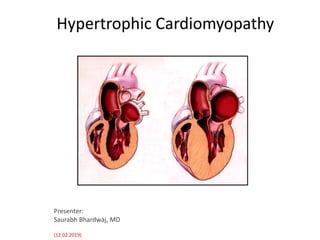
- 1. Hypertrophic Cardiomyopathy Presenter: Saurabh Bhardwaj, MD (12.02.2019)
- 2. Introduction • Hypertrophic cardiomyopathy (HCM) is the commonest of the genetic cardiovascular diseases, characterized by heterogeneous clinical expression, distinctive pathophysiologic features, and a diverse natural history. • It is caused by a multitude of mutations in genes encoding proteins of the cardiac sarcomere. • Although many patients with HCM have a normal life span, HCM has been regarded as the most common cause of sudden death (SD) in the young, including competitive athletes. • It also constitutes a risk for atrial fibrillation (AF), and it is responsible for heart failure–related disability at virtually any age.
- 3. Definition, Prevalence & Nomenclature • HCM is characterized by a thickened but nondilated left ventricle in the absence of another cardiac or systemic condition (e.g., aortic valve stenosis, systemic hypertension, and some expressions of physiologic athlete's heart) capable of producing the magnitude of left ventricular (LV) hypertrophy evident. • Global Disease: Worldwide prevalence of 1 in 500 • Estimated 2.4 million patients in India, with ∼30 000 in New Delhi alone (Maron BJ. Importance and feasibility of creating hypertrophic cardiomyopathy centers in developing countries: the experience in India. Am J Cardiol 2015;116:332- 334) • More recent estimates, which take into account genetic and imaging diagnostic modalities, place the prevalence closer to 1 : 200. • The preferred and generally accepted name for this condition is hypertrophic cardiomyopathy (HCM), with or without outflow obstruction.
- 4. • Autosomal dominant disease (transmitted as mendelian trait). • Equal frequency in men and women. • Advanced molecular studies has helped in identification of individuals with pathogenic mutations but without evidence of the disease phenotype (LV hypertrophy). • HCM is known to be caused by mutations in 11 or more genes encoding proteins of the thick and thin contractile myofilament components of the cardiac sarcomere or the adjacent Z-disc. • Two sarcomere genes, β-myosin heavy chain (MYH7) and myosin-binding protein C (MYBPC3) are by far the most common, accounting for 70% of patients for whom genotyping has been successful.
- 5. Barry J. Maron et al. JACC 2012;60:705-715
- 6. • Population-based association studies have demonstrated a more severe course in gene-positive patients than gene-negative patients, as well as in patients with thick-filament mutations (MYBPC3 and MYH7) compared with those with thin-filament mutations (TNNT2 and TNNI3) • Genetic testing is crucial for clarifying the diagnosis in patients with metabolic and storage disorders for which the clinical presentation and pattern of LV hypertrophy can mimic sarcomeric HCM but for which the pathophysiology, natural history, and management are dissimilar. • LAMP2 cardiomyopathy is associated with a lethal natural history refractory to defibrillation therapy (with survival uncommon beyond 25 years) and requires early recognition and likely heart transplant. • Fabry disease requires enzyme replacement therapy.
- 7. Phenotype & LV Hypertrophy • Cardiac imaging in clinically identified adults and children with HCM typically documents an absolute increase in LV wall thickness of 15 mm or more (21 to 22 mm on average, and up to > 50 mm), although any LV wall thicknesses (including those within normal range) are consistent with a genetically affected status. • Borderline LV wall thicknesses of 13 to 14 mm can create diagnostic ambiguity, particularly in the differential diagnosis of physiologic athlete's heart. • Diverse and myriad patterns of asymmetric LV hypertrophy are characteristic of HCM even in related patients (although identical twins share the same morphology). • No single morphologic form of HCM is considered “classic” or typical.
- 8. • Typically, one or more regions of the LV chamber are of greater thickness than other areas, often with a sharp demarcation at the point of transition in thickness, or there are noncontiguous patterns of segmental hypertrophy, as well as extension into the right ventricular (RV) wall, in some patients. • Hypertrophy is frequently extensive, involving the ventricular septum and LV free wall. • In genetically affected family members without LV hypertrophy (i.e., those who are gene positive but phenotype negative), a variety of clinical and imaging findings have been reported, including subclinical diastolic dysfunction, blood-filled myocardial crypts, mitral valve leaflet elongation, collagen precursor biomarkers and myocardial scarring, or 12-lead ECG abnormalities.
- 9. Genetic screening strategies Barry J. Maron et al. JACC 2012;60:705-715
- 10. Mitral Valve Apparatus • Primary structural abnormalities of the mitral apparatus responsible for LV outflow obstruction are part of the phenotypic expression of HCM. • The mitral valve may be more than twofold the normal size due to elongation of both leaflets, or there may be segmental enlargement of only the anterior or posterior leaflet, more frequently observed in younger patients. • In older patients, outflow obstruction often occurs in the presence of a particularly small LV outflow tract, mitral leaflets of normal length, and mitral-septal contact created by a modest anterior excursion of the valve combined with posterior motion of the septum.
- 11. Histopathology • In HCM, hypertrophied cardiac muscle cells (myocytes) in both the ventricular septum and LV free wall have bizarre shapes and are often arranged in a chaotic and disorganized architectural pattern . • Areas of cellular disarray are evident in 95% of HCM patients. • Structurally abnormal intramural coronary arterioles with thickened vessel walls caused by media smooth muscle hyperplasia. -> narrowing of the vessel lumen -> impaired vasodilator response and blunting of the coronary flow reserve-> small-vessel ischemia-> myocyte death-> myocardial fibrosis. • Interstitial (matrix) collagen compartment volume is expanded. • Predisposition to disordered patterns and increased dispersion of electrical depolarization and repolarization -> unstable electrophysiologic substrate predisposing to reentry ventricular tachyarrhythmias and a likely mechanism for SD.
- 13. Pathophysiology • HCM is predominantly a disease of mechanical obstruction majority of patients (70%) have the propensity to develop impedance to LV outflow with dynamic gradients of 30 mm Hg or more, either at rest or with physiologic exercise. • Dynamic subaortic obstruction produced by systolic anterior motion (SAM) of the mitral valve in which elongated leaflets bend sharply, contacting the ventricular septum in midsystole by means of a drag effect, i.e., pushing force of flow directly on the leaflets increased intraventricular systolic pressures increase the myocardial wall stress and oxygen demand. • The magnitude of the outflow gradient, estimated noninvasively with continuous-wave Doppler imaging, is directly related to the duration of mitral valve–septal contact, with posteriorly directed mitral regurgitation a secondary consequence.
- 14. • Subaortic gradients (and associated systolic ejection murmurs) can be reduced or abolished by interventions, which decrease myocardial contractility (e.g., beta-adrenergic blocking drugs) or increase ventricular volume or arterial pressure (e.g., squatting, isometric handgrip, phenylephrine). • Gradients can be augmented by circumstances in which the arterial pressure or ventricular volume is reduced (e.g., Valsalva maneuver, administration of nitroglycerin or amyl nitrite, blood loss, dehydration) or when LV contractility is increased (as with premature ventricular contractions, infusion of isoproterenol or dobutamine, or physiologic exercise).
- 15. Nonobstructive Hypertrophic Cardiomyopathy; Diastolic Dysfunction • Gradient < 30 m Hg at rest and with physiologic exercise. • Impaired LV relaxation and filling • Hypertrophy, replacement scarring, interstitial fibrosis, abnormal microvascular blood flow, and disorganized cellular architecture Reduced ventricular compliance • This group less likely to progress to heart failure.
- 16. The combination of abnormal cellular substrate, ischemia, ventricular anatomy, hemodynamics, known rhythm disturbances, and family history contribute to ventricular arrhythmogenesis. Ommen SR, Gersh BJ. Sudden cardiac death risk in hypertrophic cardiomyopathy. Eur Heart J 2009;30:2558–9.
- 17. Clinical Features • Symptoms of heart failure may develop at any age. • Exertional dyspnea and fatigue. • Exacerbated by large meals or ingestion of alcohol and is frequently accompanied by chest pain, either typical or atypical of angina, possibly related to structural microvasculature abnormalities. • Impaired consciousness with syncope or near-syncope and light- headedness explained by arrhythmias or outflow obstruction. • Palpitations are common and may be linked to a variety of tachyarrhythmias, most frequently supraventricular, including AF and, less commonly, ventricular ectopy. • The nature of symptoms in HCM is usually similar in patients with or without outflow obstruction.
- 18. Physical Examination • Initial clinical suspicion of HCM may occur with recognition of a heart murmur on routine or preparticipation sports examinations, although most patients are identified by virtue of symptom onset or cardiac events. • Arterial pulses may rise rapidly with the bisferiens pulse contour. • Patients with LV outflow obstruction characteristically have a medium- pitch systolic ejection murmur at the lower left sternal border and apex that varies in intensity with the magnitude of the subaortic gradient; it increases with the Valsalva maneuver, during or immediately after exercise, or on standing.
- 19. • Such variability, together with the characteristic lack of radiation of the murmur to the neck, aids in differentiating dynamic subaortic obstruction from fixed aortic stenosis. • Most HCM patients with loud murmurs of at least grade 3/6 are likely to have LV outflow gradients of more than 30 mm Hg. • Physical findings in patients without subaortic gradients are more subtle, with only a soft systolic murmur or no murmur at all, although a forceful apical systolic thrust may raise the suspicion of HCM
- 20. JVP waveform in HCM showing an augmented a wave. Carotid impulse tracing in HCM demonstrating the spike (red arrow) and dome (blue arrow) pattern. Curr Probl Cardiol. 2004;29(9):503–567.
- 21. Electrographic findings • ECG-abnormal in 90% of probands and 75% of asymptomatic relatives. • Wide variety of abnormal patterns. • The most common abnormalities-increased voltages consistent with LV hypertrophy, ST-T changes (including marked T-wave inversion in the lateral precordial leads), left atrial enlargement, deep and narrow Q waves, and diminished R waves in the lateral precordial leads. • Normal ECG- associated with mild LV hypertrophy and a favorable clinical course, but do not exclude the possibility of future SD events. • Increased voltages (tall R waves or deep S waves) are only weakly correlated with the magnitude of LV hypertrophy and do not reliably distinguish obstructive from nonobstructive HCM.
- 22. • ECG of a 51-year old patient with HCM. Note the prominent precordial voltage, widespread repolarization abnormalities, Q-wave in the lateral lead (aVL), and p-wave abnormality suggesting left atrial enlargement.
- 23. Echocardiography • Small, hyperdynamic left ventricle with a thick sigmoid septum and/or banana-shaped cavity. • Asymmetric septal hypertrophy (septal thickness ≥1.6 times the thickness of the posterior wall). • Small LVOT, elevated flow velocity in the LVOT that peaks in late systole (when the LVOT is smallest). • Systolic anterior motion of the mitral valve, and posteriorly directed MR. • The LVOT gradient (ΔP) is calculated from PW Doppler LVOT peak velocity. It reflects the degree of outflow obstruction caused by altered LV and mitral valve geometry.
- 24. Luis C. Afonso et al. JIMG 2008;1:787-800 Characteristic Echocardiographic Features of Obstructive HCM (A) Parasternal long-axis view depicting severe asymmetric septal hypertrophy and systolic anterior mitral valve motion (arrowhead); (B) M-mode across the mitral leaflets depicting prominent systolic anterior motion (thick arrows) of the anterior mitral leaflet (SAM); (C) M-mode tracing across the aortic valve demonstrating partial closure of aortic leaflets (arrowheads); and (D) Accentuation of late-peaking dynamic left ventricular outflow tract obstruction after the Valsalva maneuver. Ao = aorta; HCEM = hypertrophic cardiomyopathy; IVS = interventricular septum; LA = left atrium; SAM = systolic anterior motion; PW = posterior wall ratio
- 25. Luis C. Afonso et al. JIMG 2008;1:787-800 Echocardiographic Diagnosis of Apical Hypertrophic Cardiomyopathy (A) (B) Contrast- enhanced images a patient with apical hypertrophic cardiomyopathy in end-diastole. (C) Conventional apical 4-chamber view showing exuberant LVH (arrow) in apical HCM. (D) Two-dimensional strain (quad format) images of same patient showing paradoxical apical longitudinal strain (crimson segment and tracing) and corresponding perturbations in the color M-mode of the parametric strain map. Note loss of base-apex (strain) gradient.
- 27. • Combination of small LVOT area and motion of a relatively large, anteriorly positioned, slack mitral apparatus causes the mitral leaflets to be pushed into the LVOT in early systole by flow drag forces and, to a lesser extent, by suctioning via the LVOT gradient and Venturi effect. • A maximum wall thickness greater than 30 mm or a resting LVOT gradient greater than 30 mm Hg is associated with increased risk for SCD and progression to New York Heart Association (NYHA) Functional Class III heart failure. • In apical HCM, basal wall thickness may be normal, but the midventricular and apical portions are unusually thickened, and a midcavity gradient may exist.
- 28. Cardiac-MRI • Echocardiography missed hypertrophic segments and underestimated the magnitude of hypertrophy in the basal anterolateral wall by as much as 33% compared to CMR. And 40% apical aneurysms. • Assessing the reduction in septal thickness from surgical myectomy or alcohol septal ablation. • Markedly elevated LV mass index (males >91g/m2 and females >69g/m2) was sensitive (100%), whereas maximal wall thickness greater than 30 mm was specific (91%) for cardiac deaths. • Presence of LGE is indicative of heterogeneous fibrosis and myofibril disarray and has been associated with ventricular arrhythmias and progressive ventricular dilation. • It may allow characterization of abnormal myocardial pathophysiology secondary to coronary microvascular dysfunction, fibrosis, and hypertrophy.
- 30. (A) Asymmetric hypertrophy of ventricular septum (VS), sparing the left ventricular (LV) free wall. (B) Focal hypertrophy sharply confined to basal anterior septum (arrows). (C) Thin-walled apical aneurysm (arrowheads) with muscular mid-ventricular apposition of hypertrophied septum and LV wall (asterisks), and distinct proximal (P) and distal (D) chambers. (D) Extensive, transmural late gadolinium enhancement involving ventricular septum (arrows). (E) Massive thickening (i.e., 33 mm) confined largely to anterolateral LV wall, greatly underestimated by echocardiography (arrowheads). (F) Genotype positive-phenotype negative HCM family member with 3 myocardial crypts penetrating thickness of basal inferior wall (arrows) Maron M.S., Maron B.J., Harrigan C., et al.(2009) Hypertrophic cardiomyopathy phenotype revisited after 50 years with cardiovascular magnetic resonance. J Am Coll Cardiol 54:220–228
- 31. CMR augments diagnosis of HCM by differentiating from phenocopies and allowing visualization of anatomy obscured from echocardiography. Maron MS, Maron BJ. Clinical impact of contemporary cardiovascular magnetic resonance imaging in hypertrophic cardiomyopathy. Circulation 2015;132:292–8.
- 32. Proposed Clinical Family Screening Strategies With Echocardiography or Cardiovascular Magnetic Resonance (and 12- Lead Electrocardiography) for Detection of HCM with Left Ventricular Hypertrophy Age < 12 years • Optional unless: – Malignant family history of premature death from HCM, or other adverse complications – Competitive athlete in an intense training program – Onset of symptoms – Other clinical suspicion of early left ventricular hypertrophy Age 12 to 21 years • Every 12 to 18 months Age > 21 years • Imaging at onset of symptoms, or possibly at 5-year intervals at least through midlife; more frequent intervals for imaging are appropriate in families with malignant clinical course or history of late-onset HCM.
- 33. Invasive pressure studies • Non-invasive cardiac imaging has largely replaced cardiac catheterization in the routine assessment of cardiac function. • Invasive measurement of intra-cardiac pressures may be appropriate when non-invasive cardiac imaging is insufficient to assess the severity of LVOTO and when planning invasive therapy (e.g. treatment of valve disease) and cardiac transplantation.
- 34. • LVOTO can be assessed by placing an end-hole catheter at the LV apex and pulling it back to the base of the heart and then into the aorta. • Brockenbrough-Braunwald-Marrow sign—the hallmark of latent obstruction—refers to the phenomenon after a premature contraction, in which an increase in the contractility of the ventricle results in a marked increase in the degree of dynamic obstruction seen as increase in the outflow gradient and a decrease in the aortic pulse pressure after the pause. This is in contradistinction to a fixed obstruction in which there is an increase in gradient from the increase in stroke volume but also an increase in aortic pulse pressure.
- 35. A, In this patient with valvular aortic stenosis, the beat after the premature ventricular contraction (PVC) has an increase in pulse pressure (P-P). B, In this patient with hypertrophic cardiomyopathy, there is a reduction in the pulse pressure on the beat after the premature ventricular contraction.
- 36. Clinical Course With contemporary management strategies, a mortality rate of 0.5% per year now is achievable, in contrast to data from older eras.
- 37. Prevalence and Risk for Advanced Heart Failure in Nonobstructive HCM Patients Compared With Those With Rest or Provocable Obstruction The proportion of HCM patients who develop NYHA functional classes III/IV symptoms, as well as the rate of heart failure progression, is less among nonobstructive HCM patients than among patients with provocable or rest obstruction.
- 38. • In most patients, HCM is compatible with a normal life expectancy and a good quality of life with little or no disability; major therapeutic interventions are not necessary to achieve that outcome. • Specific adverse pathways – sudden and unexpected death; – progressive heart failure with exertional dyspnea and functional limitation (with or without obstruction) – repetitive, persistent, permanent AF, with the risk for embolic stroke.
- 40. Sudden Death (SD) • The underlying electrical substrate is unpredictably unstable, and SD may be the initial clinical manifestation of the disease unidentified in asymptomatic (or mildly symptomatic) patients. • Young adults more than older people (60 years or more).
- 41. HCM is the single most common cause of sudden death in young competitive athletes in the United States, although several other largely genetic heart diseases also account for many of these events. (Maron BJ. Historical perspectives on sudden death in young athletes with evolution over 35 years. Am J Cardiol 116:1461, 2015)
- 42. Management • Prevention of Sudden Death – ICD for aborting potentially lethal ventricular tachyarrhythmias, both for secondary prevention after cardiac arrest (12% per year) and primary prevention (4% per year). – Pharmacologic treatment with amiodarone or other antiarrhythmic drugs for primary prevention of SD in high-risk patients is an obsolete strategy. – radiofrequency ablation is an unproven treatment strategy due to a diffusely abnormal substrate, with the exception of patients with LV apical aneurysms in whom an arrhythmic focus can be abolished. • Treatment of Heart Failure – Beta-adrenergic receptor blocking drugs – Verapamil – Disopyramide – Diuretics, ACEinhibitors or ARBs, Spironolactone – Novel agents under investigation to mitigate symptoms- allosteric myosin inhibitors. – End-stage HCM, with or without systolic dysfunction, and with severely limiting heart failure symptoms - sole indication for heart transplant
- 43. Maron, BJ; N Engl J Med 2018; 379:655-668
- 45. Surgical Myectomy • Preferred and primary management option for disabled patients with severe drug-refractory symptoms (i.e., NYHA functional class III or IV disease [or the equivalent in children] due to obstruction to LV outflow under basal conditions or with physiologic exercise [i.e., gradient ≥ 50 mm Hg]) • Primary objective of surgical myectomy is reduction in heart failure symptoms and improvement in the quality of life, by virtue of relieving the outflow obstruction (and SAM) and associated mitral regurgitation, resulting in normalization of the LV pressures • Transaortic ventricular septal myectomy (Morrow procedure) involves resecting a small portion of muscle (usually 3 to 10 g) from the basal septum. • Myectomy with muscular resection. Cutting of mitral valve chordae (in association with a shallow septal resection) -to effectively achieve gradient relief. • Surgical myectomy is not recommended for asymptomatic (or mildly symptomatic) patients.
- 47. Alcohol Septal Ablation • Percutaneous alcohol septal ablation, involves injection of 1 to 3 mL of 95% alcohol into a major septal perforator coronary artery to create necrosis and a permanent transmural myocardial infarction in the proximal ventricular septum. • The scar, occupies approximately 10% of the LV wall (≤ 30% of the septum), causes progressive thinning and restricted basal septal excursion, outflow tract enlargement, and a reduced LV outflow tract gradient and mitral regurgitation. • Preferred in: ≥65 years; with significant comorbidities and potentially increased operative risk, with a strong personal aversion to surgery.
- 48. • Nonrandomized data show that gradient and symptom relief after alcohol ablation are similar to myectomy. • Complications high-degree AV block, procedural mortality, arrhythmogenecity of the scar. • Contraindications concomitant conditions, including intrinsic mitral valve disease or coronary artery disease necessitating bypass grafting, and should be discouraged in patients with extreme hypertrophy and/or complex abnormalities of the submitral valve apparatus
- 50. Gersh BJ, Nishimura RA. Management of symptomatic hypertrophic cardiomyopathy: pills, alcohol, or the scalpel? Rev Esp Cardiol (Engl Ed) 2014;67:341–4. CRUX?
- 52. Other Management Issues • Treatment of Atrial Fibrillation: – No HCM gene has been linked to AF. – Paroxysmal, persistent or permanent AF occurs in 20% to 25% of HCM patients. – the mortality rate specifically attributable to AF with HCM is low (<1% per year) and primarily due to embolic stroke in the absence of anticoagulation. – Cardioversion, anticoagulants (VKA, NOACS) – Amiodarone is regarded as the most effective drug in reducing AF recurrences; beta blockers and verapamil are usually administered to control the heart rate in patients with persistent or permanent AF. – only partial and short-term success has been achieved in controlling drug refractory recurrent paroxysmal AF with radiofrequency catheter ablation (pulmonary vein isolation)
- 53. • Dual-Chamber Pacing: exceedingly limited. • There is no evidence that HCM patients are generally at increased risk during pregnancy and delivery. • Normal (vaginal) delivery, without a necessity to consider cesarean section. • Maternal morbidity and mortality rates confined to symptomatic women with high-risk clinical profiles (e.g., severe heart failure, ventricular tachyarrhythmias, or LV outflow obstruction). • Bacterial endocarditis is an uncommon but potentially profound complication of HCM (prevalence < 1%), with vegetations most commonly involving the anterior mitral leaflet or septal endocardium at the site of mitral valve contact.
- 54. Outcomes and Future Perspectives • The emergence of contemporary treatment advances has changed the clinical course resulted in a significant reduction in deaths. • By virtue of employing contemporary treatment strategies (e.g., ICDs for primary prevention of SD, transplant for refractory heart failure in nonobstructive disease, surgical myectomy to reverse severe heart failure due to LV outflow obstruction, modern defibrillation techniques associated with therapeutic hypothermia), the mortality rates for HCM can be reduced to as low as 0.5% per year, independent of patient age at presentation. • Future investigations, development of more precise risk stratification strategies to reliably identify all patients at risk for SD.
- 55. • Use of commercial genetic testing, impact of next-generation sequencing, and further clarification of genotype-phenotype relationships. • The development of novel, targeted, disease-specific pharmacologic options to relieve heart failure symptoms, particularly in the absence of outflow obstruction. • Active research and clinical trials are ongoing to identify disease-specific drugs targeting HCM pathophysiology. • Recent data support moderate intensity exercise, although study size was insufficiently powered to evaluate safety. There are ongoing prospective studies for further evaluation (LIVE-HCM [Exercise in Genetic Cardiovascular Conditions]; NCT02549664; and RESET-HCM [Study of Exercise Training in Hypertrophic Cardiomyopathy]; NCT01127061).
- 56. Journal of the American College of Cardiology Oct 2018, 72 (16) 1898-1909
- 57. Nutshell
- 58. From: Hypertrophic cardiomyopathy in the developing world: focus on India Eur Heart J. 2014;35(36):2492-2495. doi:10.1093/eurheartj/ehu280 Proposed hierarchical model as an approach to developing dedicated hypertrophic cardiomyopathy (HCM) programmes, with components and treatment options arranged with respect to initial priority.
- 59. Thank You! Medicine is a science of uncertainty and an art of probability. ~ Sir William Osler
Editor's Notes
- Prevalence of HCM Genes in Probands Distribution of genes encoding proteins of cardiac sarcomere in hypertrophic cardiomyopathy (HCM) probands who undergo clinical genetic testing. A variety of laboratories report a wide range in mutational yield (24% to 63%), leaving a significant proportion of the HCM population genotype negative. Highest yields are in HCM probands with multiple affected family members, while lower rates are expected in probands without other affected family members (19,20).
- (A) LV apical aneurysms (arrowheads) with mid cavity muscular obstruction (Maron et al. [32], with permission of the American Heart Association); (B) “end-stage” remodeling with enlargement of LV (and atria) and wall thinning, associated with systolic dysfunction; (C) massive hypertrophy (wall thickness 34 mm) in anterolateral LV free wall (ALFW) (Maron et al. [42], with permission of Elsevier). (D to F) Morphologic abnormalities in absence of LVH: (D) primary elongation of anterior mitral leaflet (arrows) (Maron et al. [31], with permission of the American Heart Association); (E) multiple LV myocardial crypts (arrows); (F) late gadolinium enhancement indicative of replacement myocardial fibrosis (arrows). (G and G1) De novo phenotypic conversion at advanced age: (G) LVH absent at age 46 years; (G1) apical HCM (*) evident at age 51 years (Maron et al. [43], with permission of Elsevier). D = distal cavity; P = proximal cavity; other abbreviations as i
- Figure 2 AF, atrial fibrillation; ASA, alcohol septal ablation; CMR, cardiovascular magnetic resonance; ICD, implantable cardioverter defibrillator; SD, sudden death.