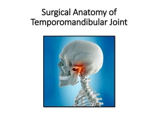
Surgical Anatomy of Temporomandibular Joint
•Download as PPTX, PDF•
7 likes•1,306 views

Detailed discussion on surgical anatomy of TMJ and its applied aspects. Surgical approaches are also discussed.

Recommended
More Related Content
What's hot
What's hot (20)
Similar to Surgical Anatomy of Temporomandibular Joint
Similar to Surgical Anatomy of Temporomandibular Joint (20)
More from Dibya Falgoon Sarkar
More from Dibya Falgoon Sarkar (18)
Recently uploaded
Recently uploaded (20)
Surgical Anatomy of Temporomandibular Joint
- 2. Introduction • Anatomy of the temporomandi- bular joint (TMJ) is highly specific and correlates with jaw function • TMJ is a ginglymoarthrodial joint, a term that is derived from ginglymus, meaning a hinge joint, allowing motion only backward and forward in one plane, and arthrodia, meaning a joint of which permits a gliding motion of the surfaces • The most important functions of the TMJ include mastication and speech
- 3. Bony Components Components of TMJ Soft-tissue components • Condyle of mandible • Articular eminence • Glenoid fossa • Articular disc • Joint capsule • Ligaments • Muscles
- 4. Unique features of TMJ •Its articular surface covered by fibrocartilage instead of hyaline cartilage. •The right and left TMJ form a bicondylar articulation and functions together as one unit. •Only joint in human body to have a rigid endpoint of closure that of the teeth making occlusal contact. •In contrast to other diarthrodial joints TMJ is last joint to start develop, in about 7th week in utero.
- 5. The Mandibular Condyle • Head of the condyle is oval in shape mediolaterally • 15-20mm long (M-L); 8-10mm wide (A-P); 8-120mm thick • Medial pole > Lateral pole • Posterior surface > Anterior surface • Articular surface is covered by fibrocartilage
- 6. Condylar Forms (According to Gray’s anatomy) Mainly 4 forms are seen 1. Convex-58% 2. Flat- 25% 3. Pointed-12% 4. Round- 3% ( mainly in children)
- 7. Cranial component of TMJ • Comprises of the articular suface of the temporal bone • Articular eminence: This is the entire transverse bony bar that forms the anterior root of zygoma. This articular surface is most heavily traveled by the condyle and disk as they ride forward and backward in normal jaw function • Articular tubercle 1. small, raised, rough, bony knob on the outer end of the articular eminence. 2. serves to attach the lateral collateral ligament of the joint. • Preglenoid plane Articular surface continuing anteriorly from the height of the articular eminence
- 9. Articular Disc • Biconcave, avascular fibro cartilaginous structure located between the mandibular condyle and glenoid fossa • functions to accommodate a hinging action as well as the gliding actions between the temporal and mandibular articular bone • Hinging movements take place in the lower compartment and gliding movements take place in the upper compartment. • The disc is attached all around the joint capsule except for the strong straps that fix the disc directly to the medial and lateral condylar poles, which ensure that the disc and condyle move together in protraction and retraction.
- 10. • Superior surface- Saddle-shaped • Inferior surface- Concave • Anteriorly – Attached to superior belly of lateral pterygoid • Posteriorly - Bilaminar region consists of two layers of fibers separated by loose connective tissue which is vascular. • Parts: 1. anterior band = 2 mm in thickness 2. posterior band = 3 mm thick, 3. Thin in the center intermediate band of 1 mm thickness. 4. posteriorly there is a bilaminar or retrodiscal region.
- 11. Ligaments related to TMJ Functions 1. Restricts and limits joint movements 2. Maintain joint spaces True ligaments: 1. Capsular ligament 2. Lateral/ Temporomandibular ligament Accessory ligaments: 1. Stylomandibular ligament 2. Sphenomandibular ligament
- 12. • Blood supply: Branches of External Carotid Artery: Mainly branches of superficial temporal artery Venous supply: 1. Maxillary vein 2. Tranverse facial vein 3. Superficial temporal vein. • Blood supply to TMJ is only superficial • TMJ takes its nourishment from Synovial fluid • Nerve supply of TMJ: 1. Auriculotemporal nerve 2. Masseteric nerve 3. Posterior deep temporal nerve
- 13. Factors maintaining stability of TMJ • Bones – Forward displacement prevented by articular tubercle and backward displacement is prevented by postglenoid tubercles • Temporomandibular Ligament • Protusion – Prevented by temporalis • Retrusion – Prevented by lat pterygoid • Occlusal intercuspation • Anterior dislocation is most common
- 14. Relations of TMJ • Anteriorly 1. Mandibular notch 2. Lateral pterygoid 3. Masseteric nerve and artery
- 15. Relations of TMJ Posteriorly 1. Parotid gland 2. Superficial temporal vessels 3. Auriculotemporal nerve
- 16. Relations of TMJ • Laterally – 1. Skin, superficial fascia, SMAS layer 2. Temporal branches of VII nerve 3. Parotid gland
- 17. Relations of TMJ • Medially – 1. Tympanic plate (separates from ICA) spine of sphenoid 2. Auriculotemporal nerve & Chorda tympani nerve 3. Middle meningeal artery &Internal maxillary artery
- 19. Important structures related to TMJ Nerves: 1. Facial nerve 2. Auriculotemporal nerve 3. Inferior alveolar nerve Vascular structures: • Middle meningeal artery • Internal maxillary artery • Superficial temporal artery • Masseteric artery
- 20. Facial Nerve • The main trunk of the facial nerve exits from the skull at the stylomastoid foramen. • Surgical landmarks for identification of facial nerve: • Tragal pointer - The main trunk of the facial nerve is located 1 cm anteroinferior and 1 cm deep to the tip of the tragal cartilage. • Digastric ridge- The main trunk is just superior to the attachment of the posterior belly of the digastric muscle to the digastric groove. This landmark also marks the approximate depth of the facial nerve. • Stylomastoid foramen - The base of the styloid process is 5 to 8 mm deep to the tympanomastoid suture line. The facial nerve can be identified as it exits the stylomastoid foramen and passes over the posterolateral aspect of the styloid process. • Tympanomastoid suture line - The nerve lies 6 to 8 mm deep to the inferior end of the tympanomastoid suture line. • Mastoid - For revision cases, extensive tumors or, as a last resort, a mastoidectomy can be performed to locate the vertical segment of the facial nerve, which can then be followed as it exits the mastoid.
- 21. Relation of VII nerve with TMJ • Approximately 1.3 cm of the facial nerve is visible to surgeon until it divides into temporofacial and cervicofacial branches • According to Alkayat & Bramley’s classic article (1979)
- 22. Radiologic study by Miloro et al
- 23. Applied Aspects • The temporal branch of the facial nerve emerges from the parotid gland and crosses the zygoma under the temporoparietal fascial to innervate the frontalis muscle (“corrugator muscle”) • Postsurgical palsy manifests as an inability to raise the eyebrow and ptosis of the brow. • Damage to the zygomatic branch results in temporary or permanent paralysis to the orbicularis oculi and may require temporary patching of the eye to prevent corneal desiccation and abrasion.
- 24. Auriculotemporal Nerve • first branch of the mandibular nerve after it exits the foramen ovale. • supplies sensation to the skin in the temporal and preauricular region, the anterior external meatus • The auriculotemporal nerve provides most of the innervation to the capsule of the temporomandibular joint itself • Branch of mandibular nerve, post division • Radiographic assessment of the exact course of this nerve through the mandible is necessary if screws will be placed through the ramus as is necessary for prosthetic joint replacement Inferior Alveolar Nerve
- 25. Vascular anatomy of TMJ • Superficial temporal artery and vein: are routinely ligated during preauricular approaches. • Internal maxillary artery: is usually not encountered unless a condylectomy is performed. If a condylectomy is performed, Dunn‐Dautrey retractors are used as the internal maxillary artery normally runs-approximately 3 mm medial from the midsigmoid notch.
- 26. Talebzadeh N, Rosenstein TP, Pogrel MA. Anatomy of the structures medial to the temporomandibular joint. Oral Surgery, Oral Medicine, Oral Pathology, Oral Radiology and Endodontics. 1999 Dec 1;88(6):674-8.
- 27. Masseteric artery A careful dissection of 16 intact human cadaveric head specimens was done. The location of the masseteric artery was then determined in relation to 3 points process: 1 ) the anterior-superior aspect of the condylar neck = 10.3 mm; 2 ) the most inferior aspect of the articular tubercle = 11.4 mm; 3 ) the inferior aspect of the sigmoid notch = 3 mm. Journal of Oral and Maxillofacial Surgery. 2009;67 (2) : 369–371
- 28. Surgical Approaches to TMJ 1. Preauricular : Dingman’s, Blair’s, Thoma’s, Al-kayat and Bramley’s, Popowitch’s 2. Postauricular 3. Endaural approach 4. Post ramal/ Hind’s approach 5. Submandibular/Risdon’s approach 6. Hemicoronal 7. Bicoronal
- 29. Preauricular Approach • Basic incision was given by Dingman (1951) • Blair & Ivy modification– “Inverted hockey stick “ incision. Facilitate exposure of arch along with condylar area. • Thoma in 1958 Angulated vertical incision. -carried out across zygomatic arch infront of ear to avoid zygomatic branch of facial nerve
- 30. Alkayat and Bramley Modified Preauricular Incision 1. Reduced haemorrhage 2. Provision of donor site for temporalis fascia 3. Better cosmetic result 4.Lesser chance of facial nerve injury
- 31. Facial nerve branches are not damaged
- 32. Post auricular approach • Introduced by Bockenheimer • Advantage: 1. Excellent exposure of the entire joint and the ability to camouflage the scar in patients who have a tendency to form keloids. 2. Better posterior exposure of TMJ • Disadvantage: 1. Auricular stenosis 2. Should not be used in the presence of joint infection or chronic otitis externa
- 34. Endaural Approach • Lempart (1938) • Short facial skin incision extending in to ext. auditory meatus • Advantage: Excellent cosmetics and posterolateral exposure • Disadvantage: 1. Meatal stenosis or chondritis 2. Injury to the branches of the facial nerve • Care must be taken not to incise the tragal cartilage because a perichondritis or poor wound healing may result.
- 35. Post-ramal/ Hind’s Approach • E.C Hinds & Girotin (1967) • Indications – 1. Condylar neck fractures 2. Condylotomy 3. Vertical ramus osteotomy • Incision- 1cm behind ramus of mand. and extends 1cm below the lobe of ear. • Advantages: Highly cosmetic, excellent visibility and accessibility. • Note: Injury may occur to posterior facial vein and main trunk of facial nerve.
- 36. Structures encountered during Hind’s approach
- 37. Combination of Retromandibular + Endaural Approach • Indication: For additional access to the temporomandibular joint for open fracture reduction, costochondral grafting, total alloplastic joint reconstruction, or tumor resection. • When combining both incisions, the surgeon must leave an intervening bridge of tissue that extends inferiorly at least 3 cm from the lowest point of the bony external auditory canal to protect VII nerve
- 38. Submandibular Risdon’s Approach •Risdon (1934) •Mainly used for neck of condyle & ramus region •Incision: Placed one‐finger breadth below the angle of the mandible ensuring avoidance of the marginal mandibular nerve
- 40. Dingman RO, Grabb WC. Surgical anatomy of the mandibular ramus of the facial nerve based on the dissection of 100 facial halves. Plastic and Reconstructive Surgery. 1962 Mar 1;29(3):266-72. • The marginal mandi- bular(MMN) branch of the facial nerve, posterior to the facial artery, passed above the inferior border of the mandible in 81% of dissections • It ran superficial to the facial vein in 98% cases • Damage to MMN: The patient is unable to depress the lower lip and show the mandibular anterior teeth.
- 41. Intraoral Approach • Incision along the external oblique ridge. A subperiosteal dissection proceeds medially and laterally along the mandibular ramus • The coronoid process and condylar neck can be reached through this incision. • Advantages: Decreased facial and auriculotemporal nerve injury and improved scar placement. • Limitations: limited access to the structures superior to the condylar neck.
- 42. Conclusion • The incisions which are chosen to approach the TMJ are established in accordance with requirements of regional anatomy and function. • Ideally, the selected approach should accomplish the following: 1. maximize exposure for the specific procedure; 2. avoid damage to the branches of the facial nerve, major vessels (e.g., internal maxillary artery and retromandibular vein), parotid gland, and ear; 3. maximize use of natural skin creases for cosmetic wound closure. • .
- 43. References • Atlas of Temporomandibular Joint Surgery (2nd edition) – Peter Quinn • Al‐Kayat A, Bramley P. (1980) A modified preauricular approach to the temporomandibular joint and malar arch. Br J Oral Surg, 17:91. • Dingman RO, Grabb WC. (1962) Surgical anatomy of the mandibular ramus of the facial nerve, based on the dissection of 100 facial halves. Plast Reconstr Sur, 29:266. • Talebzadeh N. et al. (1999) Anatomy of the structures medial to the temporomandibular joint. Oral Surg Oral Med Oral Pathol Oral Radiol Endod, 88:674. • Kreutziger KL. Surgery of the temporomandibular joint. I. Surgical anatomy and surgical incisions. Oral Surgery, Oral Medicine, Oral Pathology and Oral Radiology. 1984 Dec 1;58(6):637-46. • Textbook of Human Anatomy by A.K. Dutta
Editor's Notes
- The articular surface of the temporal bone is situated on the inferior aspect of temporal squama anterior to tympanic plate.
- Upper lamina of Disc > squamotympanic fissure Lower lamina of Disc > Neck of condyle
- Spheno liga. -The ligament is pierced by the myelohyoid nerve and vessels. This ligament is passive during jaw movements, maintaining relatively the same degree of tension during both opening and closing of the mouth. Stylo liga.- specialized dense, local concentration of deep cervical fascia……This ligament becomes tense only in extreme protrusive movements.
- . By incising the superficial layer of the temporalis fascia and the periosteum over the arch inside the 8 mm boundary, surgeons can prevent damage to the branches of the upper trunk Distance from the lowest point of the external bony auditory canal to the bifurcation was found to be 1.5–2.8 cm (mean, 2.3 cm) Distance from the postglenoid tubercle to the bifurcation was 2.4–3.5 cm (mean, 3.0 cm) Distance between the most anterior concavity of the bony external auditory canal the point on the lateral surface of the malar arch midway between its upper and lower border, where the most posterior twig of the temporal ramus of the facial nerve crosses the arch was 8-35mm
- There is minimal bleeding and less sensory loss. The posterior placement of the skin incision and its wide backwards and upwards sweep spares the main branches of vessels and nerves. Fascial places are easily identified. There is excellent visibility. This is partly due to the large flap and partly because the unyielding temporal fascia is not reflected with the skin as in the approach described by Rowe and Killey (1968). The potential complications of muscle herniation and fibrosis are avoided. The muscle is never exposed and the superficial layer of temporal fascia can be closed without tension. (5) There is remarkably little post-operative discomfort or swelling. (6) A good cosmetic result is achieved except in the very bald. (7) The technique is easily teachable and speedily execute