
Recommended
More Related Content
Similar to Activity 4 Understanding the basics of meiosisMitosis and mei.docx
Similar to Activity 4 Understanding the basics of meiosisMitosis and mei.docx (20)
More from coubroughcosta
More from coubroughcosta (20)
Recently uploaded
Recently uploaded (20)
Activity 4 Understanding the basics of meiosisMitosis and mei.docx
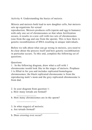
- 1. Activity 4: Understanding the basics of meiosis Mitosis and meiosis both lead to new daughter cells, but meiosis sets up organisms for sexual reproduction. Meiosis produces cells (sperm and egg in humans) with only one set of chromosomes so that when fertilization occurs, it results in a new cell with two sets of chromosomes (one from the egg and one from the sperm). This is how there is genetic recombination of DNA resulting in unique individuals. Before we talk about what can go wrong in meiosis, you need to be clear about the process itself and how genetic recombination in particular occurs. To this end, complete the following set of questions. Questions 1. In the following diagram, draw what a cell with 1 chromosome would look like in the stages of meiosis. Prophase 1 is filled in for you and includes replicated homologous chromosomes; the black replicated chromosome is from the reproducing male’s mom and the grey replicated chromosome is from dad. 2. In your diagram from question 1: a. How many tetrads are formed? _______________ b. How many chromosomes are in the sperm? _______________ 3. In what stage(s) of meiosis: a. Are tetrads formed? ________________________________ b. Does crossing over occur?
- 2. ________________________________ c. Do the chromosomes move to the poles? ________________________________ d. Do replicated chromosomes separate? ________________________________ e. Does the cytoplasm divide? ________________________________ 4. What processes in meiosis result in genetically unique daughter cells? When do these processes occur? (Note: There are two main processes; discuss both). 5. Compare and contrast meiosis with mitosis to complete the following table. Table 2. Comparison of key characteristics between meiosis and mitosis. Characteristics Mitosis Meiosis Type of organisms it occurs in # of chromosomes in human parent cell Number of times chromosomes replicate
- 3. Number of cell divisions Crossing over occurs? (Y/N) Type of daughter cells produced Number of daughter cells produced Daughter cells identical to parent cell? (Y/N) Daughter cells are: 1n or 2n? # of chromosomes in human daughter cells Activity 3: Chromosomes in mitosis and meiosis In the nucleus of the cell are the chromosomes that are composed of the hereditary material DNA. In every somatic
- 4. (body) cell of a human there are 46 chromosomes. Each species may have a different number of chromosomes than another species. Since each somatic cell of an organism contains the same number of chromosomes, there must be a duplication of material before the nucleus divides during mitosis. In each somatic cell, there are two sets of chromosomes; this is referred to as the 2n (diploid) number, in which n means number of chromosomes. In humans, 2n = 46 chromosomes. In each gamete (sex) cell, there is only one set of chromosomes; this is referred to as the 1n (haploid) number. In humans, 1n = 23 chromosomes. This means there are 23 different types of chromosomes in the nucleus of a human cell. The autosomes (non-sex chromosomes; i.e., everything but X and Y) are numbered from 1-22 according to their length and centromere position (part of the chromosome that links sister chromatids); the sex chromosomes (i.e., X and Y) are number 23. Karyotypes are used to examine the number and appearance of chromosomes. In the adjacent karyotype, the chromosomes are laid out in order by size for the autosomes followed by the sex chromosomes. There are two chromosomes of each type (2n); one came from this person’s mother through the egg (1n) and the other from her father through the sperm (1n) during fertilization. This is the karyotype of a somatic cell that is beginning mitosis; all chromosomes have been replicated. In this karyotype, the sex chromosomes are XX, so these chromosomes belong to a female (males would have XY). Karyotypes are a key tool used to detect
- 5. chromosomal abnormalities. They can be performed on a variety of tissues, including amniotic fluid (amniocentesis), placenta (chorionic villus sampling), blood (venipuncture), or bone marrow (biopsy). Monosomy refers to a condition in which there is one chromosome missing. It is abbreviated 2n-1. For example, monosomy X is a condition in which cells have only one X chromosome. Since they don’t have a Y chromosome, the individual will be female. Trisomy refers to a condition in which there is one extra chromosome. It is abbreviated 2n+1. For example, trisomy X is a condition in which cells have three X chromosomes; the individual will be female. Monosomies and trisomies usually result from nondisjunction during meiosis but can also occur during mitosis. They are more common in meiosis 1 than meiosis 2. They are generally lethal, with the exceptions of those involving sex chromosomes, chromosome 21, or, very rarely, chromosomes 13 and 18. Affected individuals have a distinctive set of physical and mental characteristics called a syndrome. Down syndrome is a developmental disorder generally caused by an extra copy of chromosome 21. Having an extra copy of this chromosome means that individuals have three copies of each of the genes on that chromosome instead of two, making it difficult for cells to properly control how much protein is made. Producing too much (or too little) protein contributes to the various symptoms of Down syndrome. Trisomy 21 is the cause in approximately 95% of individuals, with 88% coming from nondisjunction during the development of the mother’s egg, 8% from nondisjunction in the sperm, and the remainder from problems in fertilization or mitosis. Nondisjunction during meiosis 1 in the mother’s gamete results
- 6. in 67-73% of trisomy 21 humans. The following diagram depicts what could happen during meiosis 1 to cause Down syndrome. Only chromosome 21 is represented. The top two eggs would result in trisomy 21 when fertilized by a normal sperm. The bottom two eggs would result in monosomy 21 when fertilized by a normal sperm and would not develop into a viable zygote. Questions 1. How many chromosomes are in somatic cells of individuals with Down syndrome? 2. In the adjacent karyotype, a. What is the individual’s gender? b. What is the evidence of Down syndrome? 3. Maternal meiosis 2 nondisjunction results in 18-20% of trisomy 21 humans. In the following diagram, draw what could happen during meiosis 2 to cause Down syndrome. Prophase 1 is filled in with the replicated chromosomes for chromosome 21; don’t worry about drawing the other chromosomes. 4. People with Down syndrome can reproduce, and frequently their children do not have Down syndrome. Fill in the following diagram and explain why this could be. In the diagram, Prophase 1 shows the black replicated chromosomes 21 were inherited from the mother and the gray replicated chromosome 21 from the father; don’t worry about the other chromosomes. Explanation:
- 7. 6 CBIO Lab: Mitosis and Meiosis