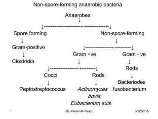
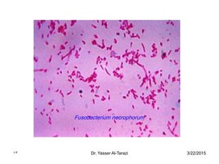
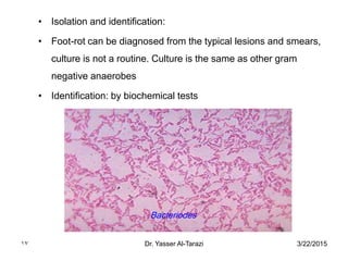
2. 3/22/2015Dr. Yasser Al-Tarazi2
• Characteristics:
• Constitute a large group of organisms that are exist in
nature, particularly in the intestinal tract of animals.
• Its isolated from many necrotic and suppurative
infections in humans and animals either alone or with
aerobic or facultative anaerobe. These bacteria live at
lower tissue oxidation-reduction potential.
• Infections caused by such bacteria are usually sporadic.
3. 3/22/2015Dr. Yasser Al-Tarazi3
• Nonspore-forming, anaerobic, gram negative rods bacteria causing
diseases in humans and animals are within the family
Bacteriodaceae, which contains many other non-pathogenic species
• Family: Bacteriodaceae
• Contains 13 genera, two of them are of veterinary importance:
• 1. Bacteriodes and 2. Fusobacterium
• All are gram negative rods that occur as normal flora of the intestinal
tract, oral cavity, pharynx, lower genitourinary tract and skin of man
and animals
• 1. Bacteriodes:
• e.g. B. fragilis, B. nodosus, B. corrodens……etc. It causes intestinal
infections, osteomyelitis, abscess and summer mastitis
4. 3/22/2015Dr. Yasser Al-Tarazi4
Fusobacterium:•
• 2. Fusobacterium
• F. necrophorum: cause affections in animals (thrush in
horses.) and in humans; sore throats, peritonsillar
abscesses, meningitis, urogenital and the
gastrointestinal tracts.
• F. nucleatum: cause affections mostly in humans
(periodontal disease, and preterm births) and
associated with colon cancer.
5. • Gram-positive anaerobic cocci:
• Contains four genera:
• Peptococcus, Peptostreptococcus and some other species of
Staphylococcus and Streptococcus.
• Peptococcus niger is rarely isolated from animals
• Peptostreptococcus anaerobius was isolated from vet. clinical cases
• Peptocus indolicus implicated in cow mastitis,
• Non of anaerobic streptococcus are consider as animal pathogens
• Aerotolerent streptocococci (2 spp.) have been recovered from vet
clinics
3/22/2015Dr. Yasser Al-Tarazi5
6. 3/22/2015Dr. Yasser Al-Tarazi6
Nonspore-forming gram positive rods
• Actinomyces pyogenes (C. pyogenes) is a facultative anaerobe
• Eubacterium suis (anaerobic) cause cystitis and pyelonephritis in
swine
• Others; Lactobacilli: frequently isolated from animals but they are
non-pathogens
• Propionic bacterium: associated with skin and dairy products and
not pathogenic
7. 3/22/2015Dr. Yasser Al-Tarazi7
• Habitat:
• Commensals on mucous membrane of upper respiratory tract,
genital and alimentary tract of animals. Make ~ 90% of the intestinal
flora (predominant in large bowel and rumen) and have vital role in
digestion.
• Infections may involved by Non spore-forming G +ve anaerobes:
• Invasion of necrotic tissues or as a secondary invader, Necrotic,
gangrenous (often with clostridia) and suppurative infections
• Abscess in the lung, liver, brain, pyometritis and less frequently
cystitis, urinary tract infections, post-surgical abscesses, diarrhea,
pneumonia, abortion, foot rot of cattle, cellulites, periodontal
abscesses ……etc
8. 3/22/2015Dr. Yasser Al-Tarazi8
• Isolation of anaerobes:
• By anaerobic jar, using different system to eliminate oxygen e.g.
gas-pack and Brewer system
• Media containing reducing agents e.g. cooked meat media,
thioglycollate semisolid media
• Specimens:
• Fluid submitted in a syringe
• Specimens not in anaerobic system should be kept at room
temperature, because oxygen absorption increased in cold
temperature and chilling is harmful to some of anaerobes
9. 3/22/2015Dr. Yasser Al-Tarazi9
• Fusobacterium necrophorum (sphaerophorus necrophorus)
• Distribution and infections:
• Worldwide distributed, commensals in the alimentary tract and
mucous membrane and the infections is endogenous
• Antigens: four biotypes
• Biotype A: isolated in pure culture from bovine liver abscess
• Biotype B: predominant in ruminal contents and lesions and isolated
from liver abscess
• Biotype AB: Not isolated from animals
• Biotype C: A virulent strain
10. 3/22/2015Dr. Yasser Al-Tarazi10
• Pathogenesis and Pathogenicity:
• Invade and multiply in the anaerobic conditions provided by
damaged tissues.
• Infections are mixed and characterized by necrotic process e.g. liver
abscess in cattle caused by F. necrophorum and actinomyces
pyogenes
• Lesions are produced by endotoxin, leukotoxin and hemolysin. In
addition to hemagglutinin, adhesin, platelets aggregation factor,
protease and DNAses
• Capsulated strains are more virulent. F. necrophorum is a
secondary invader in necrotic stomatitis, pharyngitis and enteritis of
swine. F. necrophorum infection is known as necrobacillosis
11. 3/22/2015Dr. Yasser Al-Tarazi11
• Some of diseases with which F. necrophorum is associated in
animals:
• Horses: Thrush (disease of hoof), pneumonia and septicemia
• Cattle: calf diphtheria, meteritis, cellulites, mastitis, necrotic lesions,
liver abscesses and foot-rot
• Sheep: lip and leg ulcerations, foot abscess (ovine interdigital
dermatitis) and abortion
• Swine: bull-nose and swine dysentery
• Fowl: Avian diphtheria
• Humans: Abscesses, oral lesions and thrombophlebitis
12. 3/22/2015Dr. Yasser Al-Tarazi12
• Specimens:
• Affected tissues, pus materials either cultured directly or submitted
to laboratory in a syringe
• Direct examination: Beaded Gram negative rods of variable length,
Isolation and cultivation:
• In strict anaerobic conditions for 2-4 days it reveal L – form bacteria
• Colonies are small, smooth, convex, whitish-yellow color with α or β
– haemolysis. Initial cultures may be pleomorphic, short rods and
non-branched filaments
• F. necrophorum isolated in pure culture from liver abscess
• Identifications: according to biochemical characters
14. 3/22/2015Dr. Yasser Al-Tarazi14
• Treatment and control:
• By surgery
• Sulfonamide, penicillin, tetracycline and erythromycin are effective
• Aminoglycoside (neomycin, gentamycin, tobramycin, amikacin,
kanamycin) are not effective
• Tetracycline and tylosin in feed are effective
• No vaccine available
• Toxoids provide some protection in cattle
15. 3/22/2015Dr. Yasser Al-Tarazi15
• Bacteriodes nodosus "Fusiformis nodosus"
• Characteristics:
• Large gram negative, non-motile, anaerobic rods. It’s the primary
cause of contagious foot-rot of sheep. F. necrophorum, Actinomyces
pyogenes are common secondary invaders. Its also cause infections
of the foot in goats, pigs and cattle.
• Virulence associated with production of proteolytic enzymes
resulting in breakdown of keratin. The disease is aggravated by
moist conditions. Virulent strains posses pili that attach and colonize
the epidermal matrix of ovine hoof
16. 3/22/2015Dr. Yasser Al-Tarazi16
• Antigens:
• Based on pili, ten serogroups identified "A, B, C, ………H" with
agglutination test or ELISA
• Direct examination:
• After pared the hoof, specimen taken from lesion. Gram stain
revealed gram negative rods, slightly curved, no spores with Loffler's
methylene blue, red-staining granules appear
17. 3/22/2015Dr. Yasser Al-Tarazi17
• Isolation and identification:
• Foot-rot can be diagnosed from the typical lesions and smears,
culture is not a routine. Culture is the same as other gram
negative anaerobes
• Identification: by biochemical tests
Bacteriodes
18. • Treatment and control:
• Foot trimming before treatment, formalin, copper or zinc
sulfate foot bath and 10% tincture of chloromycetin are
used.
• The organism survive not more than 2-weeks in pasture.
• Systemic antibiotics are of value accompanied with hoof
treatment. Oil-adjuvant B. nodosus vaccine is useful but
painful and form abscess. Pilus antigen vaccine provide
significant protection
3/22/2015Dr. Yasser Al-Tarazi18