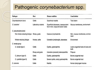
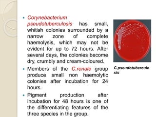
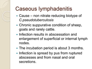
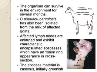
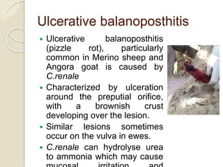
2. Introduction
Coryneform bacteria encompass several
genera, of which Corynebacterium is the
most frequently encountered in clinical
infections.
Coryneform bacteria are characterized as
irregularly shaped, non–spore-forming,
aerobic, gram-positive rods.
The following genera are covered under
coryneform bacteria:
Corynebacterium, Turicella, Arthrobacter,
Brevibacterium, Dermabacter.
6. Corynebacteria are considered to belong
to a group of organisms within the
corynebacteriaceae known as the CMN
group, which contains genera
Corynebacterium, Mycobacterium,
Nocardia and Rhodococcus.
High G+C content and their cell walls contain
mycolic acids.
Although the group is heterogenous.
Mycolic acids of the corynebacteria are
shorter than those of other genera within
the group and the carbon chains are usually
saturated.
8. Differentiation of the
corynebacteria
Most pathogenic corynebacteria are
relatively host specific and produce
identifiable clinical syndromes.
The host species and the nature of the
disease may suggest the causal agent.
Identification criteria include bacterial cell
morphology, colonial appearance and
biochemical reactions.
An enhancement of haemolysis test is used
for the identification of
C.pseudotuberculosis.
9. Colonial characteristics:
Corynebacterium bovis is a
lipophilic bacterium which
produces small, white, dry,
non-haemolytic colonies in
plates inoculated with a bovine
milk sample.
Corynebacterium kutscheri
produces whitish colonies.
Occasional isolates are
haemolytic.
10. Corynebacterium
pseudotuberculosis has small,
whitish colonies surrounded by a
narrow zone of complete
haemolysis, which may not be
evident for up to 72 hours. After
several days, the colonies become
dry, crumbly and cream-coloured.
Members of the C.renale group
produce small non haemolytic
colonies after incubation for 24
hours.
Pigment production after
incubation for 48 hours is one of
the differentiating features of the
three species in the group.
C.pseudotuberculo
sis
11. Biochemical reactions:
Conventional or commercially available
biochemical tests can be used to
differentiate the corynebacteria.
Two biotypes of C. pseudotuberculosis are
recognised.
The ovine/caprine strains lack nitrate
reducing capacity, while the equine/bovine
strains usually reduce nitrate.
Cross-infection by biotypes is thought to be
minimal.
12. The biochemical reactions used to
distinguish members of the C. renale group
are indicated in figure.
•Urease is produced by all pathogenic
corynebacteria with the exception of C.
bovis.
13. Enhancement of haemolysis test:-
The haemolysis produced by
C.pseudotuberculosis is enhanced when
the organisms are inoculated across a
streak of Rhodococcus equi
14. Virulence Factors
• Intra - host spread of C. pseudotuberculosis
depends on PLD (Phosholipase D)
production
• Potent exotoxin has vasodilatory properties.
• Dermonecrosis, phagocyte toxicity,
complement-dependent lysis of autologous
erythrocytes, and lethality at higher doses.
• Activities of PLD commonly used for
identification of C. pseudotuberculosis are
synergistic hemolysis with Rhodococcus
equi cholesterol oxidase and inhibition of
the staphylococcal β - hemolytic activity
15. Contd..
Cell wall lipids of C. pseudotuberculosis
have been considered virulence factors
They protect the bacteria from
intraphagocytic destruction.
Induce hemorrhagic necrosis and
chronic abscessation.
Iron acquisition is vital for
C.pseudotuberculosis ; the fagBCD
operon, which encodes a putative iron
uptake system, has been proposed as a
virulence factor.
16. Pathogenesis and pathogenicity
Corynebacteria, with the exception of C.
bovis, are pyogenic organisms that
cause a variety of suppurative conditions
in domestic animals.
Corynebacterium bovis, found in the teat
canal of up to 20% of apparently healthy
dairy cows, provokes a mild neutrophil
response.
C. pseudotuberculosis facultative
intracellular pathogen capable of
surviving and replicating in phagocytes.
17. CONTD..
Virulence linked to its cell wall lipid and to
the production of an exotoxin,
phospholipase D (PLD)
PLD hydrolyses sphingomyelin in
mammalian cell membranes, releasing
choline
In the early stages of infection, PLD may
enhance survival and multiplication in the
host
C. renale group are urinary tract pathogens
which cause cystitis and pyelonephritis in
cattle
produce urease, which hydrolyses urea
18. Minor trauma to the skin may allow entry
of C. kutscheri and C. pseudotuberculosis,
whereas the urinary tract pathogens avail of
diminished immunological defenses or local
tissue damage following parturition
19. Diagnostic procedures
The species of animal affected and the
clinical signs may suggest a specific
diagnosis.
Suitable specimens for laboratory
examination include pus, exudate, samples
of affected tissue and mid-stream urine.
Direct microscopic examination of Gram-
stained smears from specimens may reveal
coryneform organisms.
Culture media for routine use include blood
agar, selective blood agar and MacConkey
agar.
For primary isolation; Loeffler agar, Mueller-
20. Inoculated plates are incubated aerobically
at 37°C for 24 to 48 hours.
Identification criteria for isolates:
-Colonial characteristics
-Presence or absence of haemolysis
-Aerobic or anaerobic incubation
requirements
-Absence of growth on MacConkey agar
-Typical coryneform pleomorphism in a Gram
stained smear from culture.
-Specific tests for distinguishing members of
the C. renale group.
-Enhancement of haemolysis test for
21. Ulcerative lymphangitis
Cause -Corynebacterium
pseudotuberculosis
Ulcerative lymphangitis occurs in Africa, the
Americas, the Middle East and India. In the
USA, the disease is prevalent in autumn
and early winter and is more common in
horses than in cattle.
Infection occurs through skin wounds,
arthropod bites or by contact with
contaminated harness.
22. The condition presents either
as lymphangitis of the lower
limbs or abscessation in the
pectoral region.
Infection in cattle manifests
as lymphadenitis and
lymphangitis with abscess
formation and ulceration.
Lesions of the coronary band
with resulting lameness in
affected dairy cattle have
been reported
23. Based on isolation and identification of
C.pseudotuberculosis from lesions, since
lymphangitis can also result from infection
with other pyogenic bacteria.
CONTROL
Systemic antibiotic therapy may be
combined with topical treatment using an
iodophor shampoo.
Affected animals must be isolated and
contaminated areas should be disinfected.
Diagnosis
24. Caseous lymphadenitis
Cause – non nitrate reducing biotype of
C.pseudotuberculosis
Chronic suppurative condition of sheep,
goats and rarely cattle.
Infection results in abscessation and
enlargement of superficial or internal lymph
nodes.
The incubation period is about 3 months.
Infection is spread by pus from ruptured
abscesses and from nasal and oral
secretions.
25. The organism can survive
in the environment for
several months.
C.pseudotuberculosis
has also been isolated
from the milk of affected
goats.
Affected lymph nodes are
enlarged and exhibit
characteristic
encapsulated abscesses
which have an 'onion ring'
appearance in cross-
section.
The abscess material is
caseous, initially greenish
26. Diagnosis
The disease may be suspected on clinical
grounds or at postmortem examination.
Smears from lesions may reveal Gram-
positive bacteria.
Isolation and identification of
C.pseudotuberculosis from abscess
material is confirmatory.
A sandwich ELISA, which detects
circulating antibodies directed against the
exotoxin (PLD), has been developed for
identifying infected sheep.
27. Bovine pyelonephritis
Organisms belonging to the C. renale group
can be isolated from the vulva, vagina and
prepuce of apparently normal cattle.
The stress of parturition and the shortness
of the urethra in the cow predispose to
infection of the urinary tract.
Ascending infection from the bladder
through the ureters can result in
pyelonephritis.
28. Clinical signs
Fever, anorexia and decreased milk
production.
Restlessness and kicking at the abdomen
may indicate renal pain.
Dysuria, an arched back and blood-tinged
urine are invariably present.
Longstanding infections lead to extensive
renal damage.
29. Diagnosis
Clinical signs may suggest urinary tract
disease.
Thickened ureters and enlarged kidneys
may be detected by rectal palpation.
The condition is often unilateral.
Red blood cells and protein are present in
the urine.
Culture of C. renale from urinary deposits,
in association with characteristic clinical
signs, is confirmatory.
30. Ulcerative balanoposthitis
Ulcerative balanoposthitis
(pizzle rot), particularly
common in Merino sheep and
Angora goat is caused by
C.renale
Characterized by ulceration
around the preputial orifice,
with a brownish crust
developing over the lesion.
Similar lesions sometimes
occur on the vulva in ewes.
C.renale can hydrolyse urea
to ammonia which may cause
31. C.renale can hydrolyse urea to ammonia
which may cause mucosal irritation and
ulceration.
A high urinary urea level, a consequence of
high protein intake, may predispose to the
development of disease.
Animals grazing pastures containing high
oestrogen levels are also prone to the
condition.
Castrated sheep are affected more
frequently than rams.
A heavy wool or mohair cover around the
prepuce predisposes to infection.
Untreated cases may progress to total