SlideShare a Scribd company logo
1 of 60
Download to read offline
Cavities of body
Peritoneum
• Large serous sac
• Lines inner surface of abdomino-
pelvic wall
Why different layers?
• Invaginated by viscera from
different sides
• Invagination throws the peritoneum
into folds &
• Forms
• Parietal & visceral layers
• & potential spaces
Peritoneum - parts
• Parietal Peritoneum
• Lines the inner surface of
• Abdominal and pelvic walls and
• Lower surface
• Diaphragm
• Embryologically from somatopleuric
mesoderm
• Derives blood & nerve supply from body
wall
• Visceral Peritoneum
• Lines outer surface of viscera
• Firmly attaches with viscera
• Develops from splanchnopeuric
mesoderm
• Blood supply and nerve supply are the
same as those of underlying viscera
Interperitoneal organs
• Most part of viscera surrounded by
peritoneum
• Stomach, Liver, gallbladder
• Tail of pancreas, spleen
• First part of duodenum, jejunum, ileum
• Transverse colon, sigmoid colon
• Upper part of rectum, urinary bladder and
uterus
Retroperitoneal organs
• Organs lie on the posterior abdominal
wall are covered by peritoneum on
their anterior surfaces
• Descending and horizontal parts of
duodenum, pancreas (head, neck & body)
• Ascending & descending colon
• Kidney, suprarenal gland, ureters
• Abdominal aorta & IVC
• Middle and lower parts of rectum
Secondary retroperitoneal organs
• Duodenum
• 2nd and 3rd part
• Pancreas (except tail)
• Ascending colon
• Descending colon
• Upper rectum
Folds of Peritoneum -
types
• Many organs within abdomen are
suspended by folds of peritoneum
• Provide pathways for passage of
vessels, nerves and lymphatics
• Named after their attached organs
• Omentum
• Fold of peritoneum connecting stomach to
other organs
• Mesenteries
• Fold invests small intestine
• Mesocolon
• Fold invests large intestine
• Peritoneal ligaments
• Folds attached with other organs
Disposition – Vertical
• Start from
• Anterior abdominal wall
• At umbilical level
• Lines
• Inner aspect of anterior abdominal wall
• Trace
• Upto ant part of diaphragm (under surface)
• Right of median plane
• Falciform ligament
• In free margin it contains
• Ligamentum teres hepatis
• Obliterated left umbilical vein (extends
from umbilicus to inferior surface of
liver)
• Also it contains para umbilical veins
• Tributaries to left branch of Portal vein
Disposition – Vertical
• Inferior surface of diaphragm to upper
surface of liver
• Falciform ligament
• 2 layers diverge to right & left
• On right (right layer of falciform)
• From diaphragm to right lobe of liver forms
• Superior layer of coronary ligament
• On left (left layer of falciform lig)
• From diaphragm to left lobe of liver forms
• Left triangular ligament (anterior layer)
Disposition – Vertical
• After covering the superior and anterior
surfaces of liver
• Reaches inferior surface of liver
• Right of Gall bladder
• Peritoneum passes down to
• Suprarenal & kidney as inferior layer of
coronary ligament
• Bare area
• Between the 2 layers of coronary ligament
• Right triangular ligament
• 2 layers of coronary ligament unite to form
• Connects right lobe of liver with diaphragm
Disposition – Vertical
Ligamentum venosum - remnant of ductus venosus
(oblitrated ductus venosus), which shunted blood from
the umbilical vein to the IVC in fetal life
• On the left of gall bladder
• After covering quadrate lobe & left lobe
• Becomes posterior layer of left triangular ligament
• Then passes to
• Left margin of fissure for ligamentum venosum & anterior lip
of porta hepatis &
• From anterior lip of porta hepatis to right and then posterior lip
of porta hepatis
• Covering caudate lobe & attached to right lip of fissure for
ligamentum venosum
• These two layers form
• Lesser omentum
• Has inverted L shaped attachment
Omentum
• Peritoneal folds related to stomach
• Lesser omentum
• Greater omentum
Lesser omentum
• Connects liver to stomach
• Attachment
• Above to porta hepatis & fissure for
ligamentum venosum
• Inferiorly to lesser curvature of the
stomach & 1st inch of duodenum
• Parts
• Hepato gastric ligament
• Hepato duodenal ligament
• Forms free margin
• Contains
• Portal vein (posteriorly)
• Hepatic artery (anterior & left)
• Bile duct (anterior & right)
Lesser omentum
• Contents
• Hepatic artery, bile duct, portal vein
• In free margin -
• Rt. & lt. gastric vessels
• Along lesser curvature
Tracing lesser omentum
• Peritoneum from
• Anterior lip of porta hepatis and left lip of
fissure for ligamentum venosum
• Posterior lip of porta hepatis and right lip
of fissure for ligamentum venosum
• Continues downwards as lesser
omentum towards lesser curvature of
stomach and first one inch of
duodenum
• Forms anterior boundary for upper part
of lesser sac
Tracing lesser omentum
• After reaching lesser curvature
• Two layers separates
• And covers the surfaces
• Reaches greater curvature
• Becomes anterior two layers of greater omentum
Tracing greater
omentum
• From greater curvature
• Anterior & posterior layers
comes downwards
• After awhile it returns back
towards anterior border of
pancreas
• So 4 layers are there
• Together they form Greater
omentum
• Space between 2nd & 3rd layer is
lesser sac
• Patent in fetal life
• But obliterated - after birth
Fate of 3rd & 4th layers
• Continuation of layers of lesser
omentum
• Forms 1st & 2nd layer of greater omentum
• While returns back to pancreas
• 1st layer becomes 4th layer
• 2nd layer becomes 3rd layer
• Third layer
• Runs upwards towards anterior border of
pancreas (infront of transverse colon)
• Passes infront of
• Anterior surface of pancreas, kidney and
suprarenal gland
• Reaches the liver
• Forms posterior wall of lesser sac
Fate of 3rd & 4th layers
• 4th layer
• Runs towards anterior surface of head &
anterior border of body of Pancreas
• Then comes down to transverse colon as
upper layer of Transverse mesocolon
• Encloses transverse colon
• Then goes towards inferior surface of
pancreas – as inferior layer of
Transverse mesocolon
Fourth layer of Greater omentum and
Superior layer of Transverse mesocolon are
adherent – but separable.
Greater omentum
• Largest fold of peritoneum
• Hangs down like an apron
• In front of coils of small intestine
• Made of 4 layers
• Anterior 2 layers from Greater
curvature and 1st part of duodenum
• Extending downwards for a considerable
distance &
• Fold upon themselves to be 3rd & 4th
layer
• To anterosuperior surface of
Tranasverse colon (head & ant border
of pancreas)
Greater omentum – parts &
contents
• Parts
• Gastrophrenic ligament
• Gastrosplenic ligament
• Gastrocolic ligament
• Contents
• Rt. &Lt. gastroepiploic vessels
• Lymph nodes
• Fat
• Functions
• Store house of fat
• Limits the spread of infection
Transverse mesocolon
• Suspends transverse colon
• Attached to anterior
aspect of head & anterior
border of body of
pancreas
• Contains –Middle colic
art.
Root of mesentery
• Peritoneum from transverse mesocolon
• Covers lower part of anterior surface of
head of pancreas
• Passes to front of 3rd & 4th parts of
duodenum
• Then covers posterior abdominal wall
• Becomes folded between
• Duodenojejunal flexure(left of L2) to
ileocecal junction (rt sacroiliac joint)
• Forms root of mesentery
Mesentery
• Fan-shaped fold of peritoneum
• Suspends the coils of jejunum and
ileum from posterior abdominal wall
• Has 2 borders
• Attached
• Free
Mesentery - borders
• Attached border
• Known as root of mesentery
• Lies obliquely
• Extent
• From
• Duodenojejunal flexure (L2 – left side)
• To
• Ileocecal junction (Rt. Sacroiliac joint)
• Length - 6 inches (15 cm)
• Structures crossed by it
• 3rd part of duodenum, Abdominal aorta, IVC
• Rt. Psoas major,
• Rt. Gonadal vessels, Rt. Genitofemoral nerve &
Rt. ureter (medial to lateral)
• Rt. Sacroiliac jt
Mesentery - borders
• Free border
• Encloses jejunum and ileum
• 6 meters in length
• Contents
• Jejunum, ileum
• Lymph nodes
• Jejunal & ileal branches of superior
mesenteric artery & nerve plexus
• Fat
Meso-appendix
• Δ-gular fold
• Suspends appendix
• From post surface of lower end
of mesentery
• Appendicular artery
Peritoneum in Pelvis
• After covers the structures of
posterior abdominal wall
• Reaches pelvis
• First invests sigmoid colon from pelvic
wall
• Sigmoid mesocolon
• Then covers Rectum
• Upper third on front & sides
• Middle part on front only
Tracing the peritoneum anteriorly differs
in male & female
Sigmoid
mesocolon
• Suspends sigmoid colon
• Attached to post abdominal and
pelvic walls like inverted V
• Apex of V lies on ureter at
bifurcation of Lt. com iliac
artery
• Lt limb – along upper ½ of Lt,
Ext iliac
• Contents – Superior rectal
vessels
Peritoneum - Pelvis
• In male
• From rectum to seminal vesicle
• Then upper surface of urinary bladder
• Forms rectovesical pouch
• From apex of UB to inner surface of
anterior abdominal wall till umbilicus
• In female
• From rectum to posterior fornix of vagina
• And covers supravaginal cervix, body and
fundus
• Forms rectouterine pouch (pouch of Douglas)
• Then covers anterior surface of body
• From body to UB
• Forms uterovesical pouch
Female pelvis
• Communicates with extraperitoneal
pelvis exteriorly
• Through fallopian tubes, uterus and
vagina
Folds in anterior abdominal wall
• Median umbilical fold
• Remnant of urachus (median
umbilical ligaments)
• Medial umbilical fold
• Remnants of umbilical arteries
(medial umbilical ligaments)
• Lateral umbilical fold
• Inferior epigastric vessels
Fossae of anterior abdominal wall
Peritoneal cavity - Divisions
• Transverse mesocolon divides the
peritoneal cavity in to
• Supramesocolic space
• Also known as subphrenic spaces
• Inframesocolic space
• Bilateral paracolic & pelvic spaces
are also peritoneal spaces
Supra colic space
• Also known as subphrenic spaces
• Boundaries
• Superiorly
• Diaphragm
• Inferiorly
• Transverse colon
• In front
• Ant abdominal wall
• Behind
• Lesser omentum, stomach, greater
omentum
Supracolic compartment
• Supracolic compartment contains
• Stomach, liver, and spleen
• Infracolic compartment contains
• Small intestine and ascending & descending
colons
• Infracolic compartment is divided into
• Right and left infracolic spaces by the
mesentery of small intestine
• Communication occurs between the
supracolic and infracolic compartments
• Through paracolic gutters
• Right
• Right anterior subphrenic
(subdiaphragmatic)
• Right posterior subphrenic (hepatorenal
or Morrisons pouch)
• Right extraperitoneal
• Left
• Left anterior intraperitoneal / left
suprahepatic / subphrenic
• Left posterior intraperitoneal / left
subhepatic
• Left extraperitoneal
Supracolic compartment - Spaces
Right anterior intraperitoneal / subphrenic / suprahepatic
• Presents
• Right of falciform ligament
• Boundaries
• In front
• Ant abdominal wall & diaphragm
• Behind
• Superior anterior & right lateral surface of right
lobe of liver, superior layer of coronary
ligament
• Below
• Communicates with right posterior
intraperitoneal compartment
• Above
• Diaphragm
Morrison’s Pouch / Hepatorenal pouch /
Rt subhepatic pouch
• Right posterior subphrenic space
• Most dependent part of peritoneal cavity in
upper abdomen
• Boundaries
• In Front
• Inferior surface of rt. lobe of liver and Gall bladder
• Behind
• Anterior surface of rt. kidney & rt. supra renal gland
• Rt. Colic flexure & 2nd part of duodenum
• Above
• Inf layer of coronary ligament
• Below
• Communicate with infracolic part of peritoneal cavity
• Left
• Communicate with omental bursa
• Right
• Limited by diaphragm
Morrison’s Pouch Hepatorenal pouch Rt subhepatic pouch
• Most dependent part of abdomen
proper
• Commonest site of subphrenic abscess
• From
• Perforated peptic ulcer of stomach &
duodenum
Right extraperitoneal space
• Bare area of liver presents
• Liver directly related to diaphragm
• Boundaries
• Left
• IVC
• Right
• Rt. triangular ligament
• Above
• Superior layer of coronary ligament
• Below
• Inferior layer of coronary ligament
• Clinical anatomy
• Common site for
• Amoebic liver abscess
Left anterior intraperitoneal
• Lies on left of falciform ligament
• Boundaries
• In front
• Ant abdominal wall
• Behind
• Lt triangular ligament
• Lt. lobe of liver
• Stomach & spleen
Left posterior intraperitoneal
• It is lesser sac
Lesser Sac
• Position
• Behind the lesser omentum and stomach
walls
Lesser sac
• Boundaries
• Superior
• Peritoneum which covers the caudate lobe of liver
and diaphragm
• Anterior
• Lesser omentum
• Peritoneum of posterior wall of stomach
• And anterior two layers of greater omentum
• Posterior
• Posterior two layers of greater omentum
• Transverse colon & transverse mesocolon
• Peritoneum covering pancreas, left kidney and
suprarenalgland
• Inferior
• Continuity of anterior and posterior two layers of
greater omentum
Lesser Sac
• Right border
• From above downwards
• Deficient above
• Epiploic foramen
• Peritoneum covering
• Posterior surface of proximal part of
duodenum to the head of pancreas
• Left border
• From above downwards
• Gastrophrenic ligament
• Gastrosplenic and lienorenal
ligament
• Left border of greater omentum
Parts of lesser sac
• Body
• Lies behind lesser omentum &
stomach
• Superior recess
• Lies behind caudate lobe of liver
• Splenic recess
• Between gastrosplenic and
lienorenal ligament
• Inferior recess
• Lies between the folds of greater
omentum
Epiploic foramen
• Communication between
• Lesser and greater sac
• Location
• T -12 vertebra level
• Boundaries
• Anteriorly
• Free margin of lesser omentum
• Contains
• Front right to left
• Bile duct & hepatic artery
• Behind
• Portal vein
• Posteriorly
• IVC (covered by peritoneum)
• Superiorly
• Caudate process of liver
• Inferiorly
• 1st part of duodenum
Epiploic foramen
Boundaries
• Anteriorly
• Free margin of lesser omentum
• Contains
• Front right to left
• Bile duct & hepatic
artery
• Behind
• Portal vein
• Posteriorly
• IVC (covered by peritoneum)
• Superiorly
• Caudate process of liver
• Inferiorly
• 1st part of duodenum
Applied anatomy
• Internal abdominal hernia
• Occasionally a loop of intestine may
enter into lesser sac
• Through epiploic foramen
• None of the boundaries can be incised
to reduce the hernia
• Pushing the herniated loop out of lesser
sac by inserting the index finger into it
through ant. 2 layers of greater omentum
Infracolic Compartments
• Lies below the transverse colon &
mesocolon and behind the greater
omentum
• Divided into w by root of mesentery
• Right infracolic Compartment
• Left infracolic Compartment
Right infracolic compartmet
• Boundaries
• Above
• Transvers colon & mesocolon
• On right
• Ascending colon
• On left
• Root of Mesentery
• Below
• Closed
Left infracolic compartmet
• Lies left of root of mesentery
• Boundaries
• In front
• Anterior abdominal wall
• Above
• Transverse mesocolon
• Below
• Open with pelvis
• On right
• Root of mesentery
• On left
• Descending colon
Paracolic gutters
• Longitudinal channels
• Lined by peritoneum
• Lies along the sides of ascending &
descending colons
• Right & left paracolic gutters
• Right paracolic gutter
• Lies lateral to ascending colon
• Communication
• Beween Hepato-renal pouch (above)
& pelvis (below)
• Left paracolic gutter
• Lies lateral to descending colon
• Above it is separated from
• Spleen & lienorenal space by
Phrenico-colic ligament
• Below it is communicated with
pelvis
•
Rectouterine Pouch (Pouch of
Douglas)
• Boundaries:
• Anteriorly: uterus and posterior fornix of vagina
• Posteriorly: rectum
• Inferiorly (floor): rectovaginal fold of
peritoneum
• Clinical anatomy
• Most dependent part of peritoneal cavity
• Pus collected here
• Can be drained through rectum or posterior fornix of
vagina
Applied anatomy
• Ascites
• Excessive accumulation of peritoneal fluid
in peritoneal cavity
• Commonly due to
• Cirrhosis
• TB & Malignancy
• Paracentesis
• Draining fluid
• Peritonitis
• Infection & inflammation of peritoneum
• May result in fatal
• Minimize the damage of peritoneum

More Related Content

What's hot

Superior and inferior mediastinum
Superior and inferior  mediastinumSuperior and inferior  mediastinum
Superior and inferior mediastinumMoamer Gabsa
 
Principles of abdominal anatomy
Principles of abdominal anatomyPrinciples of abdominal anatomy
Principles of abdominal anatomyjehh87
 
Peritoneal cavity and relations
Peritoneal cavity and relationsPeritoneal cavity and relations
Peritoneal cavity and relationsAmarSaleh1
 
Anatomy superior Mesenteric artery
Anatomy superior Mesenteric artery Anatomy superior Mesenteric artery
Anatomy superior Mesenteric artery Ameer Azeez
 
Peritoneum Dr. Mehul Tandel
Peritoneum Dr. Mehul TandelPeritoneum Dr. Mehul Tandel
Peritoneum Dr. Mehul TandelMehul Tandel
 
GI development embryology & Concept of Peritoneum
GI development embryology & Concept of PeritoneumGI development embryology & Concept of Peritoneum
GI development embryology & Concept of PeritoneumProf. Dr. Hironmoy Roy
 
Inguinal canal final
Inguinal canal finalInguinal canal final
Inguinal canal finalNamo9427
 
surgical anatomy Inguinal canal anatomy dr.vishnu
surgical anatomy Inguinal canal anatomy dr.vishnusurgical anatomy Inguinal canal anatomy dr.vishnu
surgical anatomy Inguinal canal anatomy dr.vishnuvishnu mohan
 
Inguinal canal- Anatomy
Inguinal canal- AnatomyInguinal canal- Anatomy
Inguinal canal- AnatomyDINESH KUMAR D
 
Surgical Anatomy of Esophagus
Surgical Anatomy of EsophagusSurgical Anatomy of Esophagus
Surgical Anatomy of EsophagusJohn Thanakumar
 
Posterior triangle of neck
Posterior triangle of neckPosterior triangle of neck
Posterior triangle of neckAbdul Ansari
 

What's hot (20)

Portal vein
Portal vein Portal vein
Portal vein
 
Inguinal canal
Inguinal canalInguinal canal
Inguinal canal
 
Superior and inferior mediastinum
Superior and inferior  mediastinumSuperior and inferior  mediastinum
Superior and inferior mediastinum
 
Principles of abdominal anatomy
Principles of abdominal anatomyPrinciples of abdominal anatomy
Principles of abdominal anatomy
 
Peritoneal cavity and relations
Peritoneal cavity and relationsPeritoneal cavity and relations
Peritoneal cavity and relations
 
Anatomy superior Mesenteric artery
Anatomy superior Mesenteric artery Anatomy superior Mesenteric artery
Anatomy superior Mesenteric artery
 
The jejunum and ileum
The jejunum and ileumThe jejunum and ileum
The jejunum and ileum
 
Peritoneum Dr. Mehul Tandel
Peritoneum Dr. Mehul TandelPeritoneum Dr. Mehul Tandel
Peritoneum Dr. Mehul Tandel
 
GI development embryology & Concept of Peritoneum
GI development embryology & Concept of PeritoneumGI development embryology & Concept of Peritoneum
GI development embryology & Concept of Peritoneum
 
Inguinal canal final
Inguinal canal finalInguinal canal final
Inguinal canal final
 
surgical anatomy Inguinal canal anatomy dr.vishnu
surgical anatomy Inguinal canal anatomy dr.vishnusurgical anatomy Inguinal canal anatomy dr.vishnu
surgical anatomy Inguinal canal anatomy dr.vishnu
 
Inguinal canal- Anatomy
Inguinal canal- AnatomyInguinal canal- Anatomy
Inguinal canal- Anatomy
 
Anatomy of Rectum
Anatomy of RectumAnatomy of Rectum
Anatomy of Rectum
 
Abdominal aorta
Abdominal aortaAbdominal aorta
Abdominal aorta
 
Surgical Anatomy of Esophagus
Surgical Anatomy of EsophagusSurgical Anatomy of Esophagus
Surgical Anatomy of Esophagus
 
Urinary extravasation
Urinary extravasationUrinary extravasation
Urinary extravasation
 
The duodenum
The duodenumThe duodenum
The duodenum
 
Posterior triangle of neck
Posterior triangle of neckPosterior triangle of neck
Posterior triangle of neck
 
Hepatic Portal vein and portocaval anatomosis
Hepatic Portal vein and portocaval anatomosisHepatic Portal vein and portocaval anatomosis
Hepatic Portal vein and portocaval anatomosis
 
Anatomy of pharynx
Anatomy of pharynxAnatomy of pharynx
Anatomy of pharynx
 

Similar to The Cavities of the Body: An Overview of the Peritoneum

The pancrease
The pancrease The pancrease
The pancrease garvsuthar
 
Anatomy of abdomen to medicine and health student
Anatomy of abdomen to medicine and health studentAnatomy of abdomen to medicine and health student
Anatomy of abdomen to medicine and health studentosamaessa10
 
anterior abdominal wall.pptx
anterior abdominal wall.pptxanterior abdominal wall.pptx
anterior abdominal wall.pptxanatomysrs
 
Small intestine lecture slides for medical students
Small intestine lecture slides for medical studentsSmall intestine lecture slides for medical students
Small intestine lecture slides for medical studentsymusa1334
 
LARGE INTESTINE in the human body of a person
LARGE INTESTINE in the human  body of a personLARGE INTESTINE in the human  body of a person
LARGE INTESTINE in the human body of a personAtemJoshua
 
Abdomen Presentation anat.ppt
Abdomen Presentation anat.pptAbdomen Presentation anat.ppt
Abdomen Presentation anat.pptWinstonM3
 
Anatomy of genitourinary system
Anatomy of genitourinary systemAnatomy of genitourinary system
Anatomy of genitourinary systemRamanGhimire3
 
Abdomen and Perinium.powerpoint -marrah001
Abdomen and Perinium.powerpoint -marrah001Abdomen and Perinium.powerpoint -marrah001
Abdomen and Perinium.powerpoint -marrah001marrahmohamed33
 
ureter & urinary bladder -.pptx
ureter & urinary bladder -.pptxureter & urinary bladder -.pptx
ureter & urinary bladder -.pptxanatomysrs
 
Caecum, appendix inferior mesenteric artery.pptx
Caecum, appendix inferior mesenteric artery.pptxCaecum, appendix inferior mesenteric artery.pptx
Caecum, appendix inferior mesenteric artery.pptxanatomysrs
 
Diaphragm and suprarenal gland
Diaphragm and suprarenal glandDiaphragm and suprarenal gland
Diaphragm and suprarenal glandanatomysrs
 

Similar to The Cavities of the Body: An Overview of the Peritoneum (20)

The pancrease
The pancrease The pancrease
The pancrease
 
Abdomen part 2
Abdomen part 2Abdomen part 2
Abdomen part 2
 
Anatomy of abdomen to medicine and health student
Anatomy of abdomen to medicine and health studentAnatomy of abdomen to medicine and health student
Anatomy of abdomen to medicine and health student
 
anterior abdominal wall.pptx
anterior abdominal wall.pptxanterior abdominal wall.pptx
anterior abdominal wall.pptx
 
Anatomy of The Urinary System
Anatomy of The Urinary SystemAnatomy of The Urinary System
Anatomy of The Urinary System
 
Small intestine lecture slides for medical students
Small intestine lecture slides for medical studentsSmall intestine lecture slides for medical students
Small intestine lecture slides for medical students
 
LARGE INTESTINE in the human body of a person
LARGE INTESTINE in the human  body of a personLARGE INTESTINE in the human  body of a person
LARGE INTESTINE in the human body of a person
 
Intraperitoneal anatomy
Intraperitoneal anatomyIntraperitoneal anatomy
Intraperitoneal anatomy
 
Abdomen Presentation anat.ppt
Abdomen Presentation anat.pptAbdomen Presentation anat.ppt
Abdomen Presentation anat.ppt
 
Stomach.pdf
Stomach.pdfStomach.pdf
Stomach.pdf
 
Anatomy of the stomach
Anatomy of the stomach  Anatomy of the stomach
Anatomy of the stomach
 
Anatomy of genitourinary system
Anatomy of genitourinary systemAnatomy of genitourinary system
Anatomy of genitourinary system
 
Lect 3 abddomen
Lect 3 abddomenLect 3 abddomen
Lect 3 abddomen
 
Prostate.pptx
Prostate.pptxProstate.pptx
Prostate.pptx
 
Abdomen and Perinium.powerpoint -marrah001
Abdomen and Perinium.powerpoint -marrah001Abdomen and Perinium.powerpoint -marrah001
Abdomen and Perinium.powerpoint -marrah001
 
ureter & urinary bladder -.pptx
ureter & urinary bladder -.pptxureter & urinary bladder -.pptx
ureter & urinary bladder -.pptx
 
Duodenum
DuodenumDuodenum
Duodenum
 
Caecum, appendix inferior mesenteric artery.pptx
Caecum, appendix inferior mesenteric artery.pptxCaecum, appendix inferior mesenteric artery.pptx
Caecum, appendix inferior mesenteric artery.pptx
 
Diaphragm and suprarenal gland
Diaphragm and suprarenal glandDiaphragm and suprarenal gland
Diaphragm and suprarenal gland
 
Anatomy of duodenum and pancreas
Anatomy of duodenum and pancreas Anatomy of duodenum and pancreas
Anatomy of duodenum and pancreas
 

More from anatomysrs

arches of foot.pptx
arches of foot.pptxarches of foot.pptx
arches of foot.pptxanatomysrs
 
pleura 2022.pptx
pleura 2022.pptxpleura 2022.pptx
pleura 2022.pptxanatomysrs
 
cerebellum.pptx
cerebellum.pptxcerebellum.pptx
cerebellum.pptxanatomysrs
 
diaphragm.pptx
diaphragm.pptxdiaphragm.pptx
diaphragm.pptxanatomysrs
 
anal canal.pptx
anal canal.pptxanal canal.pptx
anal canal.pptxanatomysrs
 
sacroiliac .pptx
sacroiliac .pptxsacroiliac .pptx
sacroiliac .pptxanatomysrs
 
skeletal system21-22.pptx
skeletal system21-22.pptxskeletal system21-22.pptx
skeletal system21-22.pptxanatomysrs
 
Integumentary system -mbbs.pptx
Integumentary  system -mbbs.pptxIntegumentary  system -mbbs.pptx
Integumentary system -mbbs.pptxanatomysrs
 
Forearm muscles.pptx
Forearm muscles.pptxForearm muscles.pptx
Forearm muscles.pptxanatomysrs
 
Brachial plexus.pptx
Brachial  plexus.pptxBrachial  plexus.pptx
Brachial plexus.pptxanatomysrs
 
Wrist & I carpometacarpal.pptx
Wrist & I carpometacarpal.pptxWrist & I carpometacarpal.pptx
Wrist & I carpometacarpal.pptxanatomysrs
 
Anatomy of nose
Anatomy of noseAnatomy of nose
Anatomy of noseanatomysrs
 
Maxillary nerve
Maxillary nerveMaxillary nerve
Maxillary nerveanatomysrs
 
Extra ocular muscles
Extra ocular musclesExtra ocular muscles
Extra ocular musclesanatomysrs
 
Suboccipital triangle
Suboccipital triangleSuboccipital triangle
Suboccipital triangleanatomysrs
 
Cervical fascia
Cervical fasciaCervical fascia
Cervical fasciaanatomysrs
 
Autonomic nervous system
Autonomic nervous systemAutonomic nervous system
Autonomic nervous systemanatomysrs
 
Arteries of brain
Arteries of brainArteries of brain
Arteries of brainanatomysrs
 

More from anatomysrs (20)

arches of foot.pptx
arches of foot.pptxarches of foot.pptx
arches of foot.pptx
 
pleura 2022.pptx
pleura 2022.pptxpleura 2022.pptx
pleura 2022.pptx
 
cerebellum.pptx
cerebellum.pptxcerebellum.pptx
cerebellum.pptx
 
diaphragm.pptx
diaphragm.pptxdiaphragm.pptx
diaphragm.pptx
 
anal canal.pptx
anal canal.pptxanal canal.pptx
anal canal.pptx
 
sacroiliac .pptx
sacroiliac .pptxsacroiliac .pptx
sacroiliac .pptx
 
skeletal system21-22.pptx
skeletal system21-22.pptxskeletal system21-22.pptx
skeletal system21-22.pptx
 
Integumentary system -mbbs.pptx
Integumentary  system -mbbs.pptxIntegumentary  system -mbbs.pptx
Integumentary system -mbbs.pptx
 
Forearm muscles.pptx
Forearm muscles.pptxForearm muscles.pptx
Forearm muscles.pptx
 
Brachial plexus.pptx
Brachial  plexus.pptxBrachial  plexus.pptx
Brachial plexus.pptx
 
Wrist & I carpometacarpal.pptx
Wrist & I carpometacarpal.pptxWrist & I carpometacarpal.pptx
Wrist & I carpometacarpal.pptx
 
Anatomy of nose
Anatomy of noseAnatomy of nose
Anatomy of nose
 
Maxillary nerve
Maxillary nerveMaxillary nerve
Maxillary nerve
 
Extra ocular muscles
Extra ocular musclesExtra ocular muscles
Extra ocular muscles
 
Suboccipital triangle
Suboccipital triangleSuboccipital triangle
Suboccipital triangle
 
Cervical fascia
Cervical fasciaCervical fascia
Cervical fascia
 
Face
FaceFace
Face
 
Scalp
ScalpScalp
Scalp
 
Autonomic nervous system
Autonomic nervous systemAutonomic nervous system
Autonomic nervous system
 
Arteries of brain
Arteries of brainArteries of brain
Arteries of brain
 

Recently uploaded

Chandigarh Call Girls 👙 7001035870 👙 Genuine WhatsApp Number for Real Meet
Chandigarh Call Girls 👙 7001035870 👙 Genuine WhatsApp Number for Real MeetChandigarh Call Girls 👙 7001035870 👙 Genuine WhatsApp Number for Real Meet
Chandigarh Call Girls 👙 7001035870 👙 Genuine WhatsApp Number for Real Meetpriyashah722354
 
Vip Kolkata Call Girls Cossipore 👉 8250192130 ❣️💯 Available With Room 24×7
Vip Kolkata Call Girls Cossipore 👉 8250192130 ❣️💯 Available With Room 24×7Vip Kolkata Call Girls Cossipore 👉 8250192130 ❣️💯 Available With Room 24×7
Vip Kolkata Call Girls Cossipore 👉 8250192130 ❣️💯 Available With Room 24×7Miss joya
 
VIP Call Girl Sector 88 Gurgaon Delhi Just Call Me 9899900591
VIP Call Girl Sector 88 Gurgaon Delhi Just Call Me 9899900591VIP Call Girl Sector 88 Gurgaon Delhi Just Call Me 9899900591
VIP Call Girl Sector 88 Gurgaon Delhi Just Call Me 9899900591adityaroy0215
 
VIP Call Girls Sector 67 Gurgaon Just Call Me 9711199012
VIP Call Girls Sector 67 Gurgaon Just Call Me 9711199012VIP Call Girls Sector 67 Gurgaon Just Call Me 9711199012
VIP Call Girls Sector 67 Gurgaon Just Call Me 9711199012Call Girls Service Gurgaon
 
Russian Call Girls Hyderabad Saloni 9907093804 Independent Escort Service Hyd...
Russian Call Girls Hyderabad Saloni 9907093804 Independent Escort Service Hyd...Russian Call Girls Hyderabad Saloni 9907093804 Independent Escort Service Hyd...
Russian Call Girls Hyderabad Saloni 9907093804 Independent Escort Service Hyd...delhimodelshub1
 
Dehradun Call Girls Service ❤️🍑 9675010100 👄🫦Independent Escort Service Dehradun
Dehradun Call Girls Service ❤️🍑 9675010100 👄🫦Independent Escort Service DehradunDehradun Call Girls Service ❤️🍑 9675010100 👄🫦Independent Escort Service Dehradun
Dehradun Call Girls Service ❤️🍑 9675010100 👄🫦Independent Escort Service DehradunNiamh verma
 
indian Call Girl Panchkula ❤️🍑 9907093804 Low Rate Call Girls Ludhiana Tulsi
indian Call Girl Panchkula ❤️🍑 9907093804 Low Rate Call Girls Ludhiana Tulsiindian Call Girl Panchkula ❤️🍑 9907093804 Low Rate Call Girls Ludhiana Tulsi
indian Call Girl Panchkula ❤️🍑 9907093804 Low Rate Call Girls Ludhiana TulsiHigh Profile Call Girls Chandigarh Aarushi
 
Vip sexy Call Girls Service In Sector 137,9999965857 Young Female Escorts Ser...
Vip sexy Call Girls Service In Sector 137,9999965857 Young Female Escorts Ser...Vip sexy Call Girls Service In Sector 137,9999965857 Young Female Escorts Ser...
Vip sexy Call Girls Service In Sector 137,9999965857 Young Female Escorts Ser...Call Girls Noida
 
Call Girl Gurgaon Saloni 9711199012 Independent Escort Service Gurgaon
Call Girl Gurgaon Saloni 9711199012 Independent Escort Service GurgaonCall Girl Gurgaon Saloni 9711199012 Independent Escort Service Gurgaon
Call Girl Gurgaon Saloni 9711199012 Independent Escort Service GurgaonCall Girls Service Gurgaon
 
Call Girls Chandigarh 👙 7001035870 👙 Genuine WhatsApp Number for Real Meet
Call Girls Chandigarh 👙 7001035870 👙 Genuine WhatsApp Number for Real MeetCall Girls Chandigarh 👙 7001035870 👙 Genuine WhatsApp Number for Real Meet
Call Girls Chandigarh 👙 7001035870 👙 Genuine WhatsApp Number for Real Meetpriyashah722354
 
Dehradun Call Girls Service 7017441440 Real Russian Girls Looking Models
Dehradun Call Girls Service 7017441440 Real Russian Girls Looking ModelsDehradun Call Girls Service 7017441440 Real Russian Girls Looking Models
Dehradun Call Girls Service 7017441440 Real Russian Girls Looking Modelsindiancallgirl4rent
 
pOOJA sexy Call Girls In Sector 49,9999965857 Young Female Escorts Service In...
pOOJA sexy Call Girls In Sector 49,9999965857 Young Female Escorts Service In...pOOJA sexy Call Girls In Sector 49,9999965857 Young Female Escorts Service In...
pOOJA sexy Call Girls In Sector 49,9999965857 Young Female Escorts Service In...Call Girls Noida
 
Call Girls Service Chandigarh Grishma ❤️🍑 9907093804 👄🫦 Independent Escort Se...
Call Girls Service Chandigarh Grishma ❤️🍑 9907093804 👄🫦 Independent Escort Se...Call Girls Service Chandigarh Grishma ❤️🍑 9907093804 👄🫦 Independent Escort Se...
Call Girls Service Chandigarh Grishma ❤️🍑 9907093804 👄🫦 Independent Escort Se...High Profile Call Girls Chandigarh Aarushi
 
Call Girl Price Amritsar ❤️🍑 9053900678 Call Girls in Amritsar Suman
Call Girl Price Amritsar ❤️🍑 9053900678 Call Girls in Amritsar SumanCall Girl Price Amritsar ❤️🍑 9053900678 Call Girls in Amritsar Suman
Call Girl Price Amritsar ❤️🍑 9053900678 Call Girls in Amritsar SumanCall Girls Service Chandigarh Ayushi
 
Call Girls Secunderabad 7001305949 all area service COD available Any Time
Call Girls Secunderabad 7001305949 all area service COD available Any TimeCall Girls Secunderabad 7001305949 all area service COD available Any Time
Call Girls Secunderabad 7001305949 all area service COD available Any Timedelhimodelshub1
 
Call Girls Hyderabad Krisha 9907093804 Independent Escort Service Hyderabad
Call Girls Hyderabad Krisha 9907093804 Independent Escort Service HyderabadCall Girls Hyderabad Krisha 9907093804 Independent Escort Service Hyderabad
Call Girls Hyderabad Krisha 9907093804 Independent Escort Service Hyderabaddelhimodelshub1
 

Recently uploaded (20)

VIP Call Girls Lucknow Isha 🔝 9719455033 🔝 🎶 Independent Escort Service Lucknow
VIP Call Girls Lucknow Isha 🔝 9719455033 🔝 🎶 Independent Escort Service LucknowVIP Call Girls Lucknow Isha 🔝 9719455033 🔝 🎶 Independent Escort Service Lucknow
VIP Call Girls Lucknow Isha 🔝 9719455033 🔝 🎶 Independent Escort Service Lucknow
 
Chandigarh Call Girls 👙 7001035870 👙 Genuine WhatsApp Number for Real Meet
Chandigarh Call Girls 👙 7001035870 👙 Genuine WhatsApp Number for Real MeetChandigarh Call Girls 👙 7001035870 👙 Genuine WhatsApp Number for Real Meet
Chandigarh Call Girls 👙 7001035870 👙 Genuine WhatsApp Number for Real Meet
 
Vip Kolkata Call Girls Cossipore 👉 8250192130 ❣️💯 Available With Room 24×7
Vip Kolkata Call Girls Cossipore 👉 8250192130 ❣️💯 Available With Room 24×7Vip Kolkata Call Girls Cossipore 👉 8250192130 ❣️💯 Available With Room 24×7
Vip Kolkata Call Girls Cossipore 👉 8250192130 ❣️💯 Available With Room 24×7
 
VIP Call Girl Sector 88 Gurgaon Delhi Just Call Me 9899900591
VIP Call Girl Sector 88 Gurgaon Delhi Just Call Me 9899900591VIP Call Girl Sector 88 Gurgaon Delhi Just Call Me 9899900591
VIP Call Girl Sector 88 Gurgaon Delhi Just Call Me 9899900591
 
College Call Girls Dehradun Kavya 🔝 7001305949 🔝 📍 Independent Escort Service...
College Call Girls Dehradun Kavya 🔝 7001305949 🔝 📍 Independent Escort Service...College Call Girls Dehradun Kavya 🔝 7001305949 🔝 📍 Independent Escort Service...
College Call Girls Dehradun Kavya 🔝 7001305949 🔝 📍 Independent Escort Service...
 
VIP Call Girls Sector 67 Gurgaon Just Call Me 9711199012
VIP Call Girls Sector 67 Gurgaon Just Call Me 9711199012VIP Call Girls Sector 67 Gurgaon Just Call Me 9711199012
VIP Call Girls Sector 67 Gurgaon Just Call Me 9711199012
 
Russian Call Girls Hyderabad Saloni 9907093804 Independent Escort Service Hyd...
Russian Call Girls Hyderabad Saloni 9907093804 Independent Escort Service Hyd...Russian Call Girls Hyderabad Saloni 9907093804 Independent Escort Service Hyd...
Russian Call Girls Hyderabad Saloni 9907093804 Independent Escort Service Hyd...
 
Dehradun Call Girls Service ❤️🍑 9675010100 👄🫦Independent Escort Service Dehradun
Dehradun Call Girls Service ❤️🍑 9675010100 👄🫦Independent Escort Service DehradunDehradun Call Girls Service ❤️🍑 9675010100 👄🫦Independent Escort Service Dehradun
Dehradun Call Girls Service ❤️🍑 9675010100 👄🫦Independent Escort Service Dehradun
 
indian Call Girl Panchkula ❤️🍑 9907093804 Low Rate Call Girls Ludhiana Tulsi
indian Call Girl Panchkula ❤️🍑 9907093804 Low Rate Call Girls Ludhiana Tulsiindian Call Girl Panchkula ❤️🍑 9907093804 Low Rate Call Girls Ludhiana Tulsi
indian Call Girl Panchkula ❤️🍑 9907093804 Low Rate Call Girls Ludhiana Tulsi
 
Vip sexy Call Girls Service In Sector 137,9999965857 Young Female Escorts Ser...
Vip sexy Call Girls Service In Sector 137,9999965857 Young Female Escorts Ser...Vip sexy Call Girls Service In Sector 137,9999965857 Young Female Escorts Ser...
Vip sexy Call Girls Service In Sector 137,9999965857 Young Female Escorts Ser...
 
Call Girl Gurgaon Saloni 9711199012 Independent Escort Service Gurgaon
Call Girl Gurgaon Saloni 9711199012 Independent Escort Service GurgaonCall Girl Gurgaon Saloni 9711199012 Independent Escort Service Gurgaon
Call Girl Gurgaon Saloni 9711199012 Independent Escort Service Gurgaon
 
Call Girls Chandigarh 👙 7001035870 👙 Genuine WhatsApp Number for Real Meet
Call Girls Chandigarh 👙 7001035870 👙 Genuine WhatsApp Number for Real MeetCall Girls Chandigarh 👙 7001035870 👙 Genuine WhatsApp Number for Real Meet
Call Girls Chandigarh 👙 7001035870 👙 Genuine WhatsApp Number for Real Meet
 
Dehradun Call Girls Service 7017441440 Real Russian Girls Looking Models
Dehradun Call Girls Service 7017441440 Real Russian Girls Looking ModelsDehradun Call Girls Service 7017441440 Real Russian Girls Looking Models
Dehradun Call Girls Service 7017441440 Real Russian Girls Looking Models
 
pOOJA sexy Call Girls In Sector 49,9999965857 Young Female Escorts Service In...
pOOJA sexy Call Girls In Sector 49,9999965857 Young Female Escorts Service In...pOOJA sexy Call Girls In Sector 49,9999965857 Young Female Escorts Service In...
pOOJA sexy Call Girls In Sector 49,9999965857 Young Female Escorts Service In...
 
Call Girls Service Chandigarh Grishma ❤️🍑 9907093804 👄🫦 Independent Escort Se...
Call Girls Service Chandigarh Grishma ❤️🍑 9907093804 👄🫦 Independent Escort Se...Call Girls Service Chandigarh Grishma ❤️🍑 9907093804 👄🫦 Independent Escort Se...
Call Girls Service Chandigarh Grishma ❤️🍑 9907093804 👄🫦 Independent Escort Se...
 
Call Girl Price Amritsar ❤️🍑 9053900678 Call Girls in Amritsar Suman
Call Girl Price Amritsar ❤️🍑 9053900678 Call Girls in Amritsar SumanCall Girl Price Amritsar ❤️🍑 9053900678 Call Girls in Amritsar Suman
Call Girl Price Amritsar ❤️🍑 9053900678 Call Girls in Amritsar Suman
 
Call Girl Lucknow Gauri 🔝 8923113531 🔝 🎶 Independent Escort Service Lucknow
Call Girl Lucknow Gauri 🔝 8923113531  🔝 🎶 Independent Escort Service LucknowCall Girl Lucknow Gauri 🔝 8923113531  🔝 🎶 Independent Escort Service Lucknow
Call Girl Lucknow Gauri 🔝 8923113531 🔝 🎶 Independent Escort Service Lucknow
 
Call Girls Secunderabad 7001305949 all area service COD available Any Time
Call Girls Secunderabad 7001305949 all area service COD available Any TimeCall Girls Secunderabad 7001305949 all area service COD available Any Time
Call Girls Secunderabad 7001305949 all area service COD available Any Time
 
Call Girl Guwahati Aashi 👉 7001305949 👈 🔝 Independent Escort Service Guwahati
Call Girl Guwahati Aashi 👉 7001305949 👈 🔝 Independent Escort Service GuwahatiCall Girl Guwahati Aashi 👉 7001305949 👈 🔝 Independent Escort Service Guwahati
Call Girl Guwahati Aashi 👉 7001305949 👈 🔝 Independent Escort Service Guwahati
 
Call Girls Hyderabad Krisha 9907093804 Independent Escort Service Hyderabad
Call Girls Hyderabad Krisha 9907093804 Independent Escort Service HyderabadCall Girls Hyderabad Krisha 9907093804 Independent Escort Service Hyderabad
Call Girls Hyderabad Krisha 9907093804 Independent Escort Service Hyderabad
 

The Cavities of the Body: An Overview of the Peritoneum

  • 1.
  • 3.
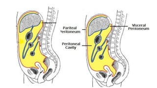
  • 4. Peritoneum • Large serous sac • Lines inner surface of abdomino- pelvic wall
  • 5. Why different layers? • Invaginated by viscera from different sides • Invagination throws the peritoneum into folds & • Forms • Parietal & visceral layers • & potential spaces
  • 6. Peritoneum - parts • Parietal Peritoneum • Lines the inner surface of • Abdominal and pelvic walls and • Lower surface • Diaphragm • Embryologically from somatopleuric mesoderm • Derives blood & nerve supply from body wall • Visceral Peritoneum • Lines outer surface of viscera • Firmly attaches with viscera • Develops from splanchnopeuric mesoderm • Blood supply and nerve supply are the same as those of underlying viscera
  • 7. Interperitoneal organs • Most part of viscera surrounded by peritoneum • Stomach, Liver, gallbladder • Tail of pancreas, spleen • First part of duodenum, jejunum, ileum • Transverse colon, sigmoid colon • Upper part of rectum, urinary bladder and uterus
  • 8. Retroperitoneal organs • Organs lie on the posterior abdominal wall are covered by peritoneum on their anterior surfaces • Descending and horizontal parts of duodenum, pancreas (head, neck & body) • Ascending & descending colon • Kidney, suprarenal gland, ureters • Abdominal aorta & IVC • Middle and lower parts of rectum
  • 9. Secondary retroperitoneal organs • Duodenum • 2nd and 3rd part • Pancreas (except tail) • Ascending colon • Descending colon • Upper rectum
  • 10. Folds of Peritoneum - types • Many organs within abdomen are suspended by folds of peritoneum • Provide pathways for passage of vessels, nerves and lymphatics • Named after their attached organs • Omentum • Fold of peritoneum connecting stomach to other organs • Mesenteries • Fold invests small intestine • Mesocolon • Fold invests large intestine • Peritoneal ligaments • Folds attached with other organs
  • 11. Disposition – Vertical • Start from • Anterior abdominal wall • At umbilical level • Lines • Inner aspect of anterior abdominal wall • Trace • Upto ant part of diaphragm (under surface) • Right of median plane • Falciform ligament • In free margin it contains • Ligamentum teres hepatis • Obliterated left umbilical vein (extends from umbilicus to inferior surface of liver) • Also it contains para umbilical veins • Tributaries to left branch of Portal vein
  • 12. Disposition – Vertical • Inferior surface of diaphragm to upper surface of liver • Falciform ligament • 2 layers diverge to right & left • On right (right layer of falciform) • From diaphragm to right lobe of liver forms • Superior layer of coronary ligament • On left (left layer of falciform lig) • From diaphragm to left lobe of liver forms • Left triangular ligament (anterior layer)
  • 13. Disposition – Vertical • After covering the superior and anterior surfaces of liver • Reaches inferior surface of liver • Right of Gall bladder • Peritoneum passes down to • Suprarenal & kidney as inferior layer of coronary ligament • Bare area • Between the 2 layers of coronary ligament • Right triangular ligament • 2 layers of coronary ligament unite to form • Connects right lobe of liver with diaphragm
  • 14. Disposition – Vertical Ligamentum venosum - remnant of ductus venosus (oblitrated ductus venosus), which shunted blood from the umbilical vein to the IVC in fetal life • On the left of gall bladder • After covering quadrate lobe & left lobe • Becomes posterior layer of left triangular ligament • Then passes to • Left margin of fissure for ligamentum venosum & anterior lip of porta hepatis & • From anterior lip of porta hepatis to right and then posterior lip of porta hepatis • Covering caudate lobe & attached to right lip of fissure for ligamentum venosum • These two layers form • Lesser omentum • Has inverted L shaped attachment
  • 15. Omentum • Peritoneal folds related to stomach • Lesser omentum • Greater omentum
  • 16. Lesser omentum • Connects liver to stomach • Attachment • Above to porta hepatis & fissure for ligamentum venosum • Inferiorly to lesser curvature of the stomach & 1st inch of duodenum • Parts • Hepato gastric ligament • Hepato duodenal ligament • Forms free margin • Contains • Portal vein (posteriorly) • Hepatic artery (anterior & left) • Bile duct (anterior & right)
  • 17. Lesser omentum • Contents • Hepatic artery, bile duct, portal vein • In free margin - • Rt. & lt. gastric vessels • Along lesser curvature
  • 18. Tracing lesser omentum • Peritoneum from • Anterior lip of porta hepatis and left lip of fissure for ligamentum venosum • Posterior lip of porta hepatis and right lip of fissure for ligamentum venosum • Continues downwards as lesser omentum towards lesser curvature of stomach and first one inch of duodenum • Forms anterior boundary for upper part of lesser sac
  • 19. Tracing lesser omentum • After reaching lesser curvature • Two layers separates • And covers the surfaces • Reaches greater curvature • Becomes anterior two layers of greater omentum
  • 20. Tracing greater omentum • From greater curvature • Anterior & posterior layers comes downwards • After awhile it returns back towards anterior border of pancreas • So 4 layers are there • Together they form Greater omentum • Space between 2nd & 3rd layer is lesser sac • Patent in fetal life • But obliterated - after birth
  • 21. Fate of 3rd & 4th layers • Continuation of layers of lesser omentum • Forms 1st & 2nd layer of greater omentum • While returns back to pancreas • 1st layer becomes 4th layer • 2nd layer becomes 3rd layer • Third layer • Runs upwards towards anterior border of pancreas (infront of transverse colon) • Passes infront of • Anterior surface of pancreas, kidney and suprarenal gland • Reaches the liver • Forms posterior wall of lesser sac
  • 22. Fate of 3rd & 4th layers • 4th layer • Runs towards anterior surface of head & anterior border of body of Pancreas • Then comes down to transverse colon as upper layer of Transverse mesocolon • Encloses transverse colon • Then goes towards inferior surface of pancreas – as inferior layer of Transverse mesocolon Fourth layer of Greater omentum and Superior layer of Transverse mesocolon are adherent – but separable.
  • 23. Greater omentum • Largest fold of peritoneum • Hangs down like an apron • In front of coils of small intestine • Made of 4 layers • Anterior 2 layers from Greater curvature and 1st part of duodenum • Extending downwards for a considerable distance & • Fold upon themselves to be 3rd & 4th layer • To anterosuperior surface of Tranasverse colon (head & ant border of pancreas)
  • 24. Greater omentum – parts & contents • Parts • Gastrophrenic ligament • Gastrosplenic ligament • Gastrocolic ligament • Contents • Rt. &Lt. gastroepiploic vessels • Lymph nodes • Fat • Functions • Store house of fat • Limits the spread of infection
  • 25. Transverse mesocolon • Suspends transverse colon • Attached to anterior aspect of head & anterior border of body of pancreas • Contains –Middle colic art.
  • 26. Root of mesentery • Peritoneum from transverse mesocolon • Covers lower part of anterior surface of head of pancreas • Passes to front of 3rd & 4th parts of duodenum • Then covers posterior abdominal wall • Becomes folded between • Duodenojejunal flexure(left of L2) to ileocecal junction (rt sacroiliac joint) • Forms root of mesentery
  • 27. Mesentery • Fan-shaped fold of peritoneum • Suspends the coils of jejunum and ileum from posterior abdominal wall • Has 2 borders • Attached • Free
  • 28. Mesentery - borders • Attached border • Known as root of mesentery • Lies obliquely • Extent • From • Duodenojejunal flexure (L2 – left side) • To • Ileocecal junction (Rt. Sacroiliac joint) • Length - 6 inches (15 cm) • Structures crossed by it • 3rd part of duodenum, Abdominal aorta, IVC • Rt. Psoas major, • Rt. Gonadal vessels, Rt. Genitofemoral nerve & Rt. ureter (medial to lateral) • Rt. Sacroiliac jt
  • 29. Mesentery - borders • Free border • Encloses jejunum and ileum • 6 meters in length • Contents • Jejunum, ileum • Lymph nodes • Jejunal & ileal branches of superior mesenteric artery & nerve plexus • Fat
  • 30. Meso-appendix • Δ-gular fold • Suspends appendix • From post surface of lower end of mesentery • Appendicular artery
  • 31. Peritoneum in Pelvis • After covers the structures of posterior abdominal wall • Reaches pelvis • First invests sigmoid colon from pelvic wall • Sigmoid mesocolon • Then covers Rectum • Upper third on front & sides • Middle part on front only Tracing the peritoneum anteriorly differs in male & female
  • 32. Sigmoid mesocolon • Suspends sigmoid colon • Attached to post abdominal and pelvic walls like inverted V • Apex of V lies on ureter at bifurcation of Lt. com iliac artery • Lt limb – along upper ½ of Lt, Ext iliac • Contents – Superior rectal vessels
  • 33. Peritoneum - Pelvis • In male • From rectum to seminal vesicle • Then upper surface of urinary bladder • Forms rectovesical pouch • From apex of UB to inner surface of anterior abdominal wall till umbilicus • In female • From rectum to posterior fornix of vagina • And covers supravaginal cervix, body and fundus • Forms rectouterine pouch (pouch of Douglas) • Then covers anterior surface of body • From body to UB • Forms uterovesical pouch
  • 34. Female pelvis • Communicates with extraperitoneal pelvis exteriorly • Through fallopian tubes, uterus and vagina
  • 35. Folds in anterior abdominal wall • Median umbilical fold • Remnant of urachus (median umbilical ligaments) • Medial umbilical fold • Remnants of umbilical arteries (medial umbilical ligaments) • Lateral umbilical fold • Inferior epigastric vessels
  • 36. Fossae of anterior abdominal wall
  • 37.
  • 38. Peritoneal cavity - Divisions • Transverse mesocolon divides the peritoneal cavity in to • Supramesocolic space • Also known as subphrenic spaces • Inframesocolic space • Bilateral paracolic & pelvic spaces are also peritoneal spaces
  • 39. Supra colic space • Also known as subphrenic spaces • Boundaries • Superiorly • Diaphragm • Inferiorly • Transverse colon • In front • Ant abdominal wall • Behind • Lesser omentum, stomach, greater omentum
  • 40. Supracolic compartment • Supracolic compartment contains • Stomach, liver, and spleen • Infracolic compartment contains • Small intestine and ascending & descending colons • Infracolic compartment is divided into • Right and left infracolic spaces by the mesentery of small intestine • Communication occurs between the supracolic and infracolic compartments • Through paracolic gutters
  • 41. • Right • Right anterior subphrenic (subdiaphragmatic) • Right posterior subphrenic (hepatorenal or Morrisons pouch) • Right extraperitoneal • Left • Left anterior intraperitoneal / left suprahepatic / subphrenic • Left posterior intraperitoneal / left subhepatic • Left extraperitoneal Supracolic compartment - Spaces
  • 42. Right anterior intraperitoneal / subphrenic / suprahepatic • Presents • Right of falciform ligament • Boundaries • In front • Ant abdominal wall & diaphragm • Behind • Superior anterior & right lateral surface of right lobe of liver, superior layer of coronary ligament • Below • Communicates with right posterior intraperitoneal compartment • Above • Diaphragm
  • 43. Morrison’s Pouch / Hepatorenal pouch / Rt subhepatic pouch • Right posterior subphrenic space • Most dependent part of peritoneal cavity in upper abdomen • Boundaries • In Front • Inferior surface of rt. lobe of liver and Gall bladder • Behind • Anterior surface of rt. kidney & rt. supra renal gland • Rt. Colic flexure & 2nd part of duodenum • Above • Inf layer of coronary ligament • Below • Communicate with infracolic part of peritoneal cavity • Left • Communicate with omental bursa • Right • Limited by diaphragm
  • 44. Morrison’s Pouch Hepatorenal pouch Rt subhepatic pouch • Most dependent part of abdomen proper • Commonest site of subphrenic abscess • From • Perforated peptic ulcer of stomach & duodenum
  • 45. Right extraperitoneal space • Bare area of liver presents • Liver directly related to diaphragm • Boundaries • Left • IVC • Right • Rt. triangular ligament • Above • Superior layer of coronary ligament • Below • Inferior layer of coronary ligament • Clinical anatomy • Common site for • Amoebic liver abscess
  • 46. Left anterior intraperitoneal • Lies on left of falciform ligament • Boundaries • In front • Ant abdominal wall • Behind • Lt triangular ligament • Lt. lobe of liver • Stomach & spleen
  • 48. Lesser Sac • Position • Behind the lesser omentum and stomach walls
  • 49. Lesser sac • Boundaries • Superior • Peritoneum which covers the caudate lobe of liver and diaphragm • Anterior • Lesser omentum • Peritoneum of posterior wall of stomach • And anterior two layers of greater omentum • Posterior • Posterior two layers of greater omentum • Transverse colon & transverse mesocolon • Peritoneum covering pancreas, left kidney and suprarenalgland • Inferior • Continuity of anterior and posterior two layers of greater omentum
  • 50. Lesser Sac • Right border • From above downwards • Deficient above • Epiploic foramen • Peritoneum covering • Posterior surface of proximal part of duodenum to the head of pancreas • Left border • From above downwards • Gastrophrenic ligament • Gastrosplenic and lienorenal ligament • Left border of greater omentum
  • 51. Parts of lesser sac • Body • Lies behind lesser omentum & stomach • Superior recess • Lies behind caudate lobe of liver • Splenic recess • Between gastrosplenic and lienorenal ligament • Inferior recess • Lies between the folds of greater omentum
  • 52. Epiploic foramen • Communication between • Lesser and greater sac • Location • T -12 vertebra level • Boundaries • Anteriorly • Free margin of lesser omentum • Contains • Front right to left • Bile duct & hepatic artery • Behind • Portal vein • Posteriorly • IVC (covered by peritoneum) • Superiorly • Caudate process of liver • Inferiorly • 1st part of duodenum
  • 53. Epiploic foramen Boundaries • Anteriorly • Free margin of lesser omentum • Contains • Front right to left • Bile duct & hepatic artery • Behind • Portal vein • Posteriorly • IVC (covered by peritoneum) • Superiorly • Caudate process of liver • Inferiorly • 1st part of duodenum
  • 54. Applied anatomy • Internal abdominal hernia • Occasionally a loop of intestine may enter into lesser sac • Through epiploic foramen • None of the boundaries can be incised to reduce the hernia • Pushing the herniated loop out of lesser sac by inserting the index finger into it through ant. 2 layers of greater omentum
  • 55. Infracolic Compartments • Lies below the transverse colon & mesocolon and behind the greater omentum • Divided into w by root of mesentery • Right infracolic Compartment • Left infracolic Compartment
  • 56. Right infracolic compartmet • Boundaries • Above • Transvers colon & mesocolon • On right • Ascending colon • On left • Root of Mesentery • Below • Closed
  • 57. Left infracolic compartmet • Lies left of root of mesentery • Boundaries • In front • Anterior abdominal wall • Above • Transverse mesocolon • Below • Open with pelvis • On right • Root of mesentery • On left • Descending colon
  • 58. Paracolic gutters • Longitudinal channels • Lined by peritoneum • Lies along the sides of ascending & descending colons • Right & left paracolic gutters • Right paracolic gutter • Lies lateral to ascending colon • Communication • Beween Hepato-renal pouch (above) & pelvis (below) • Left paracolic gutter • Lies lateral to descending colon • Above it is separated from • Spleen & lienorenal space by Phrenico-colic ligament • Below it is communicated with pelvis •
  • 59. Rectouterine Pouch (Pouch of Douglas) • Boundaries: • Anteriorly: uterus and posterior fornix of vagina • Posteriorly: rectum • Inferiorly (floor): rectovaginal fold of peritoneum • Clinical anatomy • Most dependent part of peritoneal cavity • Pus collected here • Can be drained through rectum or posterior fornix of vagina
  • 60. Applied anatomy • Ascites • Excessive accumulation of peritoneal fluid in peritoneal cavity • Commonly due to • Cirrhosis • TB & Malignancy • Paracentesis • Draining fluid • Peritonitis • Infection & inflammation of peritoneum • May result in fatal • Minimize the damage of peritoneum