Call Girls Service Nandiambakkam | 7001305949 At Low Cost Cash Payment Booking
Brachial plexus.pptx
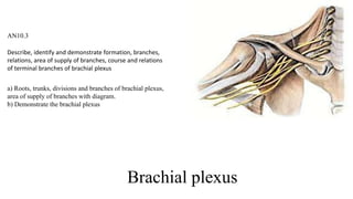
1. Brachial plexus
AN10.3
Describe, identify and demonstrate formation, branches,
relations, area of supply of branches, course and relations
of terminal branches of brachial plexus
a) Roots, trunks, divisions and branches of brachial plexus,
area of supply of branches with diagram.
b) Demonstrate the brachial plexus
2. Spinal nerve
• Anterior primary ramus supplies
• Larger area
• Skin & muscles of
• Anterior and lateral regions trunk
• Upper & lower limbs
• Supplying trunk region
• Remains in segmental pattern
• Supplying limbs
• Merge with one or more adjacent anterior
rami forming plexuses
• And gives multisegmental peripheral nerves
3. Brachial plexus
• Network of nerves which supply
brachium
• At root of neck
• Nerves form a complicated plexus
• Nerve fibres derived form different
segments of spinal cord to be
arranged and distributed efficiently
• Union of APR (anterior primary
ramus) of C5-8 & T1
4. Brachial plexus
• Limb bud appears
• 6th week of development
• By localized proliferation of
somatopleuric mesenchyme
• Causes overlying ectoderm to bulge from
the trunk
• As 2 pairs of flat paddles
• Arm bud develop first
• At the level of lower 4 cervical and
upper 2 thoracic segments
5.
6. Formation
• Mixing of nerves from different cord
levels
• By union and division of bundles
7. Advantages of plexus
• Fibres from different cord level
• Passes through a single nerve
• Distribution of a spinal segment
• Throughout the plexus
8. Roots
• Roots
• Five ventral rami (C5-
T1)
• Lies
• Between
• Scalenus anterior &
Medius
10. Trunks
• Lies in
• Posterior triangle of Neck
• 3 trunks are present
• Upper trunk
• Union of Roots C5 & C6
• Middle trunk
• Continuation of C7 root
• Lower trunk
• Union of roots C8 & T1
11. Divisions
• Lies in
• Behind the clavicle
• In cervicoaxillary canal
• Each trunk divides into
• Anterior and posterior divisions
12. Cords
• Formed within
• Axilla
• 3 cords are present
• Lateral cord
• Union of anterior divisions of
• Upper and middle trunks
• Medial cord
• Continuation of
• Inferior trunk
• Posterior cord
• Posterior divisions of
• All trunks
13. Branches
• Different parts of brachial plexus
• From Root
• Dorsal scapular
• Long thoracic nerve
• From Trunks
• Upper trunk
• Nerve to subclavius (C5 & 6)
• Suprascapular nerve
• From cords
• Lateral cord
• Lateral pectoral nerve
• Musculocutaneous nerve
• Lateral root of Median nerve
• Medial cord
• Medial pectoral nerve
• Medial cutaneous nerve of arm
• Medial cutaneous nerve of forearm
• Ulnar nerve
• Medial root of median nerve
• Posterior cord
• Upper subscapular nerve
• Thoracodorsal nerve
• Lower subscapular nerve
• Axillary nerve
• Radial nerve
14. Location of the parts of brachial plexus
• Roots
• Between
• Scalenus anterior & scalenus medius
• Posterior triangle of neck
• Trunks
• Posterior triangle of neck
• Divisions
• Behind clavicle
• In cervico axillary canal
• Cords
• Axilla
• Branches
• Starts in Axilla
15.
16.
17. Relations
• Cords
• I & II part of axillary artery
• Branches
• III part of axillary artery
18. ERB’S palsy
• Upper trunk lesion
• Nerve roots involved - C5, C6
• Erb’s point
• Where 6 nerves meet
• Causes
– Forceful separation of head from shoulder e.g.
during birth
– Fall on shoulder
• Muscles
• Biceps
• Brachialis, brchialis brachioradialis,
Supraspinatus
• Infraspinatus
• Deformity (Porter’s tip hand)
• Arm - adducted, medially rotated
• Forearm – pronated and extended
19. Klumpke’s palsy
• Lower trunk palsy
• Nerve roots involved - C 8, T1
• Cause
• Cervical rib
• Undue abduction of arm while holding something
with hands during fall from height
• Muscle Involved
– Paralysis of Intrinsic muscle of hand, ulnar
flexors of wrist and fingers
– Symptom and Sign
– Claw hand (medial two fingers more affected)
• Due to unopposed action of long flexors
fingers and extensors
• Paralysis of all interossei and medial two
lumbricals
20. Klumpke’s paralysis
• Due to involvement of T1
• Sympathetic fibres also involved
• Klumpke’s paralysis associated with
Horner’s syndrome
• Ptosis
• Drooping or falling of upper eyelid
• Miosis
• Excessive constriction (shrinking) of your
pupil
• Anhydrosis
• Decreased sweating
• Enopthalmos
• Posterior displacement of the eyeball
• Loss of ciliospinal reflex
• Dilation of ipsilateral pupil in response to
pain applied to the neck, face and upper
trunk
21. Distribution of main nerves
– Musculocutaneous
• Muscles of Anterior Compartment of arm
(flexors)
– Median
• Most Flexor muscles of forearm & Intrinsic
muscles inhand
– Ulnar
• FCU & part of FDP (forearm) and Intrinsic
muscles inhand
– Axillary
• Deltoid & Teres minor
– Radial
• Innervates all Extensor muscles of arm &
forearm
22. Long thoracic nerve
• Arises from
• Roots C5, 6, 7
• Forms on
• First digitations of serratus anterior
muscle
• Runs vertically downwards
• Just behind the mid-axillary line
• Nerve supply
• C5 – supply first two digitations
• C6 – next two digitations
• C7 – lowest four digitations
23. Lesion of long thoracic nerve
• Causes
• Sudden heavy loads on shoulder
• Carrying heavy loads on shoulder
• Symptom and sign
• Winging of scapula
• Prominence of medial border of scapula
• Loss of pushing and punching actions
• Abduction of arm affected.
• Demonstrated by
• Ask the patient to push against resistance
with the forearm
Winging of scapula
24. Upper & lower subscapular nerves
• Upper subscapular nerve
• Smaller than lower
• Enters Subscapularis at a high level.
• Frequently double
• Supplies
• Subscapularis
• Lower subscapular nerve
• Pass deep to sub scapular art.
• Supplies
• Subscapularis (lower part)
• Teres major
25. Thoracodorsal nerve
• Arises between
• Upper and lower Sub scapular
nerves
• Accompanies
• Sub scapular artery along posterior
axillary wall
• Supplies
• Latissimus dorsi