SlideShare a Scribd company logo
1 of 55
Nursing Management for patient
with Respiratory Disease
Prepared By
Dr. Zuhair Rushdi Mustafa
Lecturer at University of Duhok/
College of Nursing
• Crucial Diagnostic Tests for Respiratory disease
1. Bronchoscopy
Bronchoscopy is used to view the bronchial tree and to remove
foreign obstructions, obtain tissues for biopsy, or for
suctioning fluid.
2. Chest X-ray
Used to detect the size and position
of the heart and structural
abnormalities of the lungs.
3. Pulmonary Angiography
• Provides a view of the pulmonary circulatory system so that
the physician can determine the condition of blood flow to
the lungs.
4. Sputum Culture and Sensitivity
Sputum from the patient is cultured to determine which, if any,
bacteria is contained in the sputum and determine which
antibiotic kills the bacteria.
5. Thoracentesis
Removal of fluid from the pleural sac to drain fluid or identify
the contents of the fluid.
6. Pulmonary Function Test (PFT)
This test assesses the lungs’ ability to move air. Monitor
change from normal function; differentiate obstructive from
restrictive disease.
7. Arterial Blood Gas (ABG)
This determines the patient’s ventilation, tissue oxygenation,
and acid-base status.
HOW DOES THE TEST WORK?
Three to five milliliters of blood is sampled from an artery in a
heparinized syringe. If the sample cannot be analyzed right
away, it should be placed on ice.
• The normal results are:
• pH 7.35–7.45
• PaO2 80–100 mmHg
• PaCO2 35–45 mmHg
• HCO3 22–26 mEq/L
8. Mantoux Intradermal Skin Test (purified protein
derivative PPD)
• This determines if the patient has antibodies to the
Mycobacterium tuberculosis bacteria, which indicates that
the patient has been exposed to the bacteria.
9. Lung Biopsy
Removal of a tissue sample to be examined by the histology
lab for abnormalities.
Bronchitis
• Increased mucus production, caused by infection
and airborne irritants that block airways in the lungs,
results in the decreased ability to exchange gases.
• There are two forms of bronchitis:
1. acute bronchitis, where blockage of the airways is
reversible.
2. chronic bronchitis, where blockage is not
reversible.
• Patients with acute bronchitis are symptomatic
typically for 7 to 10 days often due to viral (but
sometimes bacterial) infection.
• Patients with chronic bronchitis will have symptoms
of a chronic productive cough for at least 3
consecutive months in 2 consecutive years.
• There is increased mucous production,
inflammatory changes, and, ultimately, fibrosis in
the airway walls.
• The patient with chronic bronchitis has an increased
incidence of respiratory infection.
Pathophysiology of Chronic bronchitis
• Chronic bronchitis characterised by an increase in mucus production
and damaged cilia in the bronchi. As a result, the bronchi become
clogged with mucus, then producing a cough. This chronic irritation
causes inflammation and the bronchial wall thickens, causing airway
obstruction. Secondary infections then occur, causing yet more
irritation and inflammation. As more and more airways become
blocked, external respiration is reduced and less oxygen is transferred
into the bloodstream.
Normal bronchiole
Narrowed and mucus-filled
bronchiole in bronchitis
• SIGNS AND SYMPTOMS of Bronchitis
1. Cough due to mucous production and irritation of
airways.
2. Shortness of breath.
3. Fever in acute episodes due to infection.
4. Accessory muscles are used for breathing
5. Productive cough due to irritation of airways.
6. Weight gain secondary to edema in chronic
bronchitis is due to right-sided heart failure.
7. Wheezing due to inflammation within the airways.
• TREATMENT
1. Acute bronchitis is treated in the short term with
symptomatic treatment and antibiotics when a
bacterial infection is present.
2. Chronic bronchitis is treated with a combination of
medications to keep the airways open, reduce
inflammation within airways, and prevent
complications or exacerbations.
3. Administer beta 2-agonists by inhaler or nebulizer
to dilate the bronchi such as albuterol
4. Administer anticholinergics which allow for
relaxation of bronchial smooth muscle such as
ipratropium inhaler
5. Administer steroids to decrease inflammation within the
airways such as hydrocortisone, methylprednisolone
systemically beclomethasone inhalers
6. prednisolone, prednisone orally.
7. Administer methylxanthines to enhance bronchodilation
such as aminophylline and theophylline.
8. Administer diuretics to reduce fluid retention in patients
who develop right sided heart failure such as furosemide.
9. Administer expectorant to help liquefy secretions such as
Mucolyte.
10. Administer antibiotics in acute exacerbation of chronic
bronchitis which is selected by culture and sensitivity study
11. Administer antacid such as antacids: aluminum
hydroxide/magnesium hydroxide, calcium carbonated, H2
blocker such as ranitidine, famotidine, cimetidine, or proton
pump inhibitor such as omeprazole, lansoprazole,
esomeprazole, rabeprazole, pantoprazole to decrease the
amount of acid in stomach, reducing possible ulcer formation
due to stress of disease or medication effects.
12. Administer vaccines—to decrease chances of infection:
• influenza
• pneumonia
13. Give 3 liters of fluid per day to help liquefy secretions
14. Oxygen: 2 liters per minute via nasal canula to help meet
body’s needs.
15. Increase protein, calories, and vitamin C in diet to meet
body’s needs.
16. Administer the incentive spirometer to encourage
coughing and expelling of mucous.
• NURSING DIAGNOSES
• Ineffective airway clearance
• Activity intolerance
• Ineffective breathing pattern
NURSING INTERVENTION
1. Monitor respirations looking at rate, effort, use of accessory
muscles, skin color; listen to breath sounds.
2. Place patient in high Fowler’s position to ease respiration.
3. Weigh the patient daily. Excess fluid due to heart failure
will increase weight. Notify physician, of weight gain of 2
pounds in 24 hours.
4. Have the patient perform the turning, coughing, and deep-
breathing exercises to enhance lung expansion and expel
mucous.
5.Monitor sputum for changes in color or amount.
6. Monitor intake and output.
7. Increase fluids to keep mucous thinner and easier to expel.
8. administer oxygen as order.
• Pneumonia
• Pneumonia is an infection of the alveoli and small
airways. Inflammation and oedema cause the alveoli
to fill with debris and exudate that commonly
impairs gas exchange.
• Etiology
• Infectious pneumonia may be due to a variety of
microorganisms and can be community-acquired or
hospital-acquired (nosocomial).
• A patient can inhale bacteria, viruses, parasites, or
irritating agents, or a patient can aspirate liquids or
foods. There may be increased mucous production
and thickening alveolar fluid as a result of impaired
gas exchange. All of these can lead to inflammation
of the lower airways.
• Organisms commonly associated with infection include
Staphylococcus aureus, Streptococcus pneumoniae, Haemophilus
influenza, Mycoplasma pneumoniae, Legionella pneumonia,
Chlamydia pneumoniae (parasite), and Pseudomonas aeruginosa
• Signs and symptoms
1. Shortness of breath due to inflammation within the lungs,
impairing gas exchange
2. Difficulty breathing (dyspnea) due to inflammation and mucus
within the lungs
3. Fever and Chills due to infectious process
4. Cough due to mucous production and irritation of the airways
5. Crackles due to fluid within the alveolar space and smaller
airways
6. wheezing due to inflammation within the larger airways
• Signs and symptoms
7. Discolored, possibly blood-tinged, sputum due to irritation
in the airways or microorganisms causing infection
8. Tachycardia and tachypnea as the body attempts to meet the
demand for oxygen
9. Pain on respiration due to pleuritic inflammation, pleural
effusion.
10. Headache, muscle aches (myalgia), joint pains, or nausea
may be present depending on the infecting organism
INTERPRETING TEST RESULTS
• Shadows on chest x-ray, indicating infiltration.
• Culture and sensitivity of the sputum to identify the infective
agent and the appropriate antibiotics.
• Elevated WBC (leukocytosis) showing sign of infection.
• Low oxygen saturation on pulse oximetry.
• Arterial blood gas may show low oxygen and elevated
carbon dioxide levels.
• Care and management
1. Administer oxygen as needed.
2. For bacterial infections, administer antibiotics such as
macrolides (azithromycin, clarithromycin), fluoroquinolones
(levofloxacin), beta-lactams (amoxicillin/clavulanate,
cefotaxime, ceftriaxone, ampicillin/sulbactam)
3. Administer antipyretics in patient with fever for comfort
such as acetaminophen, ibuprofen
4. Administer brochodilators to keep airways open, enhance
airflow if needed such as albuterol, levalbuterol via nebulizer
• Care and management
5. Increase fluid intake to help loosen secretions and
prevent dehydration.
6. Instruct the patient on how to use the incentive
spirometer to encourage deep breathing; monitor
progress.
7. placing the patient in an upright position will
promote diaphragm and intercostal muscle activity and
enhance ventilation.
Tuberculosis (TB)
An infectious disease spread by airborne route.
Infection is caused by inhalation of droplets that
contain the tuberculosis bacteria (Mycobacterium
tuberculosis).
An infected person can spread the small airborne
particles through coughing, sneezing, or talking.
Close contact with those affected increases the
chances of transmission.
Once inhaled, the organism typically settles into the
lung, but can infect any organ in the body.
The organism has an outer capsule.
• SIGNS AND SYMPTOMS
1. Weight loss and anorexia
2. Night sweats
3. Fever, possibly low-grade, due to infection
4. Productive cough with discolored, blood-tinged
sputum
5. Shortness of breath due to lung changes
6. Malaise and fatigue due to active illness affecting
lungs
INTERPRETING TEST RESULTS
1. Positive Mantoux (PPD) skin test shows exposure to
tuberculosis due to development of cell-mediated
immunity.
2. Chest x-ray may show areas of granuloma or
cavitation.
3. Sputum test identifies M. tuberculosis bacteria:
• Acid fast-staining done to initially screen for TB—
bacillus will hold stain.
• Culture confirms the diagnosis but is slow-growing
• TREATMENT
1. Initial treatment times generally range from 6 to 12
months.
2. Longer treatment plans may be necessary for those with
HIV infection or drug-resistant strains of TB.
3. Administer antitubercular medications to treat and
prevent transmission such as : isoniazid, rifampin,
pyrazinamide, ethambutol, streptomycin.
4. Respiratory isolation for in-hospital care—the bacteria is
spread by droplet.
5. Increase protein, carbohydrates, and vitamin C diet for
patients.
• NURSING DIAGNOSES
• Fatigue
• Ineffective airway clearance
NURSING INTERVENTION
1. Monitor respiration for rate, effort, use of accessory
muscles, and skin color changes.
2. Increase fluid intake to help liquefy any secretions.
3. Record fluid intake and output.
4. Explain to the patient:
A. How to prevent spreading the disease.
B. The importance of finishing all prescribed medication.
C. Plan for rest periods during the day.
Pulmonary Embolism
• Blood flow is obstructed in the lungs caused by
thrombus (blood clot), air, or fat emboli that become
stuck in an artery, causing impaired gas exchange.
• Pathophysiology
• Patients may be predisposed to clot formation, have
pooling of blood, or damage to vessel walls, or take
certain medications that increase the risk of
thrombus formation.
• Thrombus are commonly found in vessels in lower
extremities.
• When a thrombus loosens and travels in the
peripheral circulation, it is called an embolus.
• The embolus travels through the right side of the
heart and is sent to the lungs where it lodges in one
of the arteries.
SIGNS AND SYMPTOMS
1. Sudden difficulty breathing (dyspnea) happens when the
clot suddenly lodges in the artery
2. Heart rate greater than 100 beats per minute (tachycardia)
3. Respiration greater than 20 breaths per minutes (tachypnea)
as the body attempts to get more oxygen
4. Chest pain due to clot presence and area of atelectasis
5. Coughing with blood-tinged sputum (hemoptysis)
6. Crackles heard near area of clot
INTERPRETING TEST RESULTS
1. Chest x-ray may show dilated pulmonary artery or pleural
effusion.
2. CT scan will show clot in pulmonary arteries.
3. Pulmonary angiography will show presence of clot.
4. Arterial blood gases may show decreased oxygen (PaO2)
and carbon dioxide (PaCO2), depending on the size of the clot.
5. Lower extremity ultrasound is often done to test for
presence of thrombus
• TREATMENT
1. Supplemental oxygen therapy.
2. Administer thrombolytics to enhance breakdown of existing
clot such as urokinase, alteplase.
3. Administer anticoagulants to prevent further clot formation
sucha as heparin, warfarin
4. Administer analgesic for pain control such as morphine
5. Bed rest to prevent thrombus from breaking free from
lower extremities.
6. Surgical removal of the embolus may be necessary in some cases.
• NURSING INTERVENTION
• Explain to the patient:
1. To avoid sitting and standing for too long to decrease
chance of clot formation.
2. Not to cross legs to avoid constriction of vessels in the
lower extremities, decreasing the chances of clot formation.
3. How to identify side effects from using anticoagulants, such
as bleeding or bruising.
4. Explain that pulmonary embolism is an adverse effect from
using hormonal contraceptives and a different form of birth
control needs to be used in the future
• Asthma
• The airways become obstructed from either inflammation of
the lining of the airways or constriction of the bronchial
smooth muscles (bronchospasm).
• A known allergen, for example, pollen—is inhaled, causing
activation of antibodies that recognize the allergen. Mast
cells and histamine are activated, initiating a local
inflammatory response.
• Astimulus causes an inflammatory reaction, increasing the
size of the bronchial linings; this results in restriction of the
airways
• Types of Asthma
• There are two kinds of asthma:
1. Extrinsic asthma, also known as atopic, caused by
allergens such as pollen, animal dander, mold, or dust.
Often accompanied by allergic rhinitis and eczema;
this may run in families.
2. Intrinsic asthma, also known as nonatopic, caused
by a nonallergic factor such as following a respiratory
tract infection, exposure to cold air, changes in air
humidity, or respiratory irritants.
SIGNS AND SYMPTOMS
1. Wheezing initially present on expiration.
2. dyspnea as airways narrow due to inflammation.
3. Respiration greater than 20 breaths per minute (tachypnea)
4. Use of accessory muscles to breathe as the body tries
harder to get more air into the lungs.
5. Tightness in the chest due to narrowing of the airways
(bronchoconstriction).
6. Cough.
7. Tachycardia—heart rate greater than 100, as the body
attempts to get more oxygen to the tissues.
• TREATMENT
1. Administer oxygen
2. Identify and remove allergens and known triggers to avoid causing an
asthma attack.
3. Give patient 3 liters/day of fluid to help liquefy any secretions.
4. Administer short-acting beta2-adrenergic drugs to bronchodilate such
as albuterol, levalbuterol.
5. Administer steroids to decrease inflammation, which will help open
airways;these are not for acute symptoms such as hydrocortisone,
methylprednisolone intravenously, beclomethasone inhalers
prednisolone, prednisone orally
6. Administer aminophylline and theophylline
7. Administer antacid, H2 blocker, or proton pump inhibitor to decrease
the amount of acid in the stomach, reducing the possibility of ulcers due
to stress of disease or medication effects
• Emphysema
Emphysema is defined as the permanent enlargement
of airspaces beyond the terminal bronchiole and the
destruction of the alveolar wall.
Chronic inflammation reduces the flexibility of the walls of
alveoli, resulting in over-distention of the alveolar walls. This
causes air to be trapped in the lungs, impeding gas exchange.
Smoking is often linked to development of emphysema.
• SIGNS AND SYMPTOMS
1. Dyspnea due to air trapping, which retains CO2 and reduces
alveolar gas exchange.
2. Barrel chest develops over time as more air is trapped
within the distal airways.
3. Use of accessory muscles to breathe as the respiratory effort
increases.
4. Loss of weight as extra calories are needed to maintain
respiration.
5. Patients prefer a seated position which allows for greater
chest expansion.
Barrel chest
• INTERPRETING TEST RESULTS
1. Increased residual volume shown in pulmonary function
test due to air trapping.
2. Decreased O2 and increased CO2 in arterial blood gas as
gas
3. exchange is impaired due to air trapping; more pronounced
as disease progresses.
4. Chest x-ray shows over inflation of lungs and flattening of
the diaphragm.
• TREATMENT
1. Administer beta2-agonists to bronchodilate by inhaler or
nebulizer such as albuterol, levalbuterol.
2. Administer long-acting bronchodilating medications by
metered dose inhaler such as formoterol, salmeterol
3. Administer anticholinergics which allow for relaxation of
bronchial smooth muscle such as ipratropium inhaler.
4. Administer methylxanthines to dilate the bronchi such as
aminophylline and theophylline.
• TREATMENT
5. Administer steroids to decrease inflammation within the
airways such as hydrocortisone, methylprednisolone
systemically, prednisolone, prednisone orally.
6. Administer antacid such as antacids: aluminum
hydroxide/magnesium hydroxide, calcium carbonated, H2
blocker such as ranitidine, famotidine, cimetidine, or proton
pump inhibitor such as omeprazole, lansoprazole,
esomeprazole, rabeprazole, pantoprazole to decrease the
amount of acid in stomach, reducing possible ulcer formation
due to stress of disease or medication effects.
7. Administer expectorant—to loosen secretions:
8. Administer diuretics to decrease fluid retention in patients
that are developing right-sided heart failure secondary to lung
disease such as furosemide (Lasix).
9. Administer vaccines—to prevent respiratory infections:
• influenza, pneumonia
10. Administer antibiotics based on results of culture and
sensitivity.
11. Administer oxygen, 2 liters per minute.
12. Give patient 3 liters of fluids per day to help liquefy
secretions.
13. Teach patient how to use the incentive spirometer to
encourage deep breathing and enhance coughing and expelling
of mucous..
Chronic obstructive pulmonary disease
• Is the most common chronic lung disease. COPD is an
umbrella term that could refer to emphysema and chronic
bronchitis and, more commonly, a combination of these
conditions.
• It has one major cause which is smoking.
Etiology
COPD may result from:
1. cigarette smoking
2. recurrent or chronic respiratory tract infection
3. allergies
4. familial and hereditary factors such as alpha1-antitrypsin
deficiency.
Sings & Symptoms
The typical COPD patient is asymptomatic until
middle age, when the following signs and symptoms
may occur:
1. reduced ability to exercise or do strenuous work
2. productive cough
3. dyspnea with minimal exertion.
Treatment
Treatment for COPD aims to relieve symptoms and prevent
complications.
1. Administer beta-agonist bronchodilators (albuterol or
salmeterol)
2. Anticholinergic bronchodilators (ipratropium [Atrovent]),
3. Corticosteroids (beclomethasone )
These drugs are usually given by metered dose inhaler.
4.smoking cessation advice
5. education on prescribed oxygen and bronchodilator
therapies – to maximise relief of breathlessness
5. dietary advice – severe weight loss is a feature of both emphysema
and chronic bronchitis
6. pulmonary rehabilitation
• Lung Cancer
Lung cancer is the abnormal, uncontrolled cell growth in lung
tissues, resulting in a tumor.
A tumor in the lung may be primary when it develops in lung
tissue. It may be secondary when it spreads (metastasizes)
from cancer in other areas of the body, such as the liver, brain,
or kidneys.
• Repetitive exposure to inhaled irritants increases a person’s
risk for lung cancer. Cigarette smoke, occupational
exposures, air pollution containing benzopyrenes, and
hydrocarbons have all been shown to increase risk.
• There are two major categories of lung cancer:
• small cell and non-small cell.
• Small cell:
• Oat cell—fast-growing, early metastasis
• Non-small cell:
• Adenocarcinoma—moderate growth rate, early metastasis
• Squamous cell—slow-growing, late metastasis
• Large cell—fast-growing, early metastasis
• PROGNOSIS
• Lung cancer is the leading cause of cancer death. Many
patients with lung cancer are diagnosed at a later stage,
leading to the long-term (5-year) survival rate of less than
20 %. Earlier diagnosis is more beneficial for treatment and
outcome.
• The longer the cancer has been in the lungs, the greater the
likelihood of metastasis to other areas.
• SIGNS AND SYMPTOMS
1. Coughing due to irritation from mass. Presence of mucous
or exudate may not be until later in disease.
2. Coughing up blood (hemoptysis).
3. Fatigue.
4. Weight loss due to the caloric needs of the tumor, taking
away from the needs of the body.
5. Anorexia.
6. Dyspnea caused by damaged lung tissue.
7. Chest pains as mass presses on surrounding tissue; may not
be feel until late in disease.
8. Sputum production.
9. Pleural effusion.
• INTERPRETING TEST RESULTS
1. Mass in lung shown on chest x-ray.
2. CT scan shows mass, lymph node involvement.
3. Bronchoscopy may show cancer cells on bronchoscopic
4.Cancer cells seen in sputum.
5. Biopsy through chest wall or tissue biopsy from lung will
show cell type.
6. Bone scan or CT scans shows metastasis of the disease.
TREATMENT
1. Surgical removal of affected area of the lung (wedge
resection, segmental resection, lobectomy) or total lung
(pneumonectomy).
2. Radiation therapy to decrease tumor size.
3. Chemotherapy such as : cyclophosphamide, vincristine,
etoposide, cisplatin
4. Oxygen therapy to supplement the needs of the body.
5. High-protein, high-calorie diet to meet the needs of the
body.
6. Administer antiemetics to combat side effects of
chemotherapy: ondansetron (Zofran), prochlorperazine
7. Administer analgesics for pain control such as morphine,
Tramadol.
• NURSING DIAGNOSES
• Anxiety
• Activity intolerance
• Impaired gas exchange
NURSING INTERVENTION
1. Monitor respiratory status, looking at rate, effort, use of
accessory muscles, and skin color; auscultate breath sounds.
2. Monitor pain and administer analgesics appropriately.
3. Monitor vital signs for changes, elevated pulse, elevated
respiration, change in BP, and elevated temperature, which
may signal infection.
4. Monitor pulse oximetery for decrease in oxygenation levels.
5. Assist patient with turning, coughing, and deep-breathing
exercises.
6. Place patient in semi-Fowler’s position to ease respiratory
effort.
7. Explain to the patient the importance of taking rest periods.

More Related Content

What's hot (20)

Upper Respiratory Tract Infection (URTI)
Upper Respiratory Tract Infection (URTI)Upper Respiratory Tract Infection (URTI)
Upper Respiratory Tract Infection (URTI)
 
Bronchiolitis & Nursing care
Bronchiolitis & Nursing careBronchiolitis & Nursing care
Bronchiolitis & Nursing care
 
PNEUMONIA PATHOPHYSIOLOGY.pptx
PNEUMONIA PATHOPHYSIOLOGY.pptxPNEUMONIA PATHOPHYSIOLOGY.pptx
PNEUMONIA PATHOPHYSIOLOGY.pptx
 
Asthma
AsthmaAsthma
Asthma
 
Upper respiratoey diseases
Upper respiratoey diseasesUpper respiratoey diseases
Upper respiratoey diseases
 
Tb path & pathogenesis
Tb path & pathogenesisTb path & pathogenesis
Tb path & pathogenesis
 
common cold
common coldcommon cold
common cold
 
Upper respiratory tract infections
Upper respiratory tract infections Upper respiratory tract infections
Upper respiratory tract infections
 
Asthma
AsthmaAsthma
Asthma
 
Bronchitis
BronchitisBronchitis
Bronchitis
 
Pneumonia
PneumoniaPneumonia
Pneumonia
 
Bronchitis
BronchitisBronchitis
Bronchitis
 
upper respiratory tract infection
upper respiratory tract infectionupper respiratory tract infection
upper respiratory tract infection
 
Tonsillitis slideshare for medical students
Tonsillitis slideshare for medical students Tonsillitis slideshare for medical students
Tonsillitis slideshare for medical students
 
Bronchiectasis
BronchiectasisBronchiectasis
Bronchiectasis
 
Bronchitis
BronchitisBronchitis
Bronchitis
 
Pulmonary tuberculosis (1).pdf
Pulmonary tuberculosis (1).pdfPulmonary tuberculosis (1).pdf
Pulmonary tuberculosis (1).pdf
 
Bronchitis
BronchitisBronchitis
Bronchitis
 
med
medmed
med
 
Pneumonia
PneumoniaPneumonia
Pneumonia
 

Similar to Respiratory disease

Bronchiectases
BronchiectasesBronchiectases
BronchiectasesGAMANDEEP
 
Chronic Obstructive Pulmonary Disease[ COPD].pptx
Chronic Obstructive Pulmonary Disease[ COPD].pptxChronic Obstructive Pulmonary Disease[ COPD].pptx
Chronic Obstructive Pulmonary Disease[ COPD].pptxakoeljames8543
 
Acute Respiratory Distress Syndrome
Acute Respiratory Distress SyndromeAcute Respiratory Distress Syndrome
Acute Respiratory Distress SyndromeACIF ALI
 
CHRONIC OBSTRUCTIVE PULMONARY DISEASE BY AKRAM KHAN
CHRONIC OBSTRUCTIVE PULMONARY DISEASE BY AKRAM KHANCHRONIC OBSTRUCTIVE PULMONARY DISEASE BY AKRAM KHAN
CHRONIC OBSTRUCTIVE PULMONARY DISEASE BY AKRAM KHANAkram Khan
 
COPD and AE of COPD
COPD and AE of COPD COPD and AE of COPD
COPD and AE of COPD Asraf Hussain
 
Pneumonia and it's management
Pneumonia and it's managementPneumonia and it's management
Pneumonia and it's managementRakhiYadav53
 
PNEUMONIA.pptx
PNEUMONIA.pptxPNEUMONIA.pptx
PNEUMONIA.pptxSAMOEINESH
 
Pediatric pneumonia.pptx
Pediatric pneumonia.pptxPediatric pneumonia.pptx
Pediatric pneumonia.pptxDrHananTork
 
Bronchiectesis for Nurses - Easy Explanation
Bronchiectesis for Nurses - Easy ExplanationBronchiectesis for Nurses - Easy Explanation
Bronchiectesis for Nurses - Easy ExplanationSwatilekha Das
 
Chronic obstructive pulmonary disease (copd)
Chronic obstructive pulmonary disease (copd)Chronic obstructive pulmonary disease (copd)
Chronic obstructive pulmonary disease (copd)Rahil Dalal
 
Respiratory Disorders ىاغفقثصضهععا(1).pdf
Respiratory Disorders ىاغفقثصضهععا(1).pdfRespiratory Disorders ىاغفقثصضهععا(1).pdf
Respiratory Disorders ىاغفقثصضهععا(1).pdfssuser0b28a72
 
Pediatrics-Asthma. by me the one and only
Pediatrics-Asthma. by me the one and onlyPediatrics-Asthma. by me the one and only
Pediatrics-Asthma. by me the one and onlyBroski4
 

Similar to Respiratory disease (20)

Bronchiectases
BronchiectasesBronchiectases
Bronchiectases
 
Copd
CopdCopd
Copd
 
Copd , septicshock
Copd , septicshockCopd , septicshock
Copd , septicshock
 
Chronic Obstructive Pulmonary Disease[ COPD].pptx
Chronic Obstructive Pulmonary Disease[ COPD].pptxChronic Obstructive Pulmonary Disease[ COPD].pptx
Chronic Obstructive Pulmonary Disease[ COPD].pptx
 
Pneumonia
PneumoniaPneumonia
Pneumonia
 
Acute Respiratory Distress Syndrome
Acute Respiratory Distress SyndromeAcute Respiratory Distress Syndrome
Acute Respiratory Distress Syndrome
 
CHRONIC OBSTRUCTIVE PULMONARY DISEASE BY AKRAM KHAN
CHRONIC OBSTRUCTIVE PULMONARY DISEASE BY AKRAM KHANCHRONIC OBSTRUCTIVE PULMONARY DISEASE BY AKRAM KHAN
CHRONIC OBSTRUCTIVE PULMONARY DISEASE BY AKRAM KHAN
 
Copd Part 1
Copd Part 1Copd Part 1
Copd Part 1
 
COPD and AE of COPD
COPD and AE of COPD COPD and AE of COPD
COPD and AE of COPD
 
Pneumonia and it's management
Pneumonia and it's managementPneumonia and it's management
Pneumonia and it's management
 
Pneumonia
PneumoniaPneumonia
Pneumonia
 
PNEUMONIA.pptx
PNEUMONIA.pptxPNEUMONIA.pptx
PNEUMONIA.pptx
 
Pediatric pneumonia.pptx
Pediatric pneumonia.pptxPediatric pneumonia.pptx
Pediatric pneumonia.pptx
 
PNEUMONIA
PNEUMONIAPNEUMONIA
PNEUMONIA
 
Bronchitis
BronchitisBronchitis
Bronchitis
 
Bronchiectesis for Nurses - Easy Explanation
Bronchiectesis for Nurses - Easy ExplanationBronchiectesis for Nurses - Easy Explanation
Bronchiectesis for Nurses - Easy Explanation
 
Chronic obstructive pulmonary disease (copd)
Chronic obstructive pulmonary disease (copd)Chronic obstructive pulmonary disease (copd)
Chronic obstructive pulmonary disease (copd)
 
Respiratory Disorders ىاغفقثصضهععا(1).pdf
Respiratory Disorders ىاغفقثصضهععا(1).pdfRespiratory Disorders ىاغفقثصضهععا(1).pdf
Respiratory Disorders ىاغفقثصضهععا(1).pdf
 
COPD.pptx
COPD.pptxCOPD.pptx
COPD.pptx
 
Pediatrics-Asthma. by me the one and only
Pediatrics-Asthma. by me the one and onlyPediatrics-Asthma. by me the one and only
Pediatrics-Asthma. by me the one and only
 

More from Zuhair Mustafa

More from Zuhair Mustafa (20)

Performing chest physiotherapy
Performing chest physiotherapyPerforming chest physiotherapy
Performing chest physiotherapy
 
O22 & suctioning
O22 & suctioningO22 & suctioning
O22 & suctioning
 
First Aid for Wounds
First Aid for WoundsFirst Aid for Wounds
First Aid for Wounds
 
First Aid for Sting and bites
First Aid for Sting and bitesFirst Aid for Sting and bites
First Aid for Sting and bites
 
First Aid for Heat emergencies
First Aid for Heat emergenciesFirst Aid for Heat emergencies
First Aid for Heat emergencies
 
First aid CPR
First aid CPRFirst aid CPR
First aid CPR
 
First aid
First aid First aid
First aid
 
Choking
Choking Choking
Choking
 
Blood disease
Blood diseaseBlood disease
Blood disease
 
Renal disorders
Renal disordersRenal disorders
Renal disorders
 
Valvular disorders
Valvular disordersValvular disorders
Valvular disorders
 
Congenital hear defects
Congenital hear defectsCongenital hear defects
Congenital hear defects
 
Cnv disorders
Cnv disordersCnv disorders
Cnv disorders
 
Brain death
Brain deathBrain death
Brain death
 
Vital signs respiration
Vital signs respirationVital signs respiration
Vital signs respiration
 
Vital signs respiration
Vital signs respirationVital signs respiration
Vital signs respiration
 
Vital signs respiration
Vital signs respirationVital signs respiration
Vital signs respiration
 
Vital signs bp and glucose testing
Vital signs bp and glucose testingVital signs bp and glucose testing
Vital signs bp and glucose testing
 
Vital signs Blood Pressure and glucose testing
Vital signs Blood Pressure and glucose testing  Vital signs Blood Pressure and glucose testing
Vital signs Blood Pressure and glucose testing
 
Vital signs tempreture and pulse
Vital signs   tempreture and pulseVital signs   tempreture and pulse
Vital signs tempreture and pulse
 

Recently uploaded

Test bank for community public health nursing evidence for practice 4TH editi...
Test bank for community public health nursing evidence for practice 4TH editi...Test bank for community public health nursing evidence for practice 4TH editi...
Test bank for community public health nursing evidence for practice 4TH editi...robinsonayot
 
2024 PCP #IMPerative Updates in Rheumatology
2024 PCP #IMPerative Updates in Rheumatology2024 PCP #IMPerative Updates in Rheumatology
2024 PCP #IMPerative Updates in RheumatologySidney Erwin Manahan
 
Leading large scale change: a life at the interface between theory and practice
Leading large scale change: a life at the interface between theory and practiceLeading large scale change: a life at the interface between theory and practice
Leading large scale change: a life at the interface between theory and practiceHelenBevan4
 
Independent Call Girl in 😋 Goa +9316020077 Goa Call Girl
Independent Call Girl in 😋 Goa  +9316020077 Goa Call GirlIndependent Call Girl in 😋 Goa  +9316020077 Goa Call Girl
Independent Call Girl in 😋 Goa +9316020077 Goa Call GirlReal Sex Provide In Goa
 
❤️ Chandigarh Call Girls ☎️99158-51334☎️ Escort service in Chandigarh ☎️ Chan...
❤️ Chandigarh Call Girls ☎️99158-51334☎️ Escort service in Chandigarh ☎️ Chan...❤️ Chandigarh Call Girls ☎️99158-51334☎️ Escort service in Chandigarh ☎️ Chan...
❤️ Chandigarh Call Girls ☎️99158-51334☎️ Escort service in Chandigarh ☎️ Chan...rajveerescorts2022
 
❤️ Kharar Call Girls ☎️99158-51334☎️ Call Girl service in Kharar☎️ Kharar Cal...
❤️ Kharar Call Girls ☎️99158-51334☎️ Call Girl service in Kharar☎️ Kharar Cal...❤️ Kharar Call Girls ☎️99158-51334☎️ Call Girl service in Kharar☎️ Kharar Cal...
❤️ Kharar Call Girls ☎️99158-51334☎️ Call Girl service in Kharar☎️ Kharar Cal...daljeetkaur2026
 
Goa Call Girls Service +9316020077 Call GirlsGoa By Russian Call Girlsin Goa
Goa Call Girls Service  +9316020077 Call GirlsGoa By Russian Call Girlsin GoaGoa Call Girls Service  +9316020077 Call GirlsGoa By Russian Call Girlsin Goa
Goa Call Girls Service +9316020077 Call GirlsGoa By Russian Call Girlsin GoaReal Sex Provide In Goa
 
❤️ Chandigarh Call Girls Service ☎️99158-51334☎️ Escort service in Chandigarh...
❤️ Chandigarh Call Girls Service ☎️99158-51334☎️ Escort service in Chandigarh...❤️ Chandigarh Call Girls Service ☎️99158-51334☎️ Escort service in Chandigarh...
❤️ Chandigarh Call Girls Service ☎️99158-51334☎️ Escort service in Chandigarh...rajveerescorts2022
 
💚Mature Women / Aunty Call Girl Chandigarh Escorts Service 💯Call Us 🔝 9352988...
💚Mature Women / Aunty Call Girl Chandigarh Escorts Service 💯Call Us 🔝 9352988...💚Mature Women / Aunty Call Girl Chandigarh Escorts Service 💯Call Us 🔝 9352988...
💚Mature Women / Aunty Call Girl Chandigarh Escorts Service 💯Call Us 🔝 9352988...Chandigarh Call Girls
 
The Events of Cardiac Cycle - Wigger's Diagram
The Events of Cardiac Cycle - Wigger's DiagramThe Events of Cardiac Cycle - Wigger's Diagram
The Events of Cardiac Cycle - Wigger's DiagramMedicoseAcademics
 
TIME FOR ACTION: MAY 2024 Securing A Strong Nursing Workforce for North Carolina
TIME FOR ACTION: MAY 2024 Securing A Strong Nursing Workforce for North CarolinaTIME FOR ACTION: MAY 2024 Securing A Strong Nursing Workforce for North Carolina
TIME FOR ACTION: MAY 2024 Securing A Strong Nursing Workforce for North CarolinaMebane Rash
 
Call Girls In Kharar 💯Call Us 🔝 9915851334🔝 💃 Top Class ☎️ Call Girl Service ...
Call Girls In Kharar 💯Call Us 🔝 9915851334🔝 💃 Top Class ☎️ Call Girl Service ...Call Girls In Kharar 💯Call Us 🔝 9915851334🔝 💃 Top Class ☎️ Call Girl Service ...
Call Girls In Kharar 💯Call Us 🔝 9915851334🔝 💃 Top Class ☎️ Call Girl Service ...daljeetkaur2026
 
ACNE VULGARIS , ALLERGIES, ECZEMA, PEMPHIGUS.pdf
ACNE VULGARIS , ALLERGIES, ECZEMA, PEMPHIGUS.pdfACNE VULGARIS , ALLERGIES, ECZEMA, PEMPHIGUS.pdf
ACNE VULGARIS , ALLERGIES, ECZEMA, PEMPHIGUS.pdfDolisha Warbi
 
Cash Payment 😋 +9316020077 Goa Call Girl No Advance *Full Service
Cash Payment 😋  +9316020077 Goa Call Girl No Advance *Full ServiceCash Payment 😋  +9316020077 Goa Call Girl No Advance *Full Service
Cash Payment 😋 +9316020077 Goa Call Girl No Advance *Full ServiceReal Sex Provide In Goa
 
TEST BANK For Robbins & Kumar Basic Pathology, 11th Edition by Vinay Kumar, A...
TEST BANK For Robbins & Kumar Basic Pathology, 11th Edition by Vinay Kumar, A...TEST BANK For Robbins & Kumar Basic Pathology, 11th Edition by Vinay Kumar, A...
TEST BANK For Robbins & Kumar Basic Pathology, 11th Edition by Vinay Kumar, A...rightmanforbloodline
 
👉 Srinagar Call Girls Service Just Call 🍑👄6378878445 🍑👄 Top Class Call Girl S...
👉 Srinagar Call Girls Service Just Call 🍑👄6378878445 🍑👄 Top Class Call Girl S...👉 Srinagar Call Girls Service Just Call 🍑👄6378878445 🍑👄 Top Class Call Girl S...
👉 Srinagar Call Girls Service Just Call 🍑👄6378878445 🍑👄 Top Class Call Girl S...gragfaguni
 
Spauldings classification ppt by Dr C P PRINCE
Spauldings classification ppt by Dr C P PRINCESpauldings classification ppt by Dr C P PRINCE
Spauldings classification ppt by Dr C P PRINCEDR.PRINCE C P
 
Agra Call Girl 📲 ( 9084454195 ) ⏎ Independent Call Girls In Agra By Meera
Agra Call Girl 📲 ( 9084454195 ) ⏎ Independent Call Girls In Agra By MeeraAgra Call Girl 📲 ( 9084454195 ) ⏎ Independent Call Girls In Agra By Meera
Agra Call Girl 📲 ( 9084454195 ) ⏎ Independent Call Girls In Agra By MeeraInaayaeventcompany
 
Real Sex Provide In Goa ✂️ Call Girl (9316020077) Call Girl In Goa
Real Sex Provide In Goa ✂️ Call Girl   (9316020077) Call Girl In GoaReal Sex Provide In Goa ✂️ Call Girl   (9316020077) Call Girl In Goa
Real Sex Provide In Goa ✂️ Call Girl (9316020077) Call Girl In GoaReal Sex Provide In Goa
 

Recently uploaded (20)

Test bank for community public health nursing evidence for practice 4TH editi...
Test bank for community public health nursing evidence for practice 4TH editi...Test bank for community public health nursing evidence for practice 4TH editi...
Test bank for community public health nursing evidence for practice 4TH editi...
 
2024 PCP #IMPerative Updates in Rheumatology
2024 PCP #IMPerative Updates in Rheumatology2024 PCP #IMPerative Updates in Rheumatology
2024 PCP #IMPerative Updates in Rheumatology
 
Leading large scale change: a life at the interface between theory and practice
Leading large scale change: a life at the interface between theory and practiceLeading large scale change: a life at the interface between theory and practice
Leading large scale change: a life at the interface between theory and practice
 
@Safe Abortion pills IN Jeddah(+918133066128) Un_wanted kit Buy Jeddah
@Safe Abortion pills IN Jeddah(+918133066128) Un_wanted kit Buy Jeddah@Safe Abortion pills IN Jeddah(+918133066128) Un_wanted kit Buy Jeddah
@Safe Abortion pills IN Jeddah(+918133066128) Un_wanted kit Buy Jeddah
 
Independent Call Girl in 😋 Goa +9316020077 Goa Call Girl
Independent Call Girl in 😋 Goa  +9316020077 Goa Call GirlIndependent Call Girl in 😋 Goa  +9316020077 Goa Call Girl
Independent Call Girl in 😋 Goa +9316020077 Goa Call Girl
 
❤️ Chandigarh Call Girls ☎️99158-51334☎️ Escort service in Chandigarh ☎️ Chan...
❤️ Chandigarh Call Girls ☎️99158-51334☎️ Escort service in Chandigarh ☎️ Chan...❤️ Chandigarh Call Girls ☎️99158-51334☎️ Escort service in Chandigarh ☎️ Chan...
❤️ Chandigarh Call Girls ☎️99158-51334☎️ Escort service in Chandigarh ☎️ Chan...
 
❤️ Kharar Call Girls ☎️99158-51334☎️ Call Girl service in Kharar☎️ Kharar Cal...
❤️ Kharar Call Girls ☎️99158-51334☎️ Call Girl service in Kharar☎️ Kharar Cal...❤️ Kharar Call Girls ☎️99158-51334☎️ Call Girl service in Kharar☎️ Kharar Cal...
❤️ Kharar Call Girls ☎️99158-51334☎️ Call Girl service in Kharar☎️ Kharar Cal...
 
Goa Call Girls Service +9316020077 Call GirlsGoa By Russian Call Girlsin Goa
Goa Call Girls Service  +9316020077 Call GirlsGoa By Russian Call Girlsin GoaGoa Call Girls Service  +9316020077 Call GirlsGoa By Russian Call Girlsin Goa
Goa Call Girls Service +9316020077 Call GirlsGoa By Russian Call Girlsin Goa
 
❤️ Chandigarh Call Girls Service ☎️99158-51334☎️ Escort service in Chandigarh...
❤️ Chandigarh Call Girls Service ☎️99158-51334☎️ Escort service in Chandigarh...❤️ Chandigarh Call Girls Service ☎️99158-51334☎️ Escort service in Chandigarh...
❤️ Chandigarh Call Girls Service ☎️99158-51334☎️ Escort service in Chandigarh...
 
💚Mature Women / Aunty Call Girl Chandigarh Escorts Service 💯Call Us 🔝 9352988...
💚Mature Women / Aunty Call Girl Chandigarh Escorts Service 💯Call Us 🔝 9352988...💚Mature Women / Aunty Call Girl Chandigarh Escorts Service 💯Call Us 🔝 9352988...
💚Mature Women / Aunty Call Girl Chandigarh Escorts Service 💯Call Us 🔝 9352988...
 
The Events of Cardiac Cycle - Wigger's Diagram
The Events of Cardiac Cycle - Wigger's DiagramThe Events of Cardiac Cycle - Wigger's Diagram
The Events of Cardiac Cycle - Wigger's Diagram
 
TIME FOR ACTION: MAY 2024 Securing A Strong Nursing Workforce for North Carolina
TIME FOR ACTION: MAY 2024 Securing A Strong Nursing Workforce for North CarolinaTIME FOR ACTION: MAY 2024 Securing A Strong Nursing Workforce for North Carolina
TIME FOR ACTION: MAY 2024 Securing A Strong Nursing Workforce for North Carolina
 
Call Girls In Kharar 💯Call Us 🔝 9915851334🔝 💃 Top Class ☎️ Call Girl Service ...
Call Girls In Kharar 💯Call Us 🔝 9915851334🔝 💃 Top Class ☎️ Call Girl Service ...Call Girls In Kharar 💯Call Us 🔝 9915851334🔝 💃 Top Class ☎️ Call Girl Service ...
Call Girls In Kharar 💯Call Us 🔝 9915851334🔝 💃 Top Class ☎️ Call Girl Service ...
 
ACNE VULGARIS , ALLERGIES, ECZEMA, PEMPHIGUS.pdf
ACNE VULGARIS , ALLERGIES, ECZEMA, PEMPHIGUS.pdfACNE VULGARIS , ALLERGIES, ECZEMA, PEMPHIGUS.pdf
ACNE VULGARIS , ALLERGIES, ECZEMA, PEMPHIGUS.pdf
 
Cash Payment 😋 +9316020077 Goa Call Girl No Advance *Full Service
Cash Payment 😋  +9316020077 Goa Call Girl No Advance *Full ServiceCash Payment 😋  +9316020077 Goa Call Girl No Advance *Full Service
Cash Payment 😋 +9316020077 Goa Call Girl No Advance *Full Service
 
TEST BANK For Robbins & Kumar Basic Pathology, 11th Edition by Vinay Kumar, A...
TEST BANK For Robbins & Kumar Basic Pathology, 11th Edition by Vinay Kumar, A...TEST BANK For Robbins & Kumar Basic Pathology, 11th Edition by Vinay Kumar, A...
TEST BANK For Robbins & Kumar Basic Pathology, 11th Edition by Vinay Kumar, A...
 
👉 Srinagar Call Girls Service Just Call 🍑👄6378878445 🍑👄 Top Class Call Girl S...
👉 Srinagar Call Girls Service Just Call 🍑👄6378878445 🍑👄 Top Class Call Girl S...👉 Srinagar Call Girls Service Just Call 🍑👄6378878445 🍑👄 Top Class Call Girl S...
👉 Srinagar Call Girls Service Just Call 🍑👄6378878445 🍑👄 Top Class Call Girl S...
 
Spauldings classification ppt by Dr C P PRINCE
Spauldings classification ppt by Dr C P PRINCESpauldings classification ppt by Dr C P PRINCE
Spauldings classification ppt by Dr C P PRINCE
 
Agra Call Girl 📲 ( 9084454195 ) ⏎ Independent Call Girls In Agra By Meera
Agra Call Girl 📲 ( 9084454195 ) ⏎ Independent Call Girls In Agra By MeeraAgra Call Girl 📲 ( 9084454195 ) ⏎ Independent Call Girls In Agra By Meera
Agra Call Girl 📲 ( 9084454195 ) ⏎ Independent Call Girls In Agra By Meera
 
Real Sex Provide In Goa ✂️ Call Girl (9316020077) Call Girl In Goa
Real Sex Provide In Goa ✂️ Call Girl   (9316020077) Call Girl In GoaReal Sex Provide In Goa ✂️ Call Girl   (9316020077) Call Girl In Goa
Real Sex Provide In Goa ✂️ Call Girl (9316020077) Call Girl In Goa
 

Respiratory disease

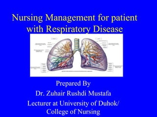
  • 1. Nursing Management for patient with Respiratory Disease Prepared By Dr. Zuhair Rushdi Mustafa Lecturer at University of Duhok/ College of Nursing
  • 2. • Crucial Diagnostic Tests for Respiratory disease 1. Bronchoscopy Bronchoscopy is used to view the bronchial tree and to remove foreign obstructions, obtain tissues for biopsy, or for suctioning fluid. 2. Chest X-ray Used to detect the size and position of the heart and structural abnormalities of the lungs. 3. Pulmonary Angiography • Provides a view of the pulmonary circulatory system so that the physician can determine the condition of blood flow to the lungs.
  • 3. 4. Sputum Culture and Sensitivity Sputum from the patient is cultured to determine which, if any, bacteria is contained in the sputum and determine which antibiotic kills the bacteria. 5. Thoracentesis Removal of fluid from the pleural sac to drain fluid or identify the contents of the fluid. 6. Pulmonary Function Test (PFT) This test assesses the lungs’ ability to move air. Monitor change from normal function; differentiate obstructive from restrictive disease.
  • 4. 7. Arterial Blood Gas (ABG) This determines the patient’s ventilation, tissue oxygenation, and acid-base status. HOW DOES THE TEST WORK? Three to five milliliters of blood is sampled from an artery in a heparinized syringe. If the sample cannot be analyzed right away, it should be placed on ice. • The normal results are: • pH 7.35–7.45 • PaO2 80–100 mmHg • PaCO2 35–45 mmHg • HCO3 22–26 mEq/L
  • 5. 8. Mantoux Intradermal Skin Test (purified protein derivative PPD) • This determines if the patient has antibodies to the Mycobacterium tuberculosis bacteria, which indicates that the patient has been exposed to the bacteria. 9. Lung Biopsy Removal of a tissue sample to be examined by the histology lab for abnormalities.
  • 6. Bronchitis • Increased mucus production, caused by infection and airborne irritants that block airways in the lungs, results in the decreased ability to exchange gases. • There are two forms of bronchitis: 1. acute bronchitis, where blockage of the airways is reversible. 2. chronic bronchitis, where blockage is not reversible.
  • 7. • Patients with acute bronchitis are symptomatic typically for 7 to 10 days often due to viral (but sometimes bacterial) infection. • Patients with chronic bronchitis will have symptoms of a chronic productive cough for at least 3 consecutive months in 2 consecutive years. • There is increased mucous production, inflammatory changes, and, ultimately, fibrosis in the airway walls. • The patient with chronic bronchitis has an increased incidence of respiratory infection.
  • 8. Pathophysiology of Chronic bronchitis • Chronic bronchitis characterised by an increase in mucus production and damaged cilia in the bronchi. As a result, the bronchi become clogged with mucus, then producing a cough. This chronic irritation causes inflammation and the bronchial wall thickens, causing airway obstruction. Secondary infections then occur, causing yet more irritation and inflammation. As more and more airways become blocked, external respiration is reduced and less oxygen is transferred into the bloodstream. Normal bronchiole Narrowed and mucus-filled bronchiole in bronchitis
  • 9. • SIGNS AND SYMPTOMS of Bronchitis 1. Cough due to mucous production and irritation of airways. 2. Shortness of breath. 3. Fever in acute episodes due to infection. 4. Accessory muscles are used for breathing 5. Productive cough due to irritation of airways. 6. Weight gain secondary to edema in chronic bronchitis is due to right-sided heart failure. 7. Wheezing due to inflammation within the airways.
  • 10. • TREATMENT 1. Acute bronchitis is treated in the short term with symptomatic treatment and antibiotics when a bacterial infection is present. 2. Chronic bronchitis is treated with a combination of medications to keep the airways open, reduce inflammation within airways, and prevent complications or exacerbations. 3. Administer beta 2-agonists by inhaler or nebulizer to dilate the bronchi such as albuterol 4. Administer anticholinergics which allow for relaxation of bronchial smooth muscle such as ipratropium inhaler
  • 11. 5. Administer steroids to decrease inflammation within the airways such as hydrocortisone, methylprednisolone systemically beclomethasone inhalers 6. prednisolone, prednisone orally. 7. Administer methylxanthines to enhance bronchodilation such as aminophylline and theophylline. 8. Administer diuretics to reduce fluid retention in patients who develop right sided heart failure such as furosemide. 9. Administer expectorant to help liquefy secretions such as Mucolyte.
  • 12. 10. Administer antibiotics in acute exacerbation of chronic bronchitis which is selected by culture and sensitivity study 11. Administer antacid such as antacids: aluminum hydroxide/magnesium hydroxide, calcium carbonated, H2 blocker such as ranitidine, famotidine, cimetidine, or proton pump inhibitor such as omeprazole, lansoprazole, esomeprazole, rabeprazole, pantoprazole to decrease the amount of acid in stomach, reducing possible ulcer formation due to stress of disease or medication effects.
  • 13. 12. Administer vaccines—to decrease chances of infection: • influenza • pneumonia 13. Give 3 liters of fluid per day to help liquefy secretions 14. Oxygen: 2 liters per minute via nasal canula to help meet body’s needs. 15. Increase protein, calories, and vitamin C in diet to meet body’s needs. 16. Administer the incentive spirometer to encourage coughing and expelling of mucous.
  • 14. • NURSING DIAGNOSES • Ineffective airway clearance • Activity intolerance • Ineffective breathing pattern
  • 15. NURSING INTERVENTION 1. Monitor respirations looking at rate, effort, use of accessory muscles, skin color; listen to breath sounds. 2. Place patient in high Fowler’s position to ease respiration. 3. Weigh the patient daily. Excess fluid due to heart failure will increase weight. Notify physician, of weight gain of 2 pounds in 24 hours. 4. Have the patient perform the turning, coughing, and deep- breathing exercises to enhance lung expansion and expel mucous. 5.Monitor sputum for changes in color or amount. 6. Monitor intake and output. 7. Increase fluids to keep mucous thinner and easier to expel. 8. administer oxygen as order.
  • 16. • Pneumonia • Pneumonia is an infection of the alveoli and small airways. Inflammation and oedema cause the alveoli to fill with debris and exudate that commonly impairs gas exchange.
  • 17. • Etiology • Infectious pneumonia may be due to a variety of microorganisms and can be community-acquired or hospital-acquired (nosocomial). • A patient can inhale bacteria, viruses, parasites, or irritating agents, or a patient can aspirate liquids or foods. There may be increased mucous production and thickening alveolar fluid as a result of impaired gas exchange. All of these can lead to inflammation of the lower airways. • Organisms commonly associated with infection include Staphylococcus aureus, Streptococcus pneumoniae, Haemophilus influenza, Mycoplasma pneumoniae, Legionella pneumonia, Chlamydia pneumoniae (parasite), and Pseudomonas aeruginosa
  • 18. • Signs and symptoms 1. Shortness of breath due to inflammation within the lungs, impairing gas exchange 2. Difficulty breathing (dyspnea) due to inflammation and mucus within the lungs 3. Fever and Chills due to infectious process 4. Cough due to mucous production and irritation of the airways 5. Crackles due to fluid within the alveolar space and smaller airways 6. wheezing due to inflammation within the larger airways
  • 19. • Signs and symptoms 7. Discolored, possibly blood-tinged, sputum due to irritation in the airways or microorganisms causing infection 8. Tachycardia and tachypnea as the body attempts to meet the demand for oxygen 9. Pain on respiration due to pleuritic inflammation, pleural effusion. 10. Headache, muscle aches (myalgia), joint pains, or nausea may be present depending on the infecting organism
  • 20. INTERPRETING TEST RESULTS • Shadows on chest x-ray, indicating infiltration. • Culture and sensitivity of the sputum to identify the infective agent and the appropriate antibiotics. • Elevated WBC (leukocytosis) showing sign of infection. • Low oxygen saturation on pulse oximetry. • Arterial blood gas may show low oxygen and elevated carbon dioxide levels.
  • 21. • Care and management 1. Administer oxygen as needed. 2. For bacterial infections, administer antibiotics such as macrolides (azithromycin, clarithromycin), fluoroquinolones (levofloxacin), beta-lactams (amoxicillin/clavulanate, cefotaxime, ceftriaxone, ampicillin/sulbactam) 3. Administer antipyretics in patient with fever for comfort such as acetaminophen, ibuprofen 4. Administer brochodilators to keep airways open, enhance airflow if needed such as albuterol, levalbuterol via nebulizer
  • 22. • Care and management 5. Increase fluid intake to help loosen secretions and prevent dehydration. 6. Instruct the patient on how to use the incentive spirometer to encourage deep breathing; monitor progress. 7. placing the patient in an upright position will promote diaphragm and intercostal muscle activity and enhance ventilation.
  • 23. Tuberculosis (TB) An infectious disease spread by airborne route. Infection is caused by inhalation of droplets that contain the tuberculosis bacteria (Mycobacterium tuberculosis). An infected person can spread the small airborne particles through coughing, sneezing, or talking. Close contact with those affected increases the chances of transmission. Once inhaled, the organism typically settles into the lung, but can infect any organ in the body. The organism has an outer capsule.
  • 24. • SIGNS AND SYMPTOMS 1. Weight loss and anorexia 2. Night sweats 3. Fever, possibly low-grade, due to infection 4. Productive cough with discolored, blood-tinged sputum 5. Shortness of breath due to lung changes 6. Malaise and fatigue due to active illness affecting lungs
  • 25. INTERPRETING TEST RESULTS 1. Positive Mantoux (PPD) skin test shows exposure to tuberculosis due to development of cell-mediated immunity. 2. Chest x-ray may show areas of granuloma or cavitation. 3. Sputum test identifies M. tuberculosis bacteria: • Acid fast-staining done to initially screen for TB— bacillus will hold stain. • Culture confirms the diagnosis but is slow-growing
  • 26. • TREATMENT 1. Initial treatment times generally range from 6 to 12 months. 2. Longer treatment plans may be necessary for those with HIV infection or drug-resistant strains of TB. 3. Administer antitubercular medications to treat and prevent transmission such as : isoniazid, rifampin, pyrazinamide, ethambutol, streptomycin. 4. Respiratory isolation for in-hospital care—the bacteria is spread by droplet. 5. Increase protein, carbohydrates, and vitamin C diet for patients.
  • 27. • NURSING DIAGNOSES • Fatigue • Ineffective airway clearance NURSING INTERVENTION 1. Monitor respiration for rate, effort, use of accessory muscles, and skin color changes. 2. Increase fluid intake to help liquefy any secretions. 3. Record fluid intake and output. 4. Explain to the patient: A. How to prevent spreading the disease. B. The importance of finishing all prescribed medication. C. Plan for rest periods during the day.
  • 28. Pulmonary Embolism • Blood flow is obstructed in the lungs caused by thrombus (blood clot), air, or fat emboli that become stuck in an artery, causing impaired gas exchange.
  • 29. • Pathophysiology • Patients may be predisposed to clot formation, have pooling of blood, or damage to vessel walls, or take certain medications that increase the risk of thrombus formation. • Thrombus are commonly found in vessels in lower extremities. • When a thrombus loosens and travels in the peripheral circulation, it is called an embolus. • The embolus travels through the right side of the heart and is sent to the lungs where it lodges in one of the arteries.
  • 30. SIGNS AND SYMPTOMS 1. Sudden difficulty breathing (dyspnea) happens when the clot suddenly lodges in the artery 2. Heart rate greater than 100 beats per minute (tachycardia) 3. Respiration greater than 20 breaths per minutes (tachypnea) as the body attempts to get more oxygen 4. Chest pain due to clot presence and area of atelectasis 5. Coughing with blood-tinged sputum (hemoptysis) 6. Crackles heard near area of clot
  • 31. INTERPRETING TEST RESULTS 1. Chest x-ray may show dilated pulmonary artery or pleural effusion. 2. CT scan will show clot in pulmonary arteries. 3. Pulmonary angiography will show presence of clot. 4. Arterial blood gases may show decreased oxygen (PaO2) and carbon dioxide (PaCO2), depending on the size of the clot. 5. Lower extremity ultrasound is often done to test for presence of thrombus
  • 32. • TREATMENT 1. Supplemental oxygen therapy. 2. Administer thrombolytics to enhance breakdown of existing clot such as urokinase, alteplase. 3. Administer anticoagulants to prevent further clot formation sucha as heparin, warfarin 4. Administer analgesic for pain control such as morphine 5. Bed rest to prevent thrombus from breaking free from lower extremities. 6. Surgical removal of the embolus may be necessary in some cases.
  • 33. • NURSING INTERVENTION • Explain to the patient: 1. To avoid sitting and standing for too long to decrease chance of clot formation. 2. Not to cross legs to avoid constriction of vessels in the lower extremities, decreasing the chances of clot formation. 3. How to identify side effects from using anticoagulants, such as bleeding or bruising. 4. Explain that pulmonary embolism is an adverse effect from using hormonal contraceptives and a different form of birth control needs to be used in the future
  • 34. • Asthma • The airways become obstructed from either inflammation of the lining of the airways or constriction of the bronchial smooth muscles (bronchospasm). • A known allergen, for example, pollen—is inhaled, causing activation of antibodies that recognize the allergen. Mast cells and histamine are activated, initiating a local inflammatory response. • Astimulus causes an inflammatory reaction, increasing the size of the bronchial linings; this results in restriction of the airways
  • 35. • Types of Asthma • There are two kinds of asthma: 1. Extrinsic asthma, also known as atopic, caused by allergens such as pollen, animal dander, mold, or dust. Often accompanied by allergic rhinitis and eczema; this may run in families. 2. Intrinsic asthma, also known as nonatopic, caused by a nonallergic factor such as following a respiratory tract infection, exposure to cold air, changes in air humidity, or respiratory irritants.
  • 36. SIGNS AND SYMPTOMS 1. Wheezing initially present on expiration. 2. dyspnea as airways narrow due to inflammation. 3. Respiration greater than 20 breaths per minute (tachypnea) 4. Use of accessory muscles to breathe as the body tries harder to get more air into the lungs. 5. Tightness in the chest due to narrowing of the airways (bronchoconstriction). 6. Cough. 7. Tachycardia—heart rate greater than 100, as the body attempts to get more oxygen to the tissues.
  • 37. • TREATMENT 1. Administer oxygen 2. Identify and remove allergens and known triggers to avoid causing an asthma attack. 3. Give patient 3 liters/day of fluid to help liquefy any secretions. 4. Administer short-acting beta2-adrenergic drugs to bronchodilate such as albuterol, levalbuterol. 5. Administer steroids to decrease inflammation, which will help open airways;these are not for acute symptoms such as hydrocortisone, methylprednisolone intravenously, beclomethasone inhalers prednisolone, prednisone orally 6. Administer aminophylline and theophylline 7. Administer antacid, H2 blocker, or proton pump inhibitor to decrease the amount of acid in the stomach, reducing the possibility of ulcers due to stress of disease or medication effects
  • 38. • Emphysema Emphysema is defined as the permanent enlargement of airspaces beyond the terminal bronchiole and the destruction of the alveolar wall. Chronic inflammation reduces the flexibility of the walls of alveoli, resulting in over-distention of the alveolar walls. This causes air to be trapped in the lungs, impeding gas exchange. Smoking is often linked to development of emphysema.
  • 39. • SIGNS AND SYMPTOMS 1. Dyspnea due to air trapping, which retains CO2 and reduces alveolar gas exchange. 2. Barrel chest develops over time as more air is trapped within the distal airways. 3. Use of accessory muscles to breathe as the respiratory effort increases. 4. Loss of weight as extra calories are needed to maintain respiration. 5. Patients prefer a seated position which allows for greater chest expansion.
  • 41. • INTERPRETING TEST RESULTS 1. Increased residual volume shown in pulmonary function test due to air trapping. 2. Decreased O2 and increased CO2 in arterial blood gas as gas 3. exchange is impaired due to air trapping; more pronounced as disease progresses. 4. Chest x-ray shows over inflation of lungs and flattening of the diaphragm.
  • 42. • TREATMENT 1. Administer beta2-agonists to bronchodilate by inhaler or nebulizer such as albuterol, levalbuterol. 2. Administer long-acting bronchodilating medications by metered dose inhaler such as formoterol, salmeterol 3. Administer anticholinergics which allow for relaxation of bronchial smooth muscle such as ipratropium inhaler. 4. Administer methylxanthines to dilate the bronchi such as aminophylline and theophylline.
  • 43. • TREATMENT 5. Administer steroids to decrease inflammation within the airways such as hydrocortisone, methylprednisolone systemically, prednisolone, prednisone orally. 6. Administer antacid such as antacids: aluminum hydroxide/magnesium hydroxide, calcium carbonated, H2 blocker such as ranitidine, famotidine, cimetidine, or proton pump inhibitor such as omeprazole, lansoprazole, esomeprazole, rabeprazole, pantoprazole to decrease the amount of acid in stomach, reducing possible ulcer formation due to stress of disease or medication effects.
  • 44. 7. Administer expectorant—to loosen secretions: 8. Administer diuretics to decrease fluid retention in patients that are developing right-sided heart failure secondary to lung disease such as furosemide (Lasix). 9. Administer vaccines—to prevent respiratory infections: • influenza, pneumonia 10. Administer antibiotics based on results of culture and sensitivity. 11. Administer oxygen, 2 liters per minute. 12. Give patient 3 liters of fluids per day to help liquefy secretions. 13. Teach patient how to use the incentive spirometer to encourage deep breathing and enhance coughing and expelling of mucous..
  • 45. Chronic obstructive pulmonary disease • Is the most common chronic lung disease. COPD is an umbrella term that could refer to emphysema and chronic bronchitis and, more commonly, a combination of these conditions. • It has one major cause which is smoking. Etiology COPD may result from: 1. cigarette smoking 2. recurrent or chronic respiratory tract infection 3. allergies 4. familial and hereditary factors such as alpha1-antitrypsin deficiency.
  • 46. Sings & Symptoms The typical COPD patient is asymptomatic until middle age, when the following signs and symptoms may occur: 1. reduced ability to exercise or do strenuous work 2. productive cough 3. dyspnea with minimal exertion.
  • 47. Treatment Treatment for COPD aims to relieve symptoms and prevent complications. 1. Administer beta-agonist bronchodilators (albuterol or salmeterol) 2. Anticholinergic bronchodilators (ipratropium [Atrovent]), 3. Corticosteroids (beclomethasone ) These drugs are usually given by metered dose inhaler. 4.smoking cessation advice 5. education on prescribed oxygen and bronchodilator therapies – to maximise relief of breathlessness 5. dietary advice – severe weight loss is a feature of both emphysema and chronic bronchitis 6. pulmonary rehabilitation
  • 48. • Lung Cancer Lung cancer is the abnormal, uncontrolled cell growth in lung tissues, resulting in a tumor. A tumor in the lung may be primary when it develops in lung tissue. It may be secondary when it spreads (metastasizes) from cancer in other areas of the body, such as the liver, brain, or kidneys. • Repetitive exposure to inhaled irritants increases a person’s risk for lung cancer. Cigarette smoke, occupational exposures, air pollution containing benzopyrenes, and hydrocarbons have all been shown to increase risk.
  • 49. • There are two major categories of lung cancer: • small cell and non-small cell. • Small cell: • Oat cell—fast-growing, early metastasis • Non-small cell: • Adenocarcinoma—moderate growth rate, early metastasis • Squamous cell—slow-growing, late metastasis • Large cell—fast-growing, early metastasis
  • 50. • PROGNOSIS • Lung cancer is the leading cause of cancer death. Many patients with lung cancer are diagnosed at a later stage, leading to the long-term (5-year) survival rate of less than 20 %. Earlier diagnosis is more beneficial for treatment and outcome. • The longer the cancer has been in the lungs, the greater the likelihood of metastasis to other areas.
  • 51. • SIGNS AND SYMPTOMS 1. Coughing due to irritation from mass. Presence of mucous or exudate may not be until later in disease. 2. Coughing up blood (hemoptysis). 3. Fatigue. 4. Weight loss due to the caloric needs of the tumor, taking away from the needs of the body. 5. Anorexia. 6. Dyspnea caused by damaged lung tissue. 7. Chest pains as mass presses on surrounding tissue; may not be feel until late in disease. 8. Sputum production. 9. Pleural effusion.
  • 52. • INTERPRETING TEST RESULTS 1. Mass in lung shown on chest x-ray. 2. CT scan shows mass, lymph node involvement. 3. Bronchoscopy may show cancer cells on bronchoscopic 4.Cancer cells seen in sputum. 5. Biopsy through chest wall or tissue biopsy from lung will show cell type. 6. Bone scan or CT scans shows metastasis of the disease.
  • 53. TREATMENT 1. Surgical removal of affected area of the lung (wedge resection, segmental resection, lobectomy) or total lung (pneumonectomy). 2. Radiation therapy to decrease tumor size. 3. Chemotherapy such as : cyclophosphamide, vincristine, etoposide, cisplatin 4. Oxygen therapy to supplement the needs of the body. 5. High-protein, high-calorie diet to meet the needs of the body. 6. Administer antiemetics to combat side effects of chemotherapy: ondansetron (Zofran), prochlorperazine 7. Administer analgesics for pain control such as morphine, Tramadol.
  • 54. • NURSING DIAGNOSES • Anxiety • Activity intolerance • Impaired gas exchange
  • 55. NURSING INTERVENTION 1. Monitor respiratory status, looking at rate, effort, use of accessory muscles, and skin color; auscultate breath sounds. 2. Monitor pain and administer analgesics appropriately. 3. Monitor vital signs for changes, elevated pulse, elevated respiration, change in BP, and elevated temperature, which may signal infection. 4. Monitor pulse oximetery for decrease in oxygenation levels. 5. Assist patient with turning, coughing, and deep-breathing exercises. 6. Place patient in semi-Fowler’s position to ease respiratory effort. 7. Explain to the patient the importance of taking rest periods.