2. Introduction.
History.
Basic principle and theory.
Sample preparation.
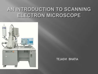
Basic set up of SEM.
Merits
Demerits
Forensic importance
3. INTRODUCTION
• SEM is one of the most versatile instrument available for
the examination and analysis of micro structural
characteristics of solid objects .
• Scanning electron microscope (SEM), is a type of
electron microscope that, images the sample surface by
scanning it with a thin high-energy beam of electrons.
• The scanning electron microscope as the name
indicates produces of highly
magnified and resolved image of the object using fine
and focused electron beam instead of light.
4. • The interaction of the electrons with the atom’s that
make up the sample produce signals that contain
information about the sample’s surface topography,
composition and other properties such as electrical
conductivity.
• When SEM interfaced with EDX or WDX, elemental
composition of the sample is obtained by analysis of
the characteristic x-rays generated.
5. The first scanning electron microscope date
back to 1935, with the work of Max Knoll.
Further pioneering on the physical principle of
SEM, and beam specimen interaction was
performed by, Manfred van Ardenne in 1977.
It was until 1965, that it was first market by the
Cambridge Instrument Company as
“Stereoscan”, after being finally worked upon
by Pro. Charles Oatley.
6. • When the high energy fine and foccussed
electron beam strikes the surface of the
specimen it would interact with the specimen
atoms.
• The energy exchange between the electron
beam and the sample results into the formation
of the following signals; -
Reflection of high-energy electrons (BSE)
Some of the electrons would be absorbed by
the specimen.
Emission of the secondary electrons.
Emission of light (Cathodoluminescence).
Emission of Characterstic X-Rays.
7.
8. . For conventional imaging in the SEM, specimen must
be electrically conductive, at least at the surface, and
electrically grounded to prevent the accumulation of
electrostatic charge at the surface.
Metallic objects require little preparation except for,
cleaning and mounting on the specimen stub.
Nonconductive.
Biological samples.
9. The essential parts of SEM are :-
• Electron source / gun
• Demagnification unit
• Scanning coil
• Specimen Platforms
• Vacuum
• Detectors
• Image processing unit
10. INSTRUMENTATION :
ELECTRON GUN :
• stable source of electrons.
• usually made up of tungsten or lanthanum hexaboride.
• Maintained at high potential voltage i.e. 1-50 kEv.
DEMAGNIFICATION UNIT:
• Electron magnetic lenses.
• The desired probe size or the beam size i.e. 1-5 nm .
• The process is carried out under vacuum condition.
11. IMAGE PROCESSING UNIT:
• The secondary Electrons so generated from
throughout the specimen surface are collected by
scintillator detectors.
• Amplified with the help of photomultiplier.
• This electronic image is built in cathode ray tube.
• Displaced on screen or a monitor.
13. MERITS
• It’s a versatile microscopy technique.
• It has much higher resolution, so closely spaced
specimens can be magnified at much higher
levels.
• Being that it uses electromagnets rather than
lenses, the researcher has much more control in
the degree of magnification.
• The production of images is strikingly clear .
• SEM produces image at a very high
magnification i.e. 2 X 10⁵ sec the image so
produced also process very high resolution i.e. 5
nm – 2.5 nm.
• SEM images are with a large depth of focus and
gives
3-dimensional appearance of the object.
14. SEM is highly sensitive and a versatile technique but at
the same time it suffers from certain drawback :-
• Only solid , nonvolatile samples can be analyzed.
• Sample should be conductive in nature if not then
sample preparation is required i.e. the sample is coated
with either gold or graphite or any other conductive
material.
• Sample should be totally devoid of moisture i.e. it should
be dry or dehydrated if not then again it has to be dried
and the moisture has to be removed in the sample in an
oven in such a way that signal and shape and
composition of sample remain unaltered.
15. It is used for the identification of gun shot residues
(GSR) by both its morphology as well as the elemental
profile.
It is used for the examination of paint in order to find out
no . of layers and elemental composition.
It is used for the analysis of various types of tool marks .
It is used for the forensic characterization of various
types of soil samples.
For the forensic analysis of various types of fiber of
plant and animal.
It is widely used for the analysis of biological evidences
such as hair , diatoms , pollen grains, and microbial
organism.
It is also used in forensic chemistry for the examination
of cannabis preparation i.e. bhang, ganja and by
studying cistolythic hairs.
16. REFERENCE
Richard Saferstein., Criminalistics., An
Introduction to Forensic Science., Eighth
Edition.
By. F.A Settle Handbook of instrumental
technique for analytical chemistry. 1997
Hearle, J.W.S., Sparrow, J.T., and Cross, P.M.,
The use of the scanning Electron
Microscope. Oxford. Pergamon.
http://www.mos.org/sln/SEM/
www.microscope. edu.
http://mse.iastate.edu/microscopy/home.htm
l