
Recommended
More Related Content
What's hot
What's hot (20)
Similar to Microscope
Similar to Microscope (20)
More from Taniya07
Recently uploaded
Recently uploaded (20)
Microscope
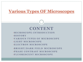
- 1. CONTENT •MICROSCOPE-INTRODUCTION •HISTORY •VARIOUS TYPES OF MICROSCOPE •LIGHT MICROSCOPE •ELECTRON MICROSCOPE •BRIGHT/DARK FIELD MICROSCOPE •PHASE CONTRAST MICROSCOPE •FLUORESCENT MICROSCOPE Various Types OF Microscopes
- 2. MICROSCOPE The word “microscope” is formed of two words: “micros”- small and ‘skipein’-to look. Microscope is an instrument which makes enlarged images of minute objects, sub cellular structures, and many more, generally hard to resolved with naked human eyes. Complexity of microscopes has increased many folds from simple lens to complex scanning electron microscope.
- 3. HISTORY Modern microscope are based on basic principle used by Zoocharia Jansen in 1590. He used second lens that enlarged imaged formed by first lens by 50-100X. Robert Hooke first made compound microscope, and also published a book “Micrographia”. His microscope had a maximum magnification of 200X. Anton van Leeuwenhoek provided improved microscope and was first to observe unicellular animal. He is called “Father Of Microbiology”. Other renowned names in history of microscopes includes C. Huygens & Abber.
- 4. VARIOUS TYPES OF MICROSCOPES Basically there are two types of microscopes: Light Microscope Electron Microscope Further classification in these basic microscopes are present. Other microscopes also present such as phase contrast microscopes, fluorescent microscope , bright field/dark field microscope.
- 5. LIGHT MICROSCOPE(LM) Most commonly used microscope. Handy in use. LM uses light source for illumination of specimen. Generally used light sources include sunlight, UV light, laser light, LEDs. Types- Simple dissecting microscope, compound microscope, stereomicroscopes.
- 9. WORKING OF LM The specimen is mounted on slide and positioned in specimen stage. Beam of light is focused on specimen by condenser. Objective lens picks up light transmitted by specimen and produce first magnified image. This image is further magnified by eyepiece lens. Eyepiece only magnifies image and brings no change in resolution.
- 10. APPLICATION OF LM 1. Study of preserved minute specimen. 2. Study activities inside the cell. 3. Identifying macromolecules of cell. 4. Medical diagnosis. 5. Histopathological studies.
- 11. ELECTRON MICROSCOPE (EM) Advance microscope. German scientist Knoll and Ruska discovered electron microscope. Electron beam is used as source of illumination. Types- Transmission electron microscope(TEM), scanning electron microscope(SEM).
- 12. TEM
- 13. SEM
- 14. WORKING OF EM Beam of electron travel through column of microscope in vacuum. Different electromagnetic lens focuses electrons into thin beam. Beam travel through specimen. Some electrons may get scattered while others transmitted(TEM), hit fluorescent screen(detector) and forms image. In SEM, reflected electron from specimen forms magnified image.
- 15. APPLICATION OF EM 1. Study causes of disease. 2. Study 3D structure of cells . 3. Analysis of surface fracture or surface contamination of cells. 4. Important part in production of silicon chips. 5. Used in industrial search centers.
- 16. BRIGHT/DARK FIELD MICROSCOPE In bright field, microscope field appears bright whereas microorganism appears dark as they absorb light. Normally micro-organism do not absorb light but absorbing ability increases due to staining. In dark field microscope, a dark background is established against a brightly illuminated object. Dark field microscopy requires additional dark field condenser and dark field object lens.
- 17. PHASE CONTRAST MICROSCOPE Phase contrast microscope was developed by Fritz Zernike, was awarded Nobel prize in Physics in 1953. By this microscopy organism can be seen alive, without staining. It requires additional specialized structure annular diaphragm and phase contrast ring. The images differences in refractive index of cellular structure. Light passes through thicker parts of cell is held up relative to the light that passes through thinner parts of cytoplasm.
- 19. FLUORESCENT MICROSCOPE Popularly used to achieve high labeling of cellular compartments. This microscope additionally requires an excitation filter, a barrier and a dichromatic mirror, fluorescent stain. A specific wavelength of light is used to excite fluorescent molecule in specimen. Light of higher wavelength is then imaged.
- 21. THE END