
Recommended
More Related Content
What's hot
What's hot (20)
Similar to The Digestive System Explained
Similar to The Digestive System Explained (20)
More from TakaleBulo
Recently uploaded
Recently uploaded (20)
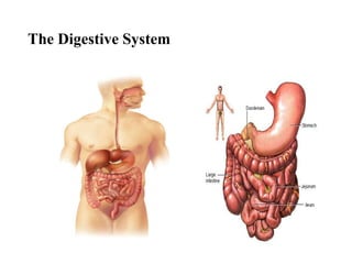
The Digestive System Explained
- 2. Introduction • Every body cell needs a constant supply of nutrients to provide energy and building blocks for the manufacture of body substances. • Food as we take it in, however, is too large to enter the cells. • It must first be broken down into particles small enough to pass through the cell membrane. This process is known as digestion. • After digestion, food must be carried to the cells in every part of the body by the circulation. • The transfer of food into the circulation is called absorption. • Digestion and absorption are the two chief functions of the digestive system. 5/10/2023 Lecture note of Human Anatomy ASU 2
- 3. Introduction • For our purpose the digestive system may be divided into two groups of organs: 1. The Gastrointestinal (GI) tract (Alimentary Canal) a continuous passageway beginning at the mouth, where food is taken in, and terminating at the anus, where the solid waste products of digestion are expelled from the body 2. The accessory organ, which are necessary for the digestive process but are not a direct part of the digestive tract. • They release substances into the digestive tract through ducts. 5/10/2023 Lecture note of Human Anatomy ASU 3
- 4. Layers of GI tract •GIT from esophagus to anus has the same basic arrangement of tissues. •The four coats or tunics of the tract from the inside out are:- 1. Mucosa •Inner lining of the tract •It is composed of layer of epithelium •It composes three layers. 4
- 5. a. Lining epithelium •Direct contact with contents of the GI tract Composed of •Non-keratinized cells serves as protection and secretion and •Simple epithelium which serves as secretion and absorption b. Lamina propria •Composed of loose connective tissue that contains many blood and lymph vessels. •c. Mascularis mucosa: containing smooth muscle 5
- 6. 2. Submucosa •Consists of areolar connective tissue •It binds the mucosa to the muscularis •It is highly vascular and contains portions of the submucosa plexus (plexus of Meissner), which is part of the autonomic nerve supply to muscularis mucosa. 6
- 7. 3. Muscularis •In the mouth, pharynx, and portion of esophagus consists of skeletal muscles that produce voluntary swallowing. •The rest consists of smooth muscles found as inner ring of circular fibers and an outer sheet of longitudinal fibers. 7
- 8. 4. Serosa •Outer most layer around organs of the GI tract below the diaphragm. •The membrane composed of simple squamous epithelium and areolar connective tissue. •The serosa secretes slippery, watery fluid that allows the tract to glide easily against other organs. 8
- 9. 9
- 10. Structure of the GI tract 10
- 11. Peritoneum •Largest serous membrane of the body. •Parietal peritoneum – lines wall of abdominal cavity. •Visceral peritoneum - covers some organs and constitutes their serosa. •Peritoneal cavity •Potential space b/n the parietal and visceral portions of peritoneum. 11
- 12. Peritoneum •Contains serous fluid •Extra fluid in the peritoneal cavity➔ results in ascites. Based on the peritoneal covering •Intraperitoneal organs→ stomach, liver, gall bladder, spleen, jejunum, ileum, transverse colon, sigmoid colon, uterus and ovaries (essentially mobile organs “slung” in mesentery) 12
- 13. •Retroperitoneal organs •Kidneys, adrenal glands, aorta, IVC, bladder, vagina, rectum, descending and ascending colon, duodenum and pancreas 13
- 14. H Ye. G 14
- 15. Omentum • Double layered peritoneum that passes from stomach and proximal duodenum to adjacent abdominal organs • Two parts 1. Greater→ greater stomach curvature inferiorly and back up to anterior surface of transverse colon. • Parts • Gastrophrenic lig→ stomach to inferior diaphragm • Gastrosplenic lig→ stomach to spleen • Gastrocolic→ stomach to transverse colon 15
- 16. Lesser omentum- extends from porta hepatis of liver to lesser curvature of stomach and first part of duodenum •hepatogastric part - from liver to lesser curvature of stomach •hepatoduodenal part - free margin of lesser omentum from liver to first part of duodenum & contains: •portal vein, bile duct & proper hepatic artery 16
- 19. The Digestive Tract • The digestive tract is a muscular tube extending through the body. • It is composed of several parts: the mouth, pharynx, esophagus, stomach, small intestine, and large intestine. • The digestive tract is sometimes called the alimentary tract, derived from a Latin word that means "food". • It is more commonly referred to as the gastrointestinal (Gl) tract because of the major importance of the stomach and intestine in the process of digestion. 5/10/2023 Lecture note of Human Anatomy ASU 19
- 20. 5/10/2023 Lecture note of Human Anatomy ASU 20 Digestive system
- 21. Mouth /Oral/ Buccal cavity Formed by Cheeks, hard & soft palates, & tongue Cheeks • Forms lateral walls of oral cavity • Muscular structures externally covered with skin • Anterior portion terminates in the superior and inferior lips Hard palate • Anterior portion of the roof of the mouth • Formed by the maxillary and palatine bones • Covered by mucous membrane, and forms a bony portion b/n the oral and nasal cavities. Soft palate • Forms posterior portion of the roof of mouth • An arch-shaped muscular portion b/n the oropharynx and nasopharynx and lined by mucus membrane 21
- 22. 22
- 23. Tongue •Forms floor of the oral cavity •Accessory structure of the Digestive System composed of skeletal muscle covered with mucus membrane. •Divided into symmetrical lateral halves by a median septum that extends through out its length and is attached inferiorly to the hyoid bone. 23
- 24. The tongue consists of Extrinsic muscles •Originate outside the tongue & insert into it. •In includes hyoglossus, genioglossus, styloglossus and palatoglossus •Moves tongue from side to side and in and out. •The movement food for chewing, shape the food, and force the food to the back of the mouth for swallowing. Intrinsic muscles •Originate and insert within the tongue. •Alters shape & size of tongue for speech & swallowing. •It includes longitudinal superior and inferior, transverses lingual, and verticalis lingual. 24
- 25. 25
- 26. Teeth/dentes •Located in sockets of alveolar processes of mandible and maxillae •Alveolar processes are covered by the gingival/gums; extend slightly into each socket forming the gingival sulcus. •Sockets are lined by the periodontal ligament, which consists of dense, fibrous connective tissue. It anchors the teeth in position. 26
- 27. Typical tooth consists of 1. Crown - portion above the level of the gums. 2. Root - 1 to 3 projections embedded in socket. 3. Neck - constricted junction lien of the crown and the root Dentin – (consist of calcified connective tissue) is a composition of teeth that gives its basic shape and rigidity. Pulp cavity - is formed by the dentin that lies in the crown and filled with pulp, a connective tissue containing blood vessels, nerves, and lymphatic. Root canals - narrow extension of the pulp cavity run through the root of teeth. 27
- 28. Apical foramen - an opening at its base of each root, where blood vessels bearing nourishment, lymphatic's affording protection, and nerves providing sensation. Enamel • Covers dentin of the crown • Consists of calcium phosphate and calcium carbonate • Hardest substance of the body • Protects the tooth from wear of chewing Cementum • Covers dentin of the root • Attach the root to the periodontal ligament 28
- 29. 29
- 30. Dentitions •Every one has two dentations or sets of teeth. The first of these are the deciduous teeth, milk teeth or baby teeth. •Erupt at about age of 6 month. They are 20 Types of teeth Incisors •These are 4 in one jaws •Closer to midline •Chisel shaped •Adapted for cutting 30
- 31. Cuspids (canines) •These 2 in one jaw •Next to incisors •Has pointed surface •Used to tear and shared food Premolar •4 in one jaws •Found next to the incisors Molars •6 in one jaw •Has four cusps •Used to crush and grind food 31
- 34. 34
- 35. 35 Salivary glands •Saliva is a fluid that is continuously secreted into the mouth for moistening, lubrication, dissolving and chemical breaking down of food. •There are three pairs of major salivary glands and minor buccal glands. 1. The parotid glands: located inferior and anterior to the ears b/n the skin and the masseter muscle. 2. The sub-mandibular glands: - found beneath the base of the tongue in the posterior part of the floor of the mouth. 3. Sablingual glands:- are located superior to the submandibular glands.
- 37. Esophagus •3rd organ involved in deglutition (swallowing) •Muscular, collapsible tube that lies behind the trachea •Begins in the neck as a continuation of the pharynx at the level if C6 and terminate at esophagogastric junction (T10), where it enters the cardial orifice of the stomach •25 cm long with a diameter of 2 cm with three parts (cervical, thoracic and abdominal) •Diaphragm act like sphincter 37
- 38. Arteries- left gastric artery, a branch of the celiac trunk, and the left inferior phrenic artery Venous drainage -portal venous system through the left gastric vein 38
- 39. Stomach •It is J-shaped enlargement of GI tract directly under the diaphragm in epigastric, umbilical and left hypochondriac regions •Superior portion of the stomach is a continuation of esophagus •Inferior portion empties into the duodenum. 39
- 40. •The stomach is divided into four areas: cardia, fundus, body and pylorus. •Cardia - surrounds the lower esophageal sphincter. •Fundus - the rounded portion above and to the left of the cardia. •Body - is largest central portion of the stomach. •Pylorus - it is the narrow, inferior region of the stomach. 40
- 41. Anatomy of the Stomach 41
- 42. Neurovasculature of the stomach 42 A) Arteries 1. Left gastric artery- from the ciliac trunk and gives esophageal artery & gastric branches 2. Right gastric artery-from the hepatic artery ◦ Both on lesser curvature. 3. Right gastro-omental artery: from gastroduodenal artery 4. Left gastro-omental artery: from splenic artery ◦ The above two are on the greater curvature 5. Short gastric artery - from the splenic artery to fundus of stomach.
- 43. Neurovasculature… 43 B) Veins • Left and right gastric veins drain directly into the portal vein • Short gastric & the Lt gastro-omental veins drain into the splenic vein • The Rt gastro-omental vein empties into the SMV C)Nerves • Parasympathetic- the anterior and posterior vagal trunks • The sympathetic - T6-T9 passes to the celiac plexus
- 44. Small Intestine •Extends from pyloric sphincter → ileocecal valve •Regions •Duodenum •Jejenum •Ileum 44
- 45. Small intestine cont… •Major portion of digestion and absorption •Begins at the pyloric sphincter of the stomach, coils through the central and lower part of the abdominal cavity, and opens into he large intestine. •Averagely 2.5cm in diameter and about 6.33m in length. 45
- 46. 46
- 47. 47
- 48. Duodenum- the widest and most fixed part. ▪begins at the pylorus on the right side and ends at the duodenojejunal junction on the left side. ▪ Four parts of the duodenum: superior (1st ) part, descending (2nd ) part, horizontal (3rd ) part and ascending (4th ) part.
- 49. Duodenum cont… Descending (2nd) part: The bile duct and main pancreatic ducts via the hepatopancreatic ampulla enter its posterior medial wall A) Arteries: • Proximal ½- by celiac trunk • Distal ½ - Superior mesenteric artery B. Veins: follow the arteries and drain into the portal vein directly/indirectly C. Nerves: vagus & sympathetic nerves from greater and lesser splanchnic
- 52. Jejunum and Ileum •The jejunum begins at the duodenojejunal junction and the ileum ends at the ileocecal junction, the union of the terminal ileum and cecum. Jejunum •The jejunum is about 2.5m long & represents proximal two-fifths. •It is mostly in the left upper quadrant of the abdomen •Is larger in diameter and has a thicker wall than the ileum. •less prominent arterial arcades & longer vasa recta (straight arteries), compared to those of ileum, are a unique characteristic of jejunum
- 53. Ileum •It is about 4m long & makes up distal three-fifths of small intestine •It is mostly in right lower quadrant. •Compared to jejunum, ileum has thinner walls, shorter vasa recta, more arterial arcades & more mesenteric fat •ileum opens into large intestine
- 54. Vessels of jejunum & ilium • The arterial supply- the superior mesenteric artery through 15 -20 Jejunal and ilial branches ▪ unite to form arterial arcades which give rise to straight vasa recta • The veins run parallel to the arteries and typically drain into the superior mesenteric vein • From the mesenteric vein, the nutrient rich venous blood from the small intestine drains into the hepatic portal vein which carries it to the liver
- 55. Distinguishing Characteristics of Jejunum and Ileum in Living Persons Characteristic Jejunum Ileum Color Deeper red Paler pink Caliber 2-4 cm 2-3 cm Wall Thick and heavy Thin and light Vascularity Greater Less Vasa recta Long Short Arcades A few large loops Many short loops Fat in mesentery Less More Circular folds (L. plicae circulares) Large, tall, and closely packed Low and sparse; absent in distal part Lymphoid nodules (Peyer patches) Few Many 55
- 56. 56
- 57. Large Intestine •Consists of the cecum, colon (ascending, transverse, descending, and sigmoid), rectum, and anal canal. •The large intestine can be distinguished from the small intestine by: ▪Teniae coli: three thickened bands of longitudinal muscle fibers. ▪Haustra: sacculations or pouches of the colon between the teniae. ▪Omental appendices: small, fatty appendices (projections) of colon. ▪Caliber: the internal diameter is much larger.
- 58. 58
- 59. The vermiform appendix •a variable position, but it is usually retrocecal (posterior to cecum). Arterial supply to cecum and appendix •ilocolic a branch of superior mesenteric artery •Appendicular a branch of iliocolic Venous drainage of caecum and appendix- iliocolic vein that drain into superior mesenteric vein Lymphatic drainage- lymph nodes in mesoappendix and iliocolic→superior mesenteric lymph nodes •Innervation- from celiac and superior mesenteric ganglia
- 60. The colon • The colon is described as having four parts ascending, transverse, descending, and sigmoid that succeed one another in an arch. 1. The ascending colon • passes superiorly from cecum to the right lobe of the liver and it turns to the left as the right colic flexure (hepatic flexure). • lies retroperitoneally along the right side of the posterior abdominal wall. • Arterial supply -iliocolic and right colic arteries from superior mesenteric artery • Venous drainage -iliocolic and right colic veins that drains to superior mesenteric vein
- 61. 2. The transverse colon- the largest and most mobile part of the large intestine ◦ crosses the abdomen from the right colic flexure to the left colic flexure, where it bends inferiorly to become the descending colon. Arterial supply- middle colic & right colic from superior mesenteric artery for the right 2/3, left colic from inferior mesenteric artery for the left 1/3 Venous drainage- superior mesenteric vein
- 62. 3. The descending colon - passes retroperitoneally from the left colic flexure into the left iliac fossa, where it is continuous with the sigmoid colon. ◦ arterial supply- left colic & sigmoid arteries from the IMA ◦ venous drainage- by IMV 4. The sigmoid colon- S-shaped; extends from the iliac fossa to the third sacral segment, where it joins the rectum ◦ termination of the teniae coli indicates the recto sigmoid junction. ◦ Arterial supply -three sigmoid artery from IMA ◦ Venous drainage- IMV
- 65. •Rectum and Anal Canal •The rectum, the fixed terminal part of the large intestine, is continuous with the sigmoid colon at the level of the S3 vertebra. •The junction is at the lower end of the mesentery of the sigmoid colon. •The rectum is continuous inferiorly with the anal canal