3. EPIDEMIOLOGY
• 3 - 5% of all fractures
• Incidence – 14.5 per 1,00,000 per year
• 2-10% - open fractures
• 60% - mid 1/3rd
• 30 % - proximal 1/3rd
• 10% - distal 1/3rd
• Bimodal age distribution :
3rd decade - men,
7th /8th decade - women
4. ANATOMY
• Extend from pectoralis major insertion to
supracondylar ridge
• Cross section – cylindrical (proximal) to
triangular (distal)
• Blood supply perforating branches of
brachial artery
• Main nutrient artery enters through medial
humerus distal to midshaft
5. NERVES
• Radial nerve :
14-15 cm proximal to lateral
epicondyle or 20cm proximal to
medial epicondyle
7. ELBOW JOINT
• Hinge joint
• Humero-ulnar joint
• Humero-radial joint
• Lower end of humerus
enlarged to form trochlea
medially and capitulum
laterally
11. MECHANISM OF INJURY
• Direct (M/C) : blow/RTA –
transverse / comminuted #
• Indirect
Fall on out stretched hand –
elderly transverse/oblique#
Throwing injuries with extreme
muscle contraction and arm
wrestling with rotational forces
spiral #
14. • Radial nerve examination before
and after # reduction – important
• Check for active MCP extension
• Active Wrist extension – often
misleading because FCRL is
sometime supplied by branch
proximal to injury
15. RADIOGRAPHY
• Xray humerus with shoulder and elbow - AP, Lateral views
• Transthoracic lateral
• Look for : site, pattern, displacement
• CT/MRI : done for pathological #
21. HANGING CAST
• Dependency traction by weight of arm
and cast
• Spiral / oblique fracture
• Relative C/I : transverse due to potiential
for distraction and healing complications
• Patient should be upright / semi upright
at all time with cast in dependant
position
26. COMPLICATIONS
1) Nerve injury
Radial nerve (M/C)
Neuropraxia at time of fracture / during
manipulation / during fracture healing (nerve
entrapment in callus)
27. HOLSTEIN LEWIS FRACTURE
• Spiral #
• Junction of mid 1/3rd and distal 1/3rd
• Associated with radial nerve palsy
• Wrist drop / finger drop
• Wrist extensors/ finger extensor/
brachioradialis / supinator – paralysed
• Sensory change in dorsal aspect of 1st web
space
28. Treatment
• Most closed # - nerve recovers spontaneously
• Open # - exploration
• Neglected cases – tendon transfer
• Modified jones transfer – popular
• Forearm muscles supplied by median and ulnar nerves – substitute
wrist and finger extension, thumb abduction-extension
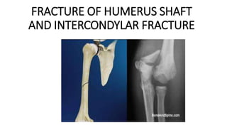
32. PATHO ANATOMY : Often badly comminuted and displaced
T-type
Y-type
33. DIAGNOSIS
• Swelling, pain, tenderness , crepitus ,echymosis around elbow
• Xray elbow – AP and Lateral view
34. TREATMENT
• Undisplaced Above elbow
slab x 3-4 weeks f/b exercise
• Displaced open reduction
and internal fixation
• Severe comminution
olecranon pin traction to reduce
and maintain reduction
35. COMPLICATIONS
• Stiffness of elbow – common because of intra articular nature
Rx : physiotherapy
• Myositis ossificans
• Malunion cubitus varus/valgus deformity
Rx : corrective osteotomy for severe deformities
• osteoarthritis