
Recommended
More Related Content
What's hot
What's hot (20)
Similar to Pulp
Similar to Pulp (20)
Recently uploaded
Recently uploaded (20)
Pulp
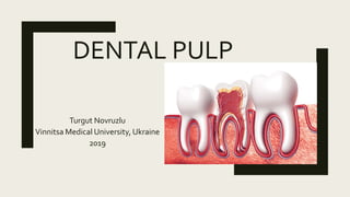
- 1. DENTAL PULP Turgut Novruzlu Vinnitsa Medical University, Ukraine 2019
- 2. What is Pulp? ■Pulp is a soft mesenchymal CT that occupies pulp cavity in the central part of the teeth ■It is a special organ, due to its unique environment
- 3. What are the parts of Pulp?
- 4. CORONAL PULP ■ Coronal – occupies the pulp chamber of the crown of the tooth, it has 6 surfaces, it has horns that correspond to cusp projections.This part of pulp constricts at the CERVICAL region, where the radicular pulp continues. ■ It has the largest mass of the pulp. It’s size directly depends on the size and and shape of the tooth.
- 5. RADICULAR ■ Radicular- occupies the PULP CANALS of the root of the tooth.This part is continuous with the periapical tissues through apical formanen. By age radicular pulp age decreases and this part might become the apical foramen itself.
- 6. APICAL FORAMEN ■ Pulp cavity terminates at the Root apex as small opening called apical foramen. ■ Radicular continuous with CT of the PERIODONTIUM through this foramen ■ Diameter in adult- Maxillary teeth – 0.4 mm , Mandibular teeth- 0.4 mm ■ During developmental periods it is open wide. ■ There may be 2-3 foramina split by cementum or dentin, so calledAPICAL DELTA
- 7. What are Anatomical and Radiographic apex ? ■ Radiographic apex- It is the HIGHEST POINT or tip of the root that is seen on the x-ray ■ AnatomicApex- Point where the NEUROVASCULAR BUNDLE enters the root apex ■ Constriction-The narrows point of the canal- usually located within 2mm of the anatomic apex.
- 10. What happens hen GP extents the ANATOMICAPEX and enters to Radiographic apex?
- 12. What does the pulp contain? ■Pulp cavity has CELLULAR, FIBROUS, NEURAL andVASCULAR components.
- 13. What is the main function of the pulp? ■ The main function of the pulp is production and maintanence of the DENTIN. ■ INDUCTIVE- induces Epithelial differention ■ FORMATIVE- involved in primary and secondary dentin formation ■ PROTECTIVE- viality of dentin by providingO2 and nutrients to odontoblasts ■ DEFENSIVE- recognition of chemical, thermal and mechanical stimuli by nerve fibres. Vasomator inntervation controls the blood volume rate and hence, interpulpal pressure ■ NUTRITIVE-
- 14. Nerve plexus of Raschkow ■ Sensory nerve fibres that originate from INFERIOR and SUPERIORALVEOLAR nerves innervate the ODONTOBLASTIC layer of the pulp cavity. ■ These nerves enter the tooth through the APICAL FORAMEN as MYELINATED NERVE BUNDLES ■ These nerve buncles form the SUBODONTOBLASTIC NERVE PLEXUS of RASCHKOW ■ In addition to the sensory nerves, SYMPHATETHIC NERVE BUNDLES also enter to tooth in order to INNERVATE BLOOD VESSELS
- 15. How well vascularised is the Pulp? ■ The pulp cavity receives blood from oneARTERY that enters the apical forman and coursed directly to the CORONAL PULP ■ Within the coronal pulp, numerous arterial branches form a INTERCONNECTED NETWORK of blood vessels. ■ The smallest capillaries LOOP around SUBODONTOBLASTIC ZONE.
- 16. Capillary network is organised in 3 layers ■ I-Terminal Capillary network- in odontoblastic layer ■ II- Capilary network next to odontoblastic layer/ consists of pre and post capillary vessels ■ Venular network of vessels ■ As the people age, their metabolism decrease and these 3 layers appear as 1 ( it sounded like I am an alien and I am talking about it to my alien friends lol)
- 17. Lymphatics ■ Lymph vessels are formed from a fine meshwork of small, thin walled lymph capillaries ■ They coalesce to form larger lymphatic vessels with valves ■ They start as blind openings nearWeil’s zone (in odontoblastic layer) ■ Larger Lymphatic vessels run along the vessels/nerves
- 18. Which other structures does the Pulp have? ■ It containsArterioles, small nerve bundles and fibroblasts.
- 19. Subodontoblastic region ■ Below the odontoblastic layer, there is the CELL-FREEZONE ofWEIL ■ After this cell free zone, pulp also contains a CELL – RICH zone which thought to provide replacement for cells for odontoblasts that die. High mitotic activity. ■ Within the these zones are the nerve plexus of Raschkow and capillary network
- 20. Pulp Core ■ It is most central region of the pulp ■ Contains major blood vessels and nerve of the pulp ■ Pulpal cells and fibroblasts can be seen as well
- 21. What are the Odontoblasts? ■ These are the cells that are responsible for forming DENTINE and PREDENTINE it initially secretes a COLLAGENOUS MATRIX then subsequently mineralized ■
- 22. Defensive cells of Pulp ■ Histocytes orTissue macrophages ■ In light microscope these cells appear irregular in shape with short blunt processes. ■ Their nuclei are small, rounded. These cells are usually within small blood vessels and capillaries
- 23. What happens in case of an Inflammation ■ In case of inflammation granules and vaculoues are exhibited from their cytoplasm ■ Plasma cells- have small concentric nuclei, their chrmatins are arranged as in a car wheel appearance. ■ They produce AntiBodies ■ Lymphocytes and Eosinophils inside pulp cavity increase in number during inflammation too
- 24. Which collagen fibres are in Pulp? ■ Type I- Present as thick striated fibrils, responsible for pulp architecture ■ Type III -Thinner, mainly in cell free and cell rich zones and give ELASTICITY of the pulp ■ TYPE IV- they rest in basement membrane of blood vessels ■ TypeV andVI- seen to form dense meshwork of thin microfibrils in stroma
Editor's Notes
- LegendA - odontoblasts B - pulp (coronal) C - predentin D - dentin