
Recommended
More Related Content
Similar to Pulp - structural components
Similar to Pulp - structural components (20)
Recently uploaded
Recently uploaded (20)
Pulp - structural components
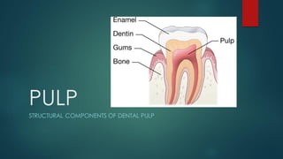
- 1. PULP STRUCTURAL COMPONENTS OF DENTAL PULP
- 2. CELLULAR EXTRACELLULAR CONNECTIVETISSUE 1.ODONTOBLASTS 1.COLLAGEN FIBERS 1.BLOOD VESSELS 2. FIBROBLASTS 2.INTERCELLULAR GS 2.LYMPHATIC CHANNELS 3.UNDIFFERENTIATED 3.NERVE FIBERS MESENCHYMAL CELLS 4.IMMUNOCOMPETENT CELLS
- 3. CELLS OF PULP 1. ODONTOBLASTS : • Ectomesenchymal origin-Dentin forming cells • Second most prominent cells of pulp • Location: adjacent to dentin (cell bodies-pulp, cell processes-dentinal tubules) • Large in coronal pulp and comparatively smaller in radicular pulp • Morphological variations – tall columnar in crown, low columnar in mid of root , flat near apex
- 4. • Shape of the cells depends upon degree of activity • More active cells – tall, oval nucleus at pulpal end and abundant cytoplasmic organelles • The cell becomes narrower at apical end and enters predentin – odontoblastic processes • Odontoblastic processes are devoid of organelles but contain microtubules, filaments and vesicles • Junctions like gap ,tight , desmosomes are seen • Based on activity odontoblastscan be classified into 1) Synthetic(active odontoblasts) 2) Intermediate(transitional odontoblasts) 3) Resting(aged odontoblasts)
- 5. TYPES OF ODONTOBLASTS IN PULP 1) ACTIVE • Elongated cells,basal nucleus & basophilic cytoplasm • Abundant synthetic cellular organelles (for secretion of dentin matrix) • Abundant secretory granules 2) TRANSITIONAL • Similar features as that of active odontoblasts • Comparatively organelles are lesser and less prominent • Less secretory granules • Condensed chromatin with organelles distributed around the nucleus 3) AGED • Little cytoplasm • Dark and close faced nucleus(transcriptio nally inactive nucleus) • Less organelles • Secretory granules and vacuolesare rare/absent
- 7. 2. FIBROBLASTS : ▪ Most numerous cells of pulp ▪ Variable shape-fusiform, stellate ▪ Numerous in younger teeth and decreases with age ▪ Function – synthesis , maintenance and degradation of pulp matrix
- 9. 3. Undifferentiated mesenchymal cells: o Polyhedral in shape with peripheral processes and a large central nucleus o Found in cell rich zone and are scattered throughout the central pulp o Reserve cells - CT cells of pulp are derivedfrom these cells o Show totipotency ( can give rise to different cells like odontoblasts, fibroblasts) o More in younger pulp o Decreases withage thus reducing the regenerative powerof pulp
- 10. 4. IMMUNOCOMPETENT CELLS : MACROPHAGES o Found in central part of pulp o Large oval/spindle shaped o RER , mitochondria, free ribosomes are found o Round and densely stained nucleus o Function – scavenger cells ( eliminate dead cells) DENDRITIC CELLS o Antigen presenting cells o Dendritic processes extend between the odontoblasts o Non phagocytotic cells o Function-Participate in cellular immunity by presenting antigens to T-cells o Increase during dental caries LYMPHOCYTES & EOSINOPHILLS o Number increases during inflammation
- 12. EXTRACELLULAR COMPONENTS : 1.FIBERS o Collagen type-I and type-III o Exhibit cross striations o In younger pulp fibrils are of smaller diameter and these aggregate in older pulp to form thicker fibers o Scattered fibers – diffusedcollagen o Bundled fibers – bundle collagen o Few reticular fibers and elasticfibers may be present 2.GROUND SUBSTANCE o Composedof mucopolysaccharides and protein polysaccharide complex( GAG’S and proteoglycans ) o Functions : 1. Medium for distribution of cells and fibers 2. Support to cells 3. Medium for transport of nutrients and catabolites o It decreases with age
- 13. Connective tissue components: a) BLOOD VESSELS o Highly vascularized – supplied by sup and inf alveolararteries o Blood vesselsenter through apical and accessory foramen o In coronal pulp they undergo extensive branching o In peripheral pulp they form sub odontoblastic capillary network o Arterioles divide into – meta arterioles, pre capillaries and capillaries o Capillariesshow fenestrations for exchange of materials o Venous drainage of pulp is by same veins
- 14. b) LYMPHATIC CHANNELS : o Lymph vessels in pulp are thin walled and irregular lumen composed of endothelial cells surrounded by an incomplete layer of smooth muscles o Anterior teeth drains into- submental lymph nodes o Posterior teeth drains into – sub mandibular lymph nodes and deep cervical lymph nodes c) Nerve supply : o The nerve bundles that enter pulp consist principally of : Sensory afferent nerves of the TRIGEMINAL NERVE Sympathetic branches from the SUPERIOR CERVICAL GANGLION The nerve fibers in pulp are non myelinated : A DELTA AND A BETA – TRANSMIT SHARP PAIN C FIBERS TRANSMIT DULL PAIN
- 15. PLEXUS OF RASCHKOW: PLEXUS OF NERVE FIBERS THAT ARE FORMED DUE TO BRANCHING AND REBRANCHING OF MYLIENATED NERVE FIBERS LOCATED IN CELL RICH ZONE OF PULP