SlideShare a Scribd company logo
1 of 9
Download to read offline
THE CELL
1
CHAPTER
CONTENTS
 Histroy
 Unicellular & Multicellular Organism
 Ultra Structure of Cell
HISTORY
In 1665, Robert Hooke observed the cell in a cork
slice with the help of a primitive microscope. He
saw that cork resembled the structure of a honey
comb consisting of many little compartments.
Robert Hooke called these boxes cells. Cell is a
Latin word for 'little room.'
UNICELLULAR & MULTICELLULAR
ORGANISM


All living organisms are made up of one or more
cells. On the basis of cell number, organism are
grouped into 2 categories -

 Unicellular organisms :
 Organism in which a single cell make the whole
body. For example, Amoeba, Paramecium, bacteria
& Chlamydomonas. 

 Multicellular organism :
 Organism in which body contains more than one
cell. For example - Plante, animals & fungi.
 Cell Size :
 The size of different cells ranges between broad
limits.
 Some plants and animals cells are visible to the
naked eye.
 Most cells are visible only with microscope.
 The prokaryotic cells usually range between
1 to 10 m.
 The eukaryotic cells usually range between 10 to
100 m.
 Amoeba proteus may reach a diameter of 0.5 mm.
 The smallest cells are those of Mycoplasma
laidlawiil (0.1µ in dimeter) or PPLO (pleuro
pneumonia like organism).
 The largest cell is egg of an Ostrich.
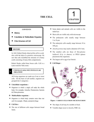
 Cell Shape :
Figure : VARIOUS CELLS FROM THE HUMAN BODY
 The shape of cell may be variable or fixed.
 Variable shape occur in Amoeba, WBC etc.
 Fixed shape occur in most plant and animals.
 Cells may be diverse shapes such as polyhedral
(8, 12 or 14 sides) spherical (e.g. eggs of mainly
animals), spindle shaped (Smooth muscle fibres),
elongated (e.g. Nerves cells) so on.
ULTRA STRUCTURE OF CELLS



Figure : AN ANIMAL CELLS AS SHOWN BY AN ELECTRON
MICROSCOPE. THIS MICROSCOPE MAGNIFIES THE OBJECTS
600,000 TIMES.

Figure : A PLANT CELL AS SHOWN BY AN
ELECTRON MICROSCOPE

 Plasma Membrane : 
Introduction : 
 Cell surface in all the cells is enclosed by a living
membrane which is called cell membrane by
C. Nageli and C. Kramer (1855).
 Historical Account :
 J.Q. Plower (1931) coined the term Plasmalemma
for cell membrane.
Ultrastructure :
 Plasma membrane forms outer covering of each
cell.
 It is present in both plant and animal cells.
 Plasma membrane is a living, thin, delicate
elastic, selectively permeable membrane.
 It separates contents of a cell from the surrounding
medium.

 Fluid Mosaic Model :
In 1972, Singer Nicolson proposed this model.
According to this, cell membrane consists-two
layers of phospholipid molecules, phospholipid &
protein molecules are arranged as a mosaic.
 Phospholipid molecules have their polar heads
directed outward non polar tail pointing inward.
 The proteins are of two types :
Peripheral and integral. Peripheral proteins are
located superficially while integral proteins are
embeded in the phospholipid matrix. The protein
monolayer have elasticity & mechanical support
to the lipid matrix.
Figure : STRUCTURAL DETAIL OF PLASMA MEMBRANE
ACCORDING TO FLUID MOSAIC MODEL
 Functions of Plasma Membrane :
 The main function of plasma membrane is to
regulate the movement of molecules inside and
outside the cell.
It allow the movement of gaseous substance from
high concentration to low concentration which
known as diffusion.
 Water also obeys the low of diffusion . The
movement of water molecule through a selectively
permeable membrane is called osmosis.
 The flexibility of cell membrane also enables the
cell to engulf in food, which is also known as
endocytosis. For example – in Amoeba
 Cell wall :
 In plants, one another rigid is called 'Cell wall'. It
is made up of cellulose which provide structural
strength to plant.
 Function of cell wall :
It maintains the shape of cell.
 It protect the cells from mechanical injury &
prevents their desication.
 It provide mechanical support against gravity. It is
due to rigid cell walls that the aerial part of plant
are able to keep erect & expose their leaves to
sunlight.
 Cell walls permit the cells to with stand very
dilute external media without bursting.


Nucleus :
 It is the most important part of cell which control
all the activities of cell.
 Structure of Nucleus :
 The nucleus has a double layered covering called
nuclear membrane.
 The nuclear membrane has pores inside the
nucleus to its outside, that is, to the cytoplasm.
 The nucleus contains chromosomes, which are
visible as rod-shaped structures only when the cell
is about to divide.
 Chromosomes are composed of DNA and protein.
 Functional segments of DNA are called genes.
 In a cell which is not dividing, this DNA is
present as part of chromatin material.

 Function of nucleus :
 It play a important role in cellular reproduction.
 DNA contain the information necessary for
constructing & organizing cells.
 Nucleoid - In some organisms nuclear region of
cell may be poorly defined due to the absence of
nuclear membrane. Such an undefined nuclear
region called nucleoid.
,
Note :
 Prokaryotic cell - Cell which do not have well
defined nuclear region . called prokaryotic cells.
Pro - Primitive

 Karyon - nucleus
 Eukaryotic cells - Cells which have well defined
nuclear region, called eukaryotic cells.
 Along with nucleus membrane, prokaryotic cells
lack most of cell organells.
 Cytoplasm :
 The fluid & semifluid matrix of a cell between
the nucleus & the plasma membrane, containing
various organelles is called cytoplasm.
 Cell organelles :
 Small membrane bound structures, which perform a
lot of chemical activities to support the function &
structure of a cell, called cell organelles -
 Some important cell organelles are following -
 Endoplasmic Reticulum :
 The endoplasmic reticulum (ER) is noticeable
only with an electron microscope.
 Structure :
 The ER is an extensive network of intracellular
membrane-bound tubes and that occupies most of
the cytoplasm in almost all eukaryotic cells. The
membranes of this system are lipoproteinic in
nature similar in structure to the plasma
membrane. The ER is more prominent in young
and dividing cells as compared to older cells. It is
absent in prokaryotic cells.
 Types. The ER is of two types :
 Rough endoplasmic reticulum (RER)
 Smooth endoplasmic reticulum (SER)
 Rough Endoplasmic reticulum (RER). These
appear rough under a microscope because of the
presence of a large number of grain-like
ribosomes over their cytoplasmic surface. The
ribosomes are the sites of protein synthesis. Thus,
RER is engaged in the synthesis and transport of
proteins. Generally, RER is more abundant in the
deeper part of cytoplasm near the nucleus where it
is connected with the outer membrane of the
nuclear envelope. RER is well developed in the
cells that synthesize and secrete proteins.
 Smooth Endoplasmic Reticulum (SER). It
consists mainly of tubules and vesicles. It is free of
ribosomes and is more abundant near the peripheral
part of the cytoplasm where it may be attached to
the plasma membrane. The SER helps in the
synthesis of fat or lipid molecules. It is, therefore,
well developed in the cells that secrete lipids.
 Functions :
 Support. The ER acts as supporting skeletal
framework of the cell and also maintains its form.
 Transport of materials. The ER facilitates
transport of materials from one part of the cell to
another.
 Exchange of materials. The ER helps in the
exchange of materials between the cytoplasm and
the nucleus.
 Localization of organelles. It keeps the cell
organelles properly stationed and distributed in
relation synthesis.
 Surface for protein synthesis. The RER offers
extensive surface on which ribosomes carry
protein synthesis.
 Surface for synthesis of other substances. The
SER provides surface for the synthesis of lipids
including phospholipids, cholesterol and steroid
hormones.
 Packaging. The proteins formed on ribosomes
pass into ER lumen where they are modified.
Then, the modified proteins move into the
transitional area where the ER buds off transport
vesicles carrying the proteins to the Golgi
apparatus. Here, they are further processed and
packaged into secretory vesicle for export by
exocytosis at the plasma membrane. Examples of
secretory proteins include, mucus, digestive
enzymes and hormones.
 Detoxification. The SER brings about detoxification
in the liver, i.e., it converts harmful materials (drugs,
insecticides, pollutants and poisons) into harmless
substances for excretion by the cell.
 Formation of organelles. The SER produces
Golgi apparatus, lysosomes and vacuoles.
 Membrane formation. Plasma membrane and
other cellular membranes are formed by ER.
 Golgi Complex :
Introduction :
 Golgi bodies are absent in prokaryotic cells. Golgi
complex is found in all eukaryotic cells except
RBCs.
Historical Account :
Camillo Golgi (1898), a zoologist, observed
Golgi bodies in the form of a network in nerve
cells of barn owl.
Ultrastructure :
 It is also called Golgi complex or Golgi
apparatus or Dictyosome (in plants cell).
 It is made up of cisternae.
 Golgi bodies are interconnected with the tubules.
Figure : GOLGI APPARATUS IN SECTION
 Functions of Golgi Apparatus :
 The main function of Golgi apparatus is secretory.
 It produces vacuoles or secretory vesicles which
contain cellular secretions like enzymes, proteins,
cellulose etc.
 Golgi apparatus is also involved in the synthesis
of cell wall, plasma membrane and lysosomes.
 Lysosomes :
Introduction :
 Lysosomes are generally found in the cytoplasm of
animal cells. Lysosomes exhibit polymorphism.
Historical Account :
 The term lysosome was introduced by De Duve in
1955.
Ultrastructure :
 It is also called demolition squads, scavengers,
cellular house keepers and suicidal bags.
 Lysosome are simple tiny spherical sac like
structures evenly distributed in the cytoplasm.
 Lysosome is small vesicle surrounded by a single
membrane and contains powerful enzymes.
 Functions of Lysosomes :
 Lysosomes serve as interacellular digestive
system, hence called digestive bags.
 Lysosomes also remove the worn out and poorly
working cellular organelles by digesting them to
make way for their new replacement.

 Mitochondria :
Introduction :
 A single mitochondrion is present in unicellular
green alga, Microsterias. Number of mitochondria
varies from 50–50,000 per cell. Mitochondria of a
cell are collectively known as chondriome.
Historical Account :
 C. Benda (1897) gave the name Mitochondria
(Mitos, thread + Chondrion, granules).
 Term ‘Bioplast’ for mitochondria was used by
Altman.
Ultrastructure :
 Mitochondria are rod shaped organelles, bounded
by a double membrane envelope.
 The outer membrane is smooth, the inner
membrane surrounds a central cavity of matrix.
Central cavity is filled with jel like substances
Ribosome Matrix
Inner membrane
DNA
Outer
membrane
Cristal inner
membrane
ATP synthase
particles
Figure : MITOCHONDRIA
 Inner membranes folds are called cristae, these
folding are tubular and called microvilli.
 Mitochondria contain electron transport systems
aggregated into compact structure. F1 particles or
oxysome, tennis racket like bodies on inner
membrane involved in oxidation & phosphorylation.
 Kreb’s cycle occurs in mitochondria.
 Each particle is made up of base, stalk and head.
 Functions of Mitochondria :
 Mitochodria are called power plants or power
houses or cellular furnaces.
Synthesis of ATP (Adenosine Tri-phosphate) in
mitochondria is called oxidative phosphorylation.
Mitochondria as place of cellular respiration was
first observed by Hogeboom.

 Plastids :
Introduction :
Plastids are organelles enclosed by a double
membrane found in all plants.
Historical Account :
 E.Heckel (1865) gave the term plastid. Plastids
are largest cell organelles.
Ultrastructure :
 Plastids occur in most plant cells and are absent in
animal cells.
 Plastids are self replicating organelles like
mitochondria i.e. they have the power to divide.
 Schimper divided plastids into three types :
(a) Chromoplast - Coloured plastids
(except green colour)
(b) Chloroplast - Green coloured plastids
(c) Leucoplast - Colourless plastid.
 Plastids also have double membrane but no cristae.
 Functions of Plastids :
 Chloroplasts trap solar energy and utilized it to
manufacture food for the plant.
 Chromoplast impart various colour of flower to
attract insect for pollination.


Vacuoles :
 Vacuoles are storage sacs for solid or liquid
contents. Vacuoles are small sized in animal cells
while plant cells have very large vacuoles. The
central vacuole of some plant cells may occupy
50-90% of the cell volume.













Function :
Vacuoles perform following functions :
 They ar storage sacs of the cell. The stored
matieral may be solid or liquid food or toxic
metabolic by-products or end products of cells.
 In some inicellular organisms, specialized
vacuoles maintain water balance of the body
(osmoregulation).
 In plants, they provide turgidity and rigidity to the
cells.
EXERCISE # 1
A.Single Choice Type Questions
Q.1 Power house of cell is -
(A) Lysosome (B) Ribosome
(C) Mitochondria (D) Vacuole
Q.2 Who discovered the cell -
(A) Robert hooke (B) Purkinje
(C) Robert brown (D) Davson
Q.3 Mitochondria are site of -
(A) Electron transport
(B) Cellular respiration
(C) ATP formation
(D) All
Q.4 Golgi body take part in -
(A) Lipid synthesis
(B) Carbodydrate synthesis
(C) Protein synthesis
(D) Oxidative phosphorylation
Q.5 Protein synthesis occurs on -
(A) Ribosome (B) Lysosome
(C) Nucleus (D) Chloroplast
Q.6 Which of the following has a single membrane -
(A) Nucleus (B) Mitochondrion
(C) Ribosome (D) Plastid
Q.7 What is the function of ER -
(A) Nucleus
(B) Mechanical support
(C) ATP formation
(D) Exchange of molecules
Q.8 Grana & Stroma lamella occur in -
(A) Ribosome (B) Chloroplast
(C) Mitochondria (D) Golgi body
Q.9 Kreb's cycle occurs in -
(A) Matrix of mitochondria
(B) Nucleoplasm
(C) Cytoplasm
(D) Protoplasm
Q.10 Organelle, which remove worn-out cell
organelle is -
(A) Lysosome
(B) Plastid
(C) Mitochondria
(D) Golgi complex
Q.11 Which of the following organelle is involved
in formation of lysosomes -
(A) SER (B) Golgi complex
(C) RER (D) Mitochondria
Q.12 Numerous membrane layer present in plastid
known as -
(A) Cisternae (B) Stroma
(C) Grana (D) Matrix
Q.13 Chromosomes are made up of -
(A) DNA (B) Protein
(C) DNA & protein (D) RNA
Q.14 Cell wall of which one of these is not made
up of cellulose -
(A) Bacteria (B) Hydrilla
(C) Mango tree (D) Cactus
Q.15 Kitchen of the cell -
(A) Mitochondria (B) ER
(C) Chloroplast (D) Golgi complex
Q.16 Membrane biogenesis is related with -
(A) Cell membrane
(B) Nuclear membrane
(C) Cell wall
(D) None
Q.17 Organelle other than nucleus, containing
DNA is -
(A) Endoplasmic reticulum
(B) Mitochondria
(C) Golgi apparatus
(D) Lysosome
Q.18 Amoeba acquires its food through a process
termed as -
(A) Exocytosis (B) Plasmolysis
(C) Endocytosis (D) Both A & B
Q.19 The outermost layer of human cheek cell is -
(A) Cell wall
(B) Nuclear membrane
(C) Plasma membrane
(D) Cytoplasm
Q.20 The diffusion of water from external solution
into dry raisins is called -
(A) Exosmosis
(B) Endosmosis
(C) Imbibition
(D) Plasmolysis
Q.21 The plasma membrane of all living cell is -
(A) Impermeable
(B) Semi permeable
(C) Permeable
(D) Selectively permeable
Q.22 Which cell organelle is not bounded by a
membrane -
(A) Nucleus (B) Lysosome
(C) Ribosome (D) ER
Q.23 In plant cells, the cell wall is -
(A) Dynamic & living
(B) Rigid & non living
(C) Dynamic & non living
(D) Rigid & living
Q.24 The outer most covering of amoeba is -
(A) Tonoplast (B) Plasma membrane
(C) Cell wall (D) Neurolemma
Q.25 Oxysomes are present in -
(A) Mitochondria (B) Peroxisomes
(C) Plastid (D) Cytoplasm



































EXERCISE # 2
A.Very Short Answer Types Questions


Q.1 What are chromosomes ?

Q.2 Name the protein factory of cell ?

Q.3 What are leucoplasts ?

Q.4 Which cell organelle is commonly called
cellular housekeeper ?

Q.5 Name any cell organelle which is non-
membranous ?

Q.6 Name the organelles having double membrane
envelope ?
Q.7 Give 2 examples of unicellular organisms ?

Q.8 Define osmosis ?

Q.9 Define diffusion.

Q.10 Name two types of Endoplasmic reticulum
present in the cell ?

B.Short Answer Types Questions


Q.11 Who discovered cell & how ? -

Q.12 Why is plasma membrane called selectively
permeable membrane ?

Q.13 Which organelle is known as the power house
of cell & why ?

Q.14 What is osmosis ?

Q.15 Why are lysosomes known as suicide bags ?
C.Long Answer Types Questions

Q.16 Draw a well labelled sketch of a ultra
structure of animal cell ?
Q.17 Explain the following -
(a) Mebrane biogenesis
(b) Diffusion
(c) Endocytosis
(d) Cell organelles

Q.18 (a) Draw a diagram of an animal cell & label
its seven parts.
(b) Mention two cell organelles which are
bounded by double membrane. Give
structural detail also.
D.Match The Following


Q.19 Column (A) Column (B)
(i) Smooth endoplasmic – Amoeba
reticulum
(ii) Lysosome – Nucleus
(iii) Food vacuoles – Bacteria
(iv) Chromatinmaterial – Detoxification
& Nucleolus
(v) Nucleoid – Suicidal bag

E.Complete the following sentences


Q.20 Transporting channels of cell is ………
Q.21 Power house of cell is…………….
Q.22 Digestive bag of cell is………………
Q.23 Kitchen of cell is …………………
Q.24 Storage sacs of the cell is ………………
Q.25 Control room of the cell is……………






More Related Content

What's hot (20)

Cells and its components(Anatomy) Easy explanation
Cells and its components(Anatomy) Easy explanationCells and its components(Anatomy) Easy explanation
Cells and its components(Anatomy) Easy explanation
 
Parts, structure and functions of nephron
Parts, structure and functions of nephronParts, structure and functions of nephron
Parts, structure and functions of nephron
 
Transport across cell membrane
Transport across cell membraneTransport across cell membrane
Transport across cell membrane
 
Introduction of tissue - Epithelial tissue
Introduction of tissue - Epithelial tissueIntroduction of tissue - Epithelial tissue
Introduction of tissue - Epithelial tissue
 
Epithelium ppt...
Epithelium ppt...Epithelium ppt...
Epithelium ppt...
 
Cell
CellCell
Cell
 
Cells structure
Cells structureCells structure
Cells structure
 
Cell Division
Cell DivisionCell Division
Cell Division
 
Cell membrane
Cell membraneCell membrane
Cell membrane
 
Structure of cell
Structure of cellStructure of cell
Structure of cell
 
functions of cell
functions of cellfunctions of cell
functions of cell
 
Cell Division
Cell DivisionCell Division
Cell Division
 
Study of a cell
Study of a cellStudy of a cell
Study of a cell
 
Lymphatic system
Lymphatic systemLymphatic system
Lymphatic system
 
Blood vessels.2.
Blood vessels.2.Blood vessels.2.
Blood vessels.2.
 
HUMAN CELL AND TISSUE ( cytology )
HUMAN CELL AND TISSUE ( cytology )HUMAN CELL AND TISSUE ( cytology )
HUMAN CELL AND TISSUE ( cytology )
 
Anatomy & Physiology Lecture Notes - Ch. 4 tissues - epithelium
Anatomy & Physiology Lecture Notes - Ch. 4 tissues - epitheliumAnatomy & Physiology Lecture Notes - Ch. 4 tissues - epithelium
Anatomy & Physiology Lecture Notes - Ch. 4 tissues - epithelium
 
Cell
CellCell
Cell
 
Cell organelles
Cell organellesCell organelles
Cell organelles
 
3. cardiac muscle tissue
3.  cardiac muscle tissue3.  cardiac muscle tissue
3. cardiac muscle tissue
 

Similar to Cell

L-5 Fundamental unit of life.pptx
L-5 Fundamental unit of life.pptxL-5 Fundamental unit of life.pptx
L-5 Fundamental unit of life.pptxPrisha20
 
Fundamental unit of life ppt
Fundamental unit of life pptFundamental unit of life ppt
Fundamental unit of life pptSimmran99
 
Lecture No 004.1 Cell_structure_function.ppt
Lecture No 004.1 Cell_structure_function.pptLecture No 004.1 Cell_structure_function.ppt
Lecture No 004.1 Cell_structure_function.pptsamiiikhan5264
 
09_science_notes_ch05_fundamental_unit_of_life.pdf
09_science_notes_ch05_fundamental_unit_of_life.pdf09_science_notes_ch05_fundamental_unit_of_life.pdf
09_science_notes_ch05_fundamental_unit_of_life.pdfaayushi926063
 
Biology holiday homework
Biology holiday homeworkBiology holiday homework
Biology holiday homeworkParidhi Patel
 
Introduction Biochemistry for BPT Students
Introduction Biochemistry for BPT StudentsIntroduction Biochemistry for BPT Students
Introduction Biochemistry for BPT StudentsKalaivanisathishr
 
3.0 Cell Structure and function (2).pptx
3.0 Cell Structure and function (2).pptx3.0 Cell Structure and function (2).pptx
3.0 Cell Structure and function (2).pptxstephenopokuasante
 
3._Cell_structure_and_function.pdf
3._Cell_structure_and_function.pdf3._Cell_structure_and_function.pdf
3._Cell_structure_and_function.pdfLakshmiKesari2
 
Cell (The function and structural unit of life) Class-8th
Cell (The function and structural unit of life) Class-8thCell (The function and structural unit of life) Class-8th
Cell (The function and structural unit of life) Class-8thAbhinav Maurya
 
Chapter 1 Introduction to Molecular Biology.ppt
Chapter 1 Introduction to Molecular Biology.pptChapter 1 Introduction to Molecular Biology.ppt
Chapter 1 Introduction to Molecular Biology.pptJeevan287994
 
Cell structure and function.pdfx
Cell structure and function.pdfxCell structure and function.pdfx
Cell structure and function.pdfxMukul Kumar
 
S C I E N C E P R O J E C T W O R K ( I T E R M )
S C I E N C E  P R O J E C T  W O R K (  I  T E R M )S C I E N C E  P R O J E C T  W O R K (  I  T E R M )
S C I E N C E P R O J E C T W O R K ( I T E R M )Nandeesh Laxetty
 
Science project work ( i term )
Science project work ( i term )Science project work ( i term )
Science project work ( i term )Nandeesh Laxetty
 
Presentation fundamental unit_of_life_class10 _biology
Presentation fundamental unit_of_life_class10 _biologyPresentation fundamental unit_of_life_class10 _biology
Presentation fundamental unit_of_life_class10 _biologyShikaShekdar1
 

Similar to Cell (20)

6920309.ppt
6920309.ppt6920309.ppt
6920309.ppt
 
L-5 Fundamental unit of life.pptx
L-5 Fundamental unit of life.pptxL-5 Fundamental unit of life.pptx
L-5 Fundamental unit of life.pptx
 
Factory Organelles
Factory OrganellesFactory Organelles
Factory Organelles
 
Fundamental unit of life ppt
Fundamental unit of life pptFundamental unit of life ppt
Fundamental unit of life ppt
 
Lecture No 004.1 Cell_structure_function.ppt
Lecture No 004.1 Cell_structure_function.pptLecture No 004.1 Cell_structure_function.ppt
Lecture No 004.1 Cell_structure_function.ppt
 
CELL ORGANELLES
CELL ORGANELLESCELL ORGANELLES
CELL ORGANELLES
 
09_science_notes_ch05_fundamental_unit_of_life.pdf
09_science_notes_ch05_fundamental_unit_of_life.pdf09_science_notes_ch05_fundamental_unit_of_life.pdf
09_science_notes_ch05_fundamental_unit_of_life.pdf
 
Cells
CellsCells
Cells
 
Biology holiday homework
Biology holiday homeworkBiology holiday homework
Biology holiday homework
 
Introduction Biochemistry for BPT Students
Introduction Biochemistry for BPT StudentsIntroduction Biochemistry for BPT Students
Introduction Biochemistry for BPT Students
 
3.0 Cell Structure and function (2).pptx
3.0 Cell Structure and function (2).pptx3.0 Cell Structure and function (2).pptx
3.0 Cell Structure and function (2).pptx
 
3._Cell_structure_and_function.pdf
3._Cell_structure_and_function.pdf3._Cell_structure_and_function.pdf
3._Cell_structure_and_function.pdf
 
Cells
CellsCells
Cells
 
Cell (The function and structural unit of life) Class-8th
Cell (The function and structural unit of life) Class-8thCell (The function and structural unit of life) Class-8th
Cell (The function and structural unit of life) Class-8th
 
Cellppt
CellpptCellppt
Cellppt
 
Chapter 1 Introduction to Molecular Biology.ppt
Chapter 1 Introduction to Molecular Biology.pptChapter 1 Introduction to Molecular Biology.ppt
Chapter 1 Introduction to Molecular Biology.ppt
 
Cell structure and function.pdfx
Cell structure and function.pdfxCell structure and function.pdfx
Cell structure and function.pdfx
 
S C I E N C E P R O J E C T W O R K ( I T E R M )
S C I E N C E  P R O J E C T  W O R K (  I  T E R M )S C I E N C E  P R O J E C T  W O R K (  I  T E R M )
S C I E N C E P R O J E C T W O R K ( I T E R M )
 
Science project work ( i term )
Science project work ( i term )Science project work ( i term )
Science project work ( i term )
 
Presentation fundamental unit_of_life_class10 _biology
Presentation fundamental unit_of_life_class10 _biologyPresentation fundamental unit_of_life_class10 _biology
Presentation fundamental unit_of_life_class10 _biology
 

Recently uploaded

Pests of safflower_Binomics_Identification_Dr.UPR.pdf
Pests of safflower_Binomics_Identification_Dr.UPR.pdfPests of safflower_Binomics_Identification_Dr.UPR.pdf
Pests of safflower_Binomics_Identification_Dr.UPR.pdfPirithiRaju
 
Best Call Girls In Sector 29 Gurgaon❤️8860477959 EscorTs Service In 24/7 Delh...
Best Call Girls In Sector 29 Gurgaon❤️8860477959 EscorTs Service In 24/7 Delh...Best Call Girls In Sector 29 Gurgaon❤️8860477959 EscorTs Service In 24/7 Delh...
Best Call Girls In Sector 29 Gurgaon❤️8860477959 EscorTs Service In 24/7 Delh...lizamodels9
 
Microphone- characteristics,carbon microphone, dynamic microphone.pptx
Microphone- characteristics,carbon microphone, dynamic microphone.pptxMicrophone- characteristics,carbon microphone, dynamic microphone.pptx
Microphone- characteristics,carbon microphone, dynamic microphone.pptxpriyankatabhane
 
LIGHT-PHENOMENA-BY-CABUALDIONALDOPANOGANCADIENTE-CONDEZA (1).pptx
LIGHT-PHENOMENA-BY-CABUALDIONALDOPANOGANCADIENTE-CONDEZA (1).pptxLIGHT-PHENOMENA-BY-CABUALDIONALDOPANOGANCADIENTE-CONDEZA (1).pptx
LIGHT-PHENOMENA-BY-CABUALDIONALDOPANOGANCADIENTE-CONDEZA (1).pptxmalonesandreagweneth
 
RESPIRATORY ADAPTATIONS TO HYPOXIA IN HUMNAS.pptx
RESPIRATORY ADAPTATIONS TO HYPOXIA IN HUMNAS.pptxRESPIRATORY ADAPTATIONS TO HYPOXIA IN HUMNAS.pptx
RESPIRATORY ADAPTATIONS TO HYPOXIA IN HUMNAS.pptxFarihaAbdulRasheed
 
preservation, maintanence and improvement of industrial organism.pptx
preservation, maintanence and improvement of industrial organism.pptxpreservation, maintanence and improvement of industrial organism.pptx
preservation, maintanence and improvement of industrial organism.pptxnoordubaliya2003
 
Analytical Profile of Coleus Forskohlii | Forskolin .pptx
Analytical Profile of Coleus Forskohlii | Forskolin .pptxAnalytical Profile of Coleus Forskohlii | Forskolin .pptx
Analytical Profile of Coleus Forskohlii | Forskolin .pptxSwapnil Therkar
 
OECD bibliometric indicators: Selected highlights, April 2024
OECD bibliometric indicators: Selected highlights, April 2024OECD bibliometric indicators: Selected highlights, April 2024
OECD bibliometric indicators: Selected highlights, April 2024innovationoecd
 
Davis plaque method.pptx recombinant DNA technology
Davis plaque method.pptx recombinant DNA technologyDavis plaque method.pptx recombinant DNA technology
Davis plaque method.pptx recombinant DNA technologycaarthichand2003
 
Pests of Blackgram, greengram, cowpea_Dr.UPR.pdf
Pests of Blackgram, greengram, cowpea_Dr.UPR.pdfPests of Blackgram, greengram, cowpea_Dr.UPR.pdf
Pests of Blackgram, greengram, cowpea_Dr.UPR.pdfPirithiRaju
 
Speech, hearing, noise, intelligibility.pptx
Speech, hearing, noise, intelligibility.pptxSpeech, hearing, noise, intelligibility.pptx
Speech, hearing, noise, intelligibility.pptxpriyankatabhane
 
BUMI DAN ANTARIKSA PROJEK IPAS SMK KELAS X.pdf
BUMI DAN ANTARIKSA PROJEK IPAS SMK KELAS X.pdfBUMI DAN ANTARIKSA PROJEK IPAS SMK KELAS X.pdf
BUMI DAN ANTARIKSA PROJEK IPAS SMK KELAS X.pdfWildaNurAmalia2
 
Base editing, prime editing, Cas13 & RNA editing and organelle base editing
Base editing, prime editing, Cas13 & RNA editing and organelle base editingBase editing, prime editing, Cas13 & RNA editing and organelle base editing
Base editing, prime editing, Cas13 & RNA editing and organelle base editingNetHelix
 
Is RISC-V ready for HPC workload? Maybe?
Is RISC-V ready for HPC workload? Maybe?Is RISC-V ready for HPC workload? Maybe?
Is RISC-V ready for HPC workload? Maybe?Patrick Diehl
 
FREE NURSING BUNDLE FOR NURSES.PDF by na
FREE NURSING BUNDLE FOR NURSES.PDF by naFREE NURSING BUNDLE FOR NURSES.PDF by na
FREE NURSING BUNDLE FOR NURSES.PDF by naJASISJULIANOELYNV
 
Citronella presentation SlideShare mani upadhyay
Citronella presentation SlideShare mani upadhyayCitronella presentation SlideShare mani upadhyay
Citronella presentation SlideShare mani upadhyayupadhyaymani499
 
User Guide: Pulsar™ Weather Station (Columbia Weather Systems)
User Guide: Pulsar™ Weather Station (Columbia Weather Systems)User Guide: Pulsar™ Weather Station (Columbia Weather Systems)
User Guide: Pulsar™ Weather Station (Columbia Weather Systems)Columbia Weather Systems
 
Microteaching on terms used in filtration .Pharmaceutical Engineering
Microteaching on terms used in filtration .Pharmaceutical EngineeringMicroteaching on terms used in filtration .Pharmaceutical Engineering
Microteaching on terms used in filtration .Pharmaceutical EngineeringPrajakta Shinde
 

Recently uploaded (20)

Pests of safflower_Binomics_Identification_Dr.UPR.pdf
Pests of safflower_Binomics_Identification_Dr.UPR.pdfPests of safflower_Binomics_Identification_Dr.UPR.pdf
Pests of safflower_Binomics_Identification_Dr.UPR.pdf
 
Best Call Girls In Sector 29 Gurgaon❤️8860477959 EscorTs Service In 24/7 Delh...
Best Call Girls In Sector 29 Gurgaon❤️8860477959 EscorTs Service In 24/7 Delh...Best Call Girls In Sector 29 Gurgaon❤️8860477959 EscorTs Service In 24/7 Delh...
Best Call Girls In Sector 29 Gurgaon❤️8860477959 EscorTs Service In 24/7 Delh...
 
Microphone- characteristics,carbon microphone, dynamic microphone.pptx
Microphone- characteristics,carbon microphone, dynamic microphone.pptxMicrophone- characteristics,carbon microphone, dynamic microphone.pptx
Microphone- characteristics,carbon microphone, dynamic microphone.pptx
 
LIGHT-PHENOMENA-BY-CABUALDIONALDOPANOGANCADIENTE-CONDEZA (1).pptx
LIGHT-PHENOMENA-BY-CABUALDIONALDOPANOGANCADIENTE-CONDEZA (1).pptxLIGHT-PHENOMENA-BY-CABUALDIONALDOPANOGANCADIENTE-CONDEZA (1).pptx
LIGHT-PHENOMENA-BY-CABUALDIONALDOPANOGANCADIENTE-CONDEZA (1).pptx
 
RESPIRATORY ADAPTATIONS TO HYPOXIA IN HUMNAS.pptx
RESPIRATORY ADAPTATIONS TO HYPOXIA IN HUMNAS.pptxRESPIRATORY ADAPTATIONS TO HYPOXIA IN HUMNAS.pptx
RESPIRATORY ADAPTATIONS TO HYPOXIA IN HUMNAS.pptx
 
preservation, maintanence and improvement of industrial organism.pptx
preservation, maintanence and improvement of industrial organism.pptxpreservation, maintanence and improvement of industrial organism.pptx
preservation, maintanence and improvement of industrial organism.pptx
 
Analytical Profile of Coleus Forskohlii | Forskolin .pptx
Analytical Profile of Coleus Forskohlii | Forskolin .pptxAnalytical Profile of Coleus Forskohlii | Forskolin .pptx
Analytical Profile of Coleus Forskohlii | Forskolin .pptx
 
OECD bibliometric indicators: Selected highlights, April 2024
OECD bibliometric indicators: Selected highlights, April 2024OECD bibliometric indicators: Selected highlights, April 2024
OECD bibliometric indicators: Selected highlights, April 2024
 
Davis plaque method.pptx recombinant DNA technology
Davis plaque method.pptx recombinant DNA technologyDavis plaque method.pptx recombinant DNA technology
Davis plaque method.pptx recombinant DNA technology
 
Pests of Blackgram, greengram, cowpea_Dr.UPR.pdf
Pests of Blackgram, greengram, cowpea_Dr.UPR.pdfPests of Blackgram, greengram, cowpea_Dr.UPR.pdf
Pests of Blackgram, greengram, cowpea_Dr.UPR.pdf
 
Speech, hearing, noise, intelligibility.pptx
Speech, hearing, noise, intelligibility.pptxSpeech, hearing, noise, intelligibility.pptx
Speech, hearing, noise, intelligibility.pptx
 
Volatile Oils Pharmacognosy And Phytochemistry -I
Volatile Oils Pharmacognosy And Phytochemistry -IVolatile Oils Pharmacognosy And Phytochemistry -I
Volatile Oils Pharmacognosy And Phytochemistry -I
 
BUMI DAN ANTARIKSA PROJEK IPAS SMK KELAS X.pdf
BUMI DAN ANTARIKSA PROJEK IPAS SMK KELAS X.pdfBUMI DAN ANTARIKSA PROJEK IPAS SMK KELAS X.pdf
BUMI DAN ANTARIKSA PROJEK IPAS SMK KELAS X.pdf
 
Base editing, prime editing, Cas13 & RNA editing and organelle base editing
Base editing, prime editing, Cas13 & RNA editing and organelle base editingBase editing, prime editing, Cas13 & RNA editing and organelle base editing
Base editing, prime editing, Cas13 & RNA editing and organelle base editing
 
Is RISC-V ready for HPC workload? Maybe?
Is RISC-V ready for HPC workload? Maybe?Is RISC-V ready for HPC workload? Maybe?
Is RISC-V ready for HPC workload? Maybe?
 
FREE NURSING BUNDLE FOR NURSES.PDF by na
FREE NURSING BUNDLE FOR NURSES.PDF by naFREE NURSING BUNDLE FOR NURSES.PDF by na
FREE NURSING BUNDLE FOR NURSES.PDF by na
 
Citronella presentation SlideShare mani upadhyay
Citronella presentation SlideShare mani upadhyayCitronella presentation SlideShare mani upadhyay
Citronella presentation SlideShare mani upadhyay
 
User Guide: Pulsar™ Weather Station (Columbia Weather Systems)
User Guide: Pulsar™ Weather Station (Columbia Weather Systems)User Guide: Pulsar™ Weather Station (Columbia Weather Systems)
User Guide: Pulsar™ Weather Station (Columbia Weather Systems)
 
Microteaching on terms used in filtration .Pharmaceutical Engineering
Microteaching on terms used in filtration .Pharmaceutical EngineeringMicroteaching on terms used in filtration .Pharmaceutical Engineering
Microteaching on terms used in filtration .Pharmaceutical Engineering
 
Hot Sexy call girls in Moti Nagar,🔝 9953056974 🔝 escort Service
Hot Sexy call girls in  Moti Nagar,🔝 9953056974 🔝 escort ServiceHot Sexy call girls in  Moti Nagar,🔝 9953056974 🔝 escort Service
Hot Sexy call girls in Moti Nagar,🔝 9953056974 🔝 escort Service
 

Cell

  • 1. THE CELL 1 CHAPTER CONTENTS  Histroy  Unicellular & Multicellular Organism  Ultra Structure of Cell HISTORY In 1665, Robert Hooke observed the cell in a cork slice with the help of a primitive microscope. He saw that cork resembled the structure of a honey comb consisting of many little compartments. Robert Hooke called these boxes cells. Cell is a Latin word for 'little room.' UNICELLULAR & MULTICELLULAR ORGANISM   All living organisms are made up of one or more cells. On the basis of cell number, organism are grouped into 2 categories -   Unicellular organisms :  Organism in which a single cell make the whole body. For example, Amoeba, Paramecium, bacteria & Chlamydomonas.    Multicellular organism :  Organism in which body contains more than one cell. For example - Plante, animals & fungi.  Cell Size :  The size of different cells ranges between broad limits.  Some plants and animals cells are visible to the naked eye.  Most cells are visible only with microscope.  The prokaryotic cells usually range between 1 to 10 m.  The eukaryotic cells usually range between 10 to 100 m.  Amoeba proteus may reach a diameter of 0.5 mm.  The smallest cells are those of Mycoplasma laidlawiil (0.1µ in dimeter) or PPLO (pleuro pneumonia like organism).  The largest cell is egg of an Ostrich.  Cell Shape : Figure : VARIOUS CELLS FROM THE HUMAN BODY  The shape of cell may be variable or fixed.  Variable shape occur in Amoeba, WBC etc.
  • 2.  Fixed shape occur in most plant and animals.  Cells may be diverse shapes such as polyhedral (8, 12 or 14 sides) spherical (e.g. eggs of mainly animals), spindle shaped (Smooth muscle fibres), elongated (e.g. Nerves cells) so on. ULTRA STRUCTURE OF CELLS    Figure : AN ANIMAL CELLS AS SHOWN BY AN ELECTRON MICROSCOPE. THIS MICROSCOPE MAGNIFIES THE OBJECTS 600,000 TIMES.  Figure : A PLANT CELL AS SHOWN BY AN ELECTRON MICROSCOPE   Plasma Membrane :  Introduction :   Cell surface in all the cells is enclosed by a living membrane which is called cell membrane by C. Nageli and C. Kramer (1855).  Historical Account :  J.Q. Plower (1931) coined the term Plasmalemma for cell membrane. Ultrastructure :  Plasma membrane forms outer covering of each cell.  It is present in both plant and animal cells.  Plasma membrane is a living, thin, delicate elastic, selectively permeable membrane.  It separates contents of a cell from the surrounding medium.   Fluid Mosaic Model : In 1972, Singer Nicolson proposed this model. According to this, cell membrane consists-two layers of phospholipid molecules, phospholipid & protein molecules are arranged as a mosaic.  Phospholipid molecules have their polar heads directed outward non polar tail pointing inward.  The proteins are of two types : Peripheral and integral. Peripheral proteins are located superficially while integral proteins are embeded in the phospholipid matrix. The protein monolayer have elasticity & mechanical support to the lipid matrix. Figure : STRUCTURAL DETAIL OF PLASMA MEMBRANE ACCORDING TO FLUID MOSAIC MODEL  Functions of Plasma Membrane :  The main function of plasma membrane is to regulate the movement of molecules inside and outside the cell.
  • 3. It allow the movement of gaseous substance from high concentration to low concentration which known as diffusion.  Water also obeys the low of diffusion . The movement of water molecule through a selectively permeable membrane is called osmosis.  The flexibility of cell membrane also enables the cell to engulf in food, which is also known as endocytosis. For example – in Amoeba  Cell wall :  In plants, one another rigid is called 'Cell wall'. It is made up of cellulose which provide structural strength to plant.  Function of cell wall : It maintains the shape of cell.  It protect the cells from mechanical injury & prevents their desication.  It provide mechanical support against gravity. It is due to rigid cell walls that the aerial part of plant are able to keep erect & expose their leaves to sunlight.  Cell walls permit the cells to with stand very dilute external media without bursting.   Nucleus :  It is the most important part of cell which control all the activities of cell.  Structure of Nucleus :  The nucleus has a double layered covering called nuclear membrane.  The nuclear membrane has pores inside the nucleus to its outside, that is, to the cytoplasm.  The nucleus contains chromosomes, which are visible as rod-shaped structures only when the cell is about to divide.  Chromosomes are composed of DNA and protein.  Functional segments of DNA are called genes.  In a cell which is not dividing, this DNA is present as part of chromatin material.   Function of nucleus :  It play a important role in cellular reproduction.  DNA contain the information necessary for constructing & organizing cells.  Nucleoid - In some organisms nuclear region of cell may be poorly defined due to the absence of nuclear membrane. Such an undefined nuclear region called nucleoid. , Note :  Prokaryotic cell - Cell which do not have well defined nuclear region . called prokaryotic cells. Pro - Primitive   Karyon - nucleus  Eukaryotic cells - Cells which have well defined nuclear region, called eukaryotic cells.  Along with nucleus membrane, prokaryotic cells lack most of cell organells.  Cytoplasm :  The fluid & semifluid matrix of a cell between the nucleus & the plasma membrane, containing various organelles is called cytoplasm.  Cell organelles :  Small membrane bound structures, which perform a lot of chemical activities to support the function & structure of a cell, called cell organelles -  Some important cell organelles are following -  Endoplasmic Reticulum :  The endoplasmic reticulum (ER) is noticeable only with an electron microscope.  Structure :  The ER is an extensive network of intracellular membrane-bound tubes and that occupies most of the cytoplasm in almost all eukaryotic cells. The membranes of this system are lipoproteinic in nature similar in structure to the plasma membrane. The ER is more prominent in young and dividing cells as compared to older cells. It is absent in prokaryotic cells.
  • 4.  Types. The ER is of two types :  Rough endoplasmic reticulum (RER)  Smooth endoplasmic reticulum (SER)  Rough Endoplasmic reticulum (RER). These appear rough under a microscope because of the presence of a large number of grain-like ribosomes over their cytoplasmic surface. The ribosomes are the sites of protein synthesis. Thus, RER is engaged in the synthesis and transport of proteins. Generally, RER is more abundant in the deeper part of cytoplasm near the nucleus where it is connected with the outer membrane of the nuclear envelope. RER is well developed in the cells that synthesize and secrete proteins.  Smooth Endoplasmic Reticulum (SER). It consists mainly of tubules and vesicles. It is free of ribosomes and is more abundant near the peripheral part of the cytoplasm where it may be attached to the plasma membrane. The SER helps in the synthesis of fat or lipid molecules. It is, therefore, well developed in the cells that secrete lipids.  Functions :  Support. The ER acts as supporting skeletal framework of the cell and also maintains its form.  Transport of materials. The ER facilitates transport of materials from one part of the cell to another.  Exchange of materials. The ER helps in the exchange of materials between the cytoplasm and the nucleus.  Localization of organelles. It keeps the cell organelles properly stationed and distributed in relation synthesis.  Surface for protein synthesis. The RER offers extensive surface on which ribosomes carry protein synthesis.  Surface for synthesis of other substances. The SER provides surface for the synthesis of lipids including phospholipids, cholesterol and steroid hormones.  Packaging. The proteins formed on ribosomes pass into ER lumen where they are modified. Then, the modified proteins move into the transitional area where the ER buds off transport vesicles carrying the proteins to the Golgi apparatus. Here, they are further processed and packaged into secretory vesicle for export by exocytosis at the plasma membrane. Examples of secretory proteins include, mucus, digestive enzymes and hormones.  Detoxification. The SER brings about detoxification in the liver, i.e., it converts harmful materials (drugs, insecticides, pollutants and poisons) into harmless substances for excretion by the cell.  Formation of organelles. The SER produces Golgi apparatus, lysosomes and vacuoles.  Membrane formation. Plasma membrane and other cellular membranes are formed by ER.  Golgi Complex : Introduction :  Golgi bodies are absent in prokaryotic cells. Golgi complex is found in all eukaryotic cells except RBCs. Historical Account : Camillo Golgi (1898), a zoologist, observed Golgi bodies in the form of a network in nerve cells of barn owl. Ultrastructure :  It is also called Golgi complex or Golgi apparatus or Dictyosome (in plants cell).  It is made up of cisternae.  Golgi bodies are interconnected with the tubules. Figure : GOLGI APPARATUS IN SECTION
  • 5.  Functions of Golgi Apparatus :  The main function of Golgi apparatus is secretory.  It produces vacuoles or secretory vesicles which contain cellular secretions like enzymes, proteins, cellulose etc.  Golgi apparatus is also involved in the synthesis of cell wall, plasma membrane and lysosomes.  Lysosomes : Introduction :  Lysosomes are generally found in the cytoplasm of animal cells. Lysosomes exhibit polymorphism. Historical Account :  The term lysosome was introduced by De Duve in 1955. Ultrastructure :  It is also called demolition squads, scavengers, cellular house keepers and suicidal bags.  Lysosome are simple tiny spherical sac like structures evenly distributed in the cytoplasm.  Lysosome is small vesicle surrounded by a single membrane and contains powerful enzymes.  Functions of Lysosomes :  Lysosomes serve as interacellular digestive system, hence called digestive bags.  Lysosomes also remove the worn out and poorly working cellular organelles by digesting them to make way for their new replacement.   Mitochondria : Introduction :  A single mitochondrion is present in unicellular green alga, Microsterias. Number of mitochondria varies from 50–50,000 per cell. Mitochondria of a cell are collectively known as chondriome. Historical Account :  C. Benda (1897) gave the name Mitochondria (Mitos, thread + Chondrion, granules).  Term ‘Bioplast’ for mitochondria was used by Altman. Ultrastructure :  Mitochondria are rod shaped organelles, bounded by a double membrane envelope.  The outer membrane is smooth, the inner membrane surrounds a central cavity of matrix. Central cavity is filled with jel like substances Ribosome Matrix Inner membrane DNA Outer membrane Cristal inner membrane ATP synthase particles Figure : MITOCHONDRIA  Inner membranes folds are called cristae, these folding are tubular and called microvilli.  Mitochondria contain electron transport systems aggregated into compact structure. F1 particles or oxysome, tennis racket like bodies on inner membrane involved in oxidation & phosphorylation.  Kreb’s cycle occurs in mitochondria.  Each particle is made up of base, stalk and head.  Functions of Mitochondria :  Mitochodria are called power plants or power houses or cellular furnaces. Synthesis of ATP (Adenosine Tri-phosphate) in mitochondria is called oxidative phosphorylation. Mitochondria as place of cellular respiration was first observed by Hogeboom. 
  • 6.  Plastids : Introduction : Plastids are organelles enclosed by a double membrane found in all plants. Historical Account :  E.Heckel (1865) gave the term plastid. Plastids are largest cell organelles. Ultrastructure :  Plastids occur in most plant cells and are absent in animal cells.  Plastids are self replicating organelles like mitochondria i.e. they have the power to divide.  Schimper divided plastids into three types : (a) Chromoplast - Coloured plastids (except green colour) (b) Chloroplast - Green coloured plastids (c) Leucoplast - Colourless plastid.  Plastids also have double membrane but no cristae.  Functions of Plastids :  Chloroplasts trap solar energy and utilized it to manufacture food for the plant.  Chromoplast impart various colour of flower to attract insect for pollination.   Vacuoles :  Vacuoles are storage sacs for solid or liquid contents. Vacuoles are small sized in animal cells while plant cells have very large vacuoles. The central vacuole of some plant cells may occupy 50-90% of the cell volume.              Function : Vacuoles perform following functions :  They ar storage sacs of the cell. The stored matieral may be solid or liquid food or toxic metabolic by-products or end products of cells.  In some inicellular organisms, specialized vacuoles maintain water balance of the body (osmoregulation).  In plants, they provide turgidity and rigidity to the cells.
  • 7. EXERCISE # 1 A.Single Choice Type Questions Q.1 Power house of cell is - (A) Lysosome (B) Ribosome (C) Mitochondria (D) Vacuole Q.2 Who discovered the cell - (A) Robert hooke (B) Purkinje (C) Robert brown (D) Davson Q.3 Mitochondria are site of - (A) Electron transport (B) Cellular respiration (C) ATP formation (D) All Q.4 Golgi body take part in - (A) Lipid synthesis (B) Carbodydrate synthesis (C) Protein synthesis (D) Oxidative phosphorylation Q.5 Protein synthesis occurs on - (A) Ribosome (B) Lysosome (C) Nucleus (D) Chloroplast Q.6 Which of the following has a single membrane - (A) Nucleus (B) Mitochondrion (C) Ribosome (D) Plastid Q.7 What is the function of ER - (A) Nucleus (B) Mechanical support (C) ATP formation (D) Exchange of molecules Q.8 Grana & Stroma lamella occur in - (A) Ribosome (B) Chloroplast (C) Mitochondria (D) Golgi body Q.9 Kreb's cycle occurs in - (A) Matrix of mitochondria (B) Nucleoplasm (C) Cytoplasm (D) Protoplasm Q.10 Organelle, which remove worn-out cell organelle is - (A) Lysosome (B) Plastid (C) Mitochondria (D) Golgi complex Q.11 Which of the following organelle is involved in formation of lysosomes - (A) SER (B) Golgi complex (C) RER (D) Mitochondria Q.12 Numerous membrane layer present in plastid known as - (A) Cisternae (B) Stroma (C) Grana (D) Matrix Q.13 Chromosomes are made up of - (A) DNA (B) Protein (C) DNA & protein (D) RNA Q.14 Cell wall of which one of these is not made up of cellulose - (A) Bacteria (B) Hydrilla (C) Mango tree (D) Cactus Q.15 Kitchen of the cell - (A) Mitochondria (B) ER (C) Chloroplast (D) Golgi complex Q.16 Membrane biogenesis is related with - (A) Cell membrane (B) Nuclear membrane (C) Cell wall (D) None Q.17 Organelle other than nucleus, containing DNA is - (A) Endoplasmic reticulum (B) Mitochondria (C) Golgi apparatus (D) Lysosome Q.18 Amoeba acquires its food through a process termed as - (A) Exocytosis (B) Plasmolysis (C) Endocytosis (D) Both A & B
  • 8. Q.19 The outermost layer of human cheek cell is - (A) Cell wall (B) Nuclear membrane (C) Plasma membrane (D) Cytoplasm Q.20 The diffusion of water from external solution into dry raisins is called - (A) Exosmosis (B) Endosmosis (C) Imbibition (D) Plasmolysis Q.21 The plasma membrane of all living cell is - (A) Impermeable (B) Semi permeable (C) Permeable (D) Selectively permeable Q.22 Which cell organelle is not bounded by a membrane - (A) Nucleus (B) Lysosome (C) Ribosome (D) ER Q.23 In plant cells, the cell wall is - (A) Dynamic & living (B) Rigid & non living (C) Dynamic & non living (D) Rigid & living Q.24 The outer most covering of amoeba is - (A) Tonoplast (B) Plasma membrane (C) Cell wall (D) Neurolemma Q.25 Oxysomes are present in - (A) Mitochondria (B) Peroxisomes (C) Plastid (D) Cytoplasm                                   
  • 9. EXERCISE # 2 A.Very Short Answer Types Questions   Q.1 What are chromosomes ?  Q.2 Name the protein factory of cell ?  Q.3 What are leucoplasts ?  Q.4 Which cell organelle is commonly called cellular housekeeper ?  Q.5 Name any cell organelle which is non- membranous ?  Q.6 Name the organelles having double membrane envelope ? Q.7 Give 2 examples of unicellular organisms ?  Q.8 Define osmosis ?  Q.9 Define diffusion.  Q.10 Name two types of Endoplasmic reticulum present in the cell ?  B.Short Answer Types Questions   Q.11 Who discovered cell & how ? -  Q.12 Why is plasma membrane called selectively permeable membrane ?  Q.13 Which organelle is known as the power house of cell & why ?  Q.14 What is osmosis ?  Q.15 Why are lysosomes known as suicide bags ? C.Long Answer Types Questions  Q.16 Draw a well labelled sketch of a ultra structure of animal cell ? Q.17 Explain the following - (a) Mebrane biogenesis (b) Diffusion (c) Endocytosis (d) Cell organelles  Q.18 (a) Draw a diagram of an animal cell & label its seven parts. (b) Mention two cell organelles which are bounded by double membrane. Give structural detail also. D.Match The Following   Q.19 Column (A) Column (B) (i) Smooth endoplasmic – Amoeba reticulum (ii) Lysosome – Nucleus (iii) Food vacuoles – Bacteria (iv) Chromatinmaterial – Detoxification & Nucleolus (v) Nucleoid – Suicidal bag  E.Complete the following sentences   Q.20 Transporting channels of cell is ……… Q.21 Power house of cell is……………. Q.22 Digestive bag of cell is……………… Q.23 Kitchen of cell is ………………… Q.24 Storage sacs of the cell is ……………… Q.25 Control room of the cell is……………     