1. Braze Ring Failure
Investigator: Samuel Gates, Woodward Inc. Greenville, SC
Background Electron Microprobe
Problem: This summer, Woodward, Inc. Greenville was having a large dropout
rate with a braze weld joint on an oil lance. The braze joint failed both visual
inspection and XRI, exhibiting a large number of voids and lack of wetting.
Goal: A systematic investigation of causes of failure was undertaken in order to
curb the high dropout rate. Specifically, it needed to be determined that the
braze ring material composition was what was specified (AMS 4787: 82%Au
& 18% Ni) and that no contaminants were present.
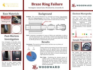
X-ray investigation of the failure shows
little to no wetting into the flange and only
minor penetration of the tube. A
significant amount of braze filler material
is remaining in the groove. Ideally, this
cavity would be evacuated, suggesting an
optimal use of the braze filler material.
The lack of braze material residue
indicates minimal capillary action and
interface adherence necessary for a
successful braze weld.
Base Materials
Flange 321 SS with
Ni coating
Tube Hastelloy X
Braze Ring 82% Au 18%
Ni
Post-Mortem
Investigation
Results
Using an electron microprobe, the post-braze material
was found to be the specified braze ring material
(AMS 4787: 82% Au & 18% Ni). Notice trace diffusion
elements of chromium (Cr) and Iron (Fe) from the
flange and the tube, which is normal considering the
high temperature operation
Next Steps: Considering the minimal amount of
capillary action, it is likely that the groove is too
large for the specified amount of filler material A
reinvestigation on the amount of filler material
required for the specified diametric tolerances
should be undertaken.
Conclusion: The material used was correct, and there was no evidence of
abnormal contamination.
Analysis of braze material residue
was done using an electron
microprobe, which employs energy
dispersive x-ray spectroscopy (EDS).
Energy dispersive X-ray
spectroscopy excites measureable X-
ray lines from the elements allowing
for determination of the elements
present.
Sample Preparation
The sample was prepared using
standard metallographic techniques.
The metallographic mount was then
prepared for analysis by evaporative
carbon coating to provide a uniformly
conductive surface and to prevent
electrical charging by the electron
beam. The prepared mount was
placed in the sample chamber of the
electron microprobe for elemental
analysis by EDS.