3. WHO DEFINITION OF STROKE
A NEUROLOGICAL DEFICIT OF
• Sudden onset
• With focal rather than global dysfunction
• In which, after adequate investigations, symptoms are presumed to be of
non-traumatic vascular origin, and
• lasts for >24 hours
4. Transient Ischemic Attack (TIA)
An acute focal neurological deficit resulting from cerebrovascular
disease
• With resolution of signs and symptoms within 24 hours
• No evidence of ischemia / infarction on imaging studies
6. Stroke Risk Factors: Non modifiable
1. AGE
2. Gender - male
3. Race – Blacks > Asians or Hispanics> Whites
4. Family History.
5. Coagulation Disorders
6. Cardiac Disease
16. Endovascular options
Endovascular intervention
a. I.A. thrombolysis
b. Angioplasty and stenting
c. Mechanical clot disruption
d. Clot extraction (Mechanical
thrombectomy)
• Carotid end artrectomy
19. Perform neurological examination
Every 15 min during the infusion thereafter for the next 6 hrs then hourly until 24 hour
aftr treatment
If pt develop severe headache, acute HTN, nausea , vomiting, discontinue the process and
obtain an emergency CT scan
21. Mechanical thrombectomy
CRITERIA FOR MECHANICAL THROMBECTOMY
1. mRS (modified Rankin score) of 0-1
2. Large vessel Occlusion (LVO): Internal carotid artery or MCA segment 1
3. Age >18yr
4. NIHSS score > 6
5. ASPECT (Alberta Stroke Program Early CT) Score of >6
6. Treatment can be initiated (groin puncture) within 6 hours of symptom onset
22.
23.
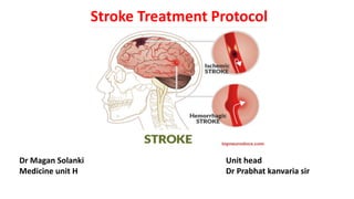
24. Hemorrhagic stroke
Types
1. Intracerebral Hemorrhage(15%)
• Caused by bleeding within the brain tissue itself
2. Sub Arachnoid Hemorrhage(5%)
• Caused by extravasation of blood into the subarachnoid space
25. Intracerebral Hemorrhage
1. An acute and spontaneous extravasation of blood in to the brain
parenchyma that may extend into ventricles and subarachnoid space.
2. 10-15% of all cases of stroke.
3. Classification:
• Primary ICH: Hemorrhage originate from spontaneous rupture of small
arteries or arterioles damaged by chronic HTN or amyloid angiopathy.
• Secondary ICH: Haemorrhage results from trauma, rupture of Aneurysm,
vascular malformation, coagulopathy, haemorrhagic transformation of
cerebral infarct, intracranial neoplasm, venous angioma, dural sinus thrombosis
26. Clinical features- ICH
1. Onset of a sudden focal neurological deficit while the patient is
active, which progresses over minutes to hours, as Weakness or
paresis, Facial droop , blindness, Dysarthria ;Seizure
2. Headache is more common in ICH
3. Vomiting
4. Increased systolic BP and impaired level of consciousness
30. 2. Intracranial Pressure:
• Place ICP monitor or EVD drain in patients with GCS < 8.
• GOAL: Maintain ICP < 20mmHg
• Minimal Cerebral Perfusion Pressure > 60mmHg
3. Haemostatic therapy: Eptacog alpha (Recombinant fac VII)
4. Anticonvulsant therapy:
• Lorazepam, Phenytoin, Fosphenytoin, valproic acid, phenobarbital
5. Fever control
6. Management of Hypergylcemia:
• Insulin if Blood sugar > 185mg/dl
7. Nutrition
8. DVT prophylaxis
31. Surgical Management- ICH
1. Aims:
• Decompression to reduce or prevent elevated ICP
• Removal of acute haematoma to reduce mass effect
• Minimise toxicity from blood breakdown products to surrounding brain
2. Options:
• Ventriculostomy
• Stereotactic aspiration of haematoma
• Endoscopic haematoma evacuation
• Craniotomy
• Hemicraniectomy
32. SUBARACHNOID HAEMORRHAGE
• Neurological emergency characterised by haemorrhage
into the subarachnoid space.
• One of the most important cause of sudden, acute severe
headache.
• 85 % of non traumatic cases are due to ruptured
cerebral aneurysm
• Incidence: F > M (3:2)
• Risk higher in blacks than in whites
• Incidence increases with age and peaks at 50
33. Causes of SAH
1. Trauma: Most common cause
2. Vascular:
• Ruptured intracranial aneurysm, AVM, Tumors with
hemorrhage
3. Vasculopathy
• Collagen vascular disease, Amyloid angiopathy, Arterial
dissection
4. Haematological
• Anticoagulant,leukemia, Hepatic or renal induced
coagulopathy
5. Drugs : Cocaine, Amphetamine
34. Clinical Presentation of SAH
1. Prodormal events:
• Symptoms:Headache, dizziness, orbital pain, diplopia, visual
loss
• Signs: Sensory or motor disturbance, seizure, ptosis,
dysphasia
2. Focal neurological findings
3. CLASSIC presentation:
• Sudden onset severe headache(Thunderclap headache)
• Nausea/vomiting
36. Management of SAH
Medical management of SAH focuses on
• Protecting the airway
• Managing the BP
• Preventing rebleeding prior to treatment
• Managing vasospasm(Calcium channel antagonist Nimodipine)
• Treating Hydrocephalus(EVD or permanent ventricular shunting)
• Preventing Pulmonary embolus
• Bed rest in quiet room and stool softner,if needed, to prevent
straining.
• If headache or neck pain is severe, Mild sedation and analgesia.
• Adequate Hydration
37. Managing raised ICP
For stupurous patient, Emergency Ventriculostomy to measure ICP
.
Medical therapies: Mild hyperventilation, Mannitol and sedation
Maintain adequate cerebral perfusion pressure(60-70mmHg) while
avoiding excessive elevation/fall of arterial pressure
38. Surgical mx of SAH
1. Clipping of the aneurysm
2. Coiling of the aneurysm