
Recommended
More Related Content
Similar to MICROSCOPE.pptx
Similar to MICROSCOPE.pptx (20)
Recently uploaded
Recently uploaded (20)
MICROSCOPE.pptx
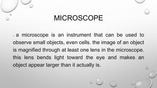
- 1. MICROSCOPE - a microscope is an instrument that can be used to observe small objects, even cells. the image of an object is magnified through at least one lens in the microscope. this lens bends light toward the eye and makes an object appear larger than it actually is.
- 2. 1. OPTICALS MICROSCOPE - consists of only one lens the simplest form of optical microscope consists of one double convex lens with a short focal length. the lens is capable of magnifying an object from 15x up to 2000x. example: magnifying glass. 2. COMPOUND MICROSCOPE – uses visible light to produce a magnified image of an object. this is composed of an objective lens and ocular lens mounted at opposite ends of a closed tube TYPES OF MICROSCOPE
- 3. a) MONOCULAR COMPOUND MICROSCOPE – composed of one eyepiece. b) BINOCULAR COMPOUND MICROSCOPE – composed of two eyepieces. c) TRINOCULAR COMPOUND MICROSCOPE – composed of three eyepieces where one of the eyepieces is a camera that takes the picture or projects that picture on a screen.
- 4. SPECIAL PURPOSE OPTICAL MICROSCOPES • 1. STEREOSCOPIC MICROSCOPES – consists of two low-powered microscopes arranged so that they converge on the specimen. these produce a 3-dimensional image that has its right side up. • 2. ULTRAVIOLET MICROSCOPE – uses the ultraviolet wavelength of the spectrum instead of the visible wavelength. lenses used may be quartz or fluorine instead of glass.
- 5. 3. PETROGRAPHIC MICROSCOPE – this is a compound microscope which uses plane polarized light and is used to identify and estimate the mineral content of igneous and metamorphic rocks. 4. DARK FIELD MICROSCOPE – uses lighting in the form of hollow, very intense cone of light concentrated at the specimen
- 6. 5. BRIGHT FIELD MICROSCOPE – operates through the reflection of light from objects to the eyes
- 7. TYPES OF ELECTRON MICROSCOPES 1. SCANNING ELECTRON MICROSCOPE (SEM) – using electrons to light objects, they can magnify from 100 000 up to a million times. the scanning probe microscope can make atoms visible and capable of 3-dimensional image. other types are scanning transmission electron microscope, electron probe micro analyzer, and atomic force microscope. 2. TRANSMISSION ELECTRON MICROSCOPES (TEM) – capable of up to 1 million times magnification.
- 9. MECHANICAL PARTS • STAND – made of heavy foot and a horshoe-shaped based that supports th microscope • BODY TUDE – cylindrical part where the lenses are attached and can be raised of lowered for better focusing. • COARSE ADJUSTMENT KNOB – this is done first after the objective lens is lowered near the object. • FINE ADJUSTMENT KNOB – used sparingly for further focusing of the object. • STAGE – a platform with an opening to let the light pass where the specimen in a glass silde is placed. a mechanical stage has a device for controlled shifting of the slide.
- 10. OPTICAL PART • STAGE CLIPS – keep the glass slide in place so that it will not move. • ARM – connects the base and the stage with the tube. • DRAW TUBE –holds the eyepiece • OBJECTIVES – lens located near the object, magnifies up to 90 times. May be low or high powered. • EYEPIECE– located at the top of the body tube. It is the part where you peep during observation.
- 11. • REVOLVING NOSEPIECE – MAY BE SINGLE, DOUBLE, OR TRIPLE-LOCATED AT THE BOTTOM OF THE BODY TUBE WHICH CARRIES THE MAGNIFYING LENSES OR OBJECTIVES. • MIRROR – USED TO FOCUS THE RAYS OF THE LIGHT SOURCE TOWARD THE OBJECT. • DIAPHRAGM – CONTROLS THE LIGHT THAT ENTERS THE STAGE OPENING. • CONDENSER LENS – USED TO FURTHER CONTROL THE LIGHT AS NEEDED.