
Recommended
More Related Content
What's hot
What's hot (20)
Similar to Nematodes and Helminths: Enterobius vermicularis and Trichuris trichiura
Similar to Nematodes and Helminths: Enterobius vermicularis and Trichuris trichiura (20)
More from Ragya Bharadwaj
More from Ragya Bharadwaj (12)
Recently uploaded
Recently uploaded (20)
Nematodes and Helminths: Enterobius vermicularis and Trichuris trichiura
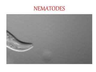
- 1. NEMATODES
- 3. HELMINTH
- 6. ENTEROBIUS VERMICULARIS • Introduction • Habitat • Morphology • Life Cycle • Pathogenicity • Clinical Features • Laboratory Diagnosis • Treatment
- 7. INTRODUCTION • Previously Oxyuris vermicularis • Humans – The only hosts • Pinworm (“Threadworm”) • Person sleeps, female pinworms leave the intestine through the anus and deposit their eggs
- 8. HABITAT • Caecum • Appendix • In submucosa
- 9. MORPHOLOGY • ADULT White, Spindle shaped Anteriorly - Cervical alae ( wing like expansion) Posteriorly – Globular bulb ( esophagus) MALE FEMALE 2-4mm * 0.1-02mm 8-12 * 0.3-0.5mm Post 1/3 – sharply truncated Posterior – long tapering tail Rarely visualised -? After laying eggs dies 2-3weeks
- 10. • Floats in saturated salt solution - Planoconvex! - Transparent shell
- 13. LIFE CYCLE • Newly laid eggs in perianal skin • Contains larva • Completes development in 24-36 hours • Ingestion of these eggs by next human • Egg shells dissolved by juices, larva goes to small intestine • Small intestine – Adolescent worm • Sexually mature – Male fertilizes female and dies • Gravid female migrate to caecum and appendix • Lays eggs in perianal skin
- 15. PATHOGENICITY • At Risk – school-aged and preschool-aged children, – institutionalized persons – household members and caretakers of persons with pinworm infection. • Mode of Infection Ingestion of eggs with larva ( fecal-oral route ) Autoinfection – laying of eggs - itching – fingers – mouth Retroinfection – Larva hatched in perianal skin travel back to rectum
- 16. CLINICAL FEATURES • Pruritis periani and perineum • Salpingitis • Nocturnal enuresis • Inflammation of appendix
- 18. LABORATORY DIAGNOSIS • Divided into 2 – Detection of Adult worm Demonstration of Eggs • Detection of Adult Worms - Self detection by self / parents - Passage in stools normally / after enema - Inspection of anal region
- 19. DEMONSTRATION OF EGGS • Direct smear • Concentration method • Sample collection – NIH swab – early morning
- 21. TREATMENT • Pyrantel Palmoate – 10mg/kg SD • Albendazole - 400 mg SD • Mebendazole - 100mg SD
- 22. TRICHURIS
- 24. INTRODUCTION • Linnaeus in 1771, AKA – Whipworm • Trichuris trichiura • Trichuriasis • Large intestine – caecum, appendix • Tropics
- 25. MORPHOLOGY • ADULT Whip shaped Male – 30 – 45 micron Ant 3/5th – Thin, Post 2/5th – Thick coiled Post end – Males – coiled Females – comma shaped Life – 5-10yrs
- 27. • EGGS Barrel shaped Projecting mucus plugs – Each Pole Bile stained Plugs – colourless Float – sat salt Freshly passed eggs in stool – NOT INFECTIOUS
- 30. LIFE CYCLE • Single host , No INTERMEDIATE • Adult worm live in LARGE intestine • Eggs passed in soil • Development - Rhabditiform larva in eggs develops (Tropical >> Temperate) • Embryonated eggs – Infective • Ingested • Egg shell dissolves • Larva comes out • Caecum • Sexually mature • Lay eggs
- 33. PATHOGENESIS • Whipworm infection • Asymptomatic • Massive Infection – Mechanical obstruction – Allergic – Rectum – edematous – difficult to defecate – Rectal bleeding – Abdominal cramps
- 34. LABORATORY DIAGNOSIS • Stool microscopy • Eggs +
- 35. TREATMENT AND PROPHYLAXIS • Metronidazole • Proper disposal of feces • Avoid eating raw vegetables • Identifying infected patients