1. Design of Prosthetic Heart Valves
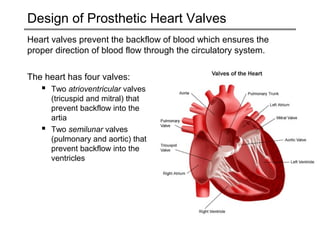
Heart valves prevent the backflow of blood which ensures the
proper direction of blood flow through the circulatory system.
The heart has four valves:
Two atrioventricular valves
(tricuspid and mitral) that
prevent backflow into the
artia
Two semilunar valves
(pulmonary and aortic) that
prevent backflow into the
ventricles
2. Heart Valve Disease
There are numerous complications
and diseases of the heart valves that
prevent the proper flow of blood.
Essentially, heart valve diseases fall
into two categories, stenosis and
incompetence.
A stenotic heart valve prevents the
valve from opening fully, due to
stiffened valve tissue. Thus, more
work is required to push blood
through the valve.
An incompetent valves cause
inefficient blood circulation by
permitting backflow of blood in the
heart.
3. Treatment Options
On a large scale, medication is the best alternative.
Although in some cases defective valves have to be replaced with
a prosthetic valve in order for the patient to lead a normal life. An
enormous amount of research and development has proven to be
beneficial, as prosthetic heart valve technology has saved
hundreds of thousands of lives.
The two main prosthetic valve designs are:
Mechanical Heart Valves
Bioprosthetic Heart Valves
4. Evolution of Mechanical Heart Valves
The first prosthetic heart valve was implanted in 1952 by Charles
Hufnagel. The device was an acrylic ball valve inserted into the
descending aorta. As the valve only prevented regurgitant flow
from the lower body, cardiac work was only partially relieved and
coronary flow was not improved. In addition, embolization and
thrombosis of the valve frequently occurred, and the noise
generated by the valve was disconcerting — reminiscent,
according to some, of a ticking time bomb.
5. Evolution of Mechanical Heart Valves
In spite of its generally poor success, others recognized the
importance of the approach which led to the development of over
30 different mechanical designs worldwide.
These valves have progressed from simple caged ball valves, to
modern bileaflet valves. Heart valves are designed to fit the
peculiar requirements of blood flow through the specific chambers
of the heart, with emphasis on producing more central flow and
reducing thrombosis.
6. Caged-Ball Design
The caged ball design is one of the early designs, that uses a small
ball that is held in place by a welded metal cage.
Although effective, caged-ball valves completely block central flow
(blood to flow through the valve centre) and the heart must work
harder to compensate for the momentum lost to the change of
direction of the fluid.
In addition, the ball causes
damage to blood cells due to
collisions. These damaged
blood cells release blood clotting
agents, thereby requiring
patients to take anticoagulants.
7. Tilting Disc Design
In the mid-1960s, a new class of mechanical valves were designed
that utilized a tilting disc to better mimic the natural patterns of
blood flow. Tilting-disc valves have a floating polymer disc held in
place by two welded struts. The tilting motion provides improved
central flow while preventing backflow and also reduce mechanical
damage to blood cells.
Although vastly superior
to the caged-ball design,
tilting discs valves have
a tendency for the outlet
struts to fracture as a
result of fatigue.
8. Bileaflet Design
In 1979, a new mechanical heart valve was introduced: bileaflet
valves. These valves consisted of two semicircular leaflets that
pivot on hinges. The leaflets swing open completely, parallel to the
direction of the blood flow. The result is the closest approximation
to central flow achieved in a natural heart valve. For this reason,
the bileaflet valve is the most popular of the modern designs.
The problem with these
valves is that the leaflets do
not close completely, which
permits some backflow.
Since backflow is a property
of a defective valve, the
bileaflet design is still not
ideal.
9. Materials
Current designs use materials that do not induce clotting in the
blood stream.
Most commonly used materials include:
stainless steel alloys
molybdenum alloys
pyrolitic carbon for the valve housings and leaflets
silicone, polytetrafluoroethylene (teflon®)
polyester (Dacron®) for sewing rings
A new generation of mechanical valves made of materials with
improved blood contact properties, better wear characteristics and
resistance to infection are currently under development.
10. Advantages and Disadvantages
Advantages
The main advantage of mechanical valves is their high durability.
Mechanical heart valves placed in young patients can typically last for their
lifetime
Disadvantages
The major problem with all mechanical
valves is the increased risk of blood
clotting. As a preventative measure,
mechanical valve recipients must take
anticoagulantants. These anticoagulants
can cause birth defects in the first
trimester of fetal development, rendering
mechanical valves unsuitable for women
of child-bearing age.
11. Future of Mechanical Heart Valves
To develop the next generation of mechanical heart valves, new
age tools that are being used to improve valve design, which
include:
accelerated wear testing
advanced blood contact property testing
computer assisted design and manufacturing
coatings to reduce the chance of infection and improve healing
12. Bioprosthetic Heart Valves
Bioprosthetic heart valves are valves made from actual valve tissue
(animal or human).
These valves hold many advantages over mechanical valves:
design is closer to the natural valve
better hemodynamics
do not cause damage to blood cells
patients do not require long-term
anticoagulants
do not suffer from structural problems
(e.g. fatigue)
13. Bioprosthetic Heart Valves
Animal tissue valves are often referred to as xenograft valves.
These valves are constructed from recovered heart tissue at the
time of commercial meat processing. After fabrication, they are
chemically crosslinked to limit degradation.
The most commonly used animal tissues are porcine aortic valves
(explanted valve with an attached Dacron cloth sewing skirt) and
bovine pericardial valves (sewn leaflets from pericardial tissue with
an attached Dacron cloth sewing skirt). Bioprosthetic valves have
good durability and usually last for 10-15 years.
The common cause of failure in these valves is due to tissue
calcification. Calcification stiffens the valve tissue leading to the
restriction of blood flow through the valve (stenosis) and/or
generation of tears (from stress concentrations) in the valve
leaflets.