SlideShare a Scribd company logo
1 of 7
Download to read offline
ARTICLES
PUBLISHED ONLINE: 12 OCTOBER 2015 | DOI: 10.1038/NMAT4448
Enhanced energy transport in genetically
engineered excitonic networks
Heechul Park1,2†
, Nimrod Heldman1,2,3†
, Patrick Rebentrost4
, Luigi Abbondanza5
,
Alessandro Iagatti6,7
, Andrea Alessi5
, Barbara Patrizi6
, Mario Salvalaggio5
, Laura Bussotti6
,
Masoud Mohseni4
, Filippo Caruso6,8
, Hannah C. Johnsen3
, Roberto Fusco5
, Paolo Foggi6,7,9
,
Petra F. Scudo5
*, Seth Lloyd4,10
* and Angela M. Belcher1,2,3
*
One of the challenges for achieving efficient exciton transport in solar energy conversion systems is precise structural control
of the light-harvesting building blocks. Here, we create a tunable material consisting of a connected chromophore network on
an ordered biological virus template. Using genetic engineering, we establish a link between the inter-chromophoric distances
and emerging transport properties. The combination of spectroscopy measurements and dynamic modelling enables us to
elucidate quantum coherent and classical incoherent energy transport at room temperature. Through genetic modifications,
we obtain a significant enhancement of exciton diffusion length of about 68% in an intermediate quantum-classical regime.
L
ight-harvesting materials with efficient exciton transport
are of great interest for practical solar energy conversion
applications. Organic light-harvesting materials, such as cyclic
dye systems1
, dendritic structures2
and J-aggregate nanotubes3
, have
been investigated for such applications. Biological materials can
serve as templates for chromophore assemblies, as for example
shown using DNA (ref. 4), tobacco mosaic viruses5,6
and the
M13 virus7
. The M13 virus has been shown to be a versatile
and tunable engineering tool with a large range of applications
such as batteries8
, solar cells9
, water splitting10
, electrochromic
devices11
, and cancer imaging12
. In particular, for chromophore
assemblies of zinc porphyrins on the M13 virus it was proposed
that both Förster resonance energy transfer (FRET) and the Dexter
mechanism are involved in the transport process7
. For designing
efficient energy transport, the selection and structural arrangement
of chromophores, and inter-chromophoric couplings play a crucial
role. In addition, quantum transport in contrast to a semi-classical
hopping mechanism can contribute to the high efficiency, as has
been suggested in biological chromophore systems13–19
. Some of
the challenges are precisely controlling structures on the level of
individual light-harvesting chromophores and engineering a regime
of beyond-Förster energy transport.
Here, we create a light-harvesting antenna system that shows
genetically enhanced exciton transport. We demonstrate that the
M13 virus can act as a tunable chromophore scaffold carrying
donors and acceptors, and explore the underlying mechanism of
the efficient transfer of energy in this light-harvesting antenna
system. The use of programmable genes allows us to manipulate the
positioning of the binding sites, thus multiplying the possibilities
for creating intricate chromophore networks and for controlling
the energy transfer. Two types of M13 viruses are genetically
engineered: one virus is shown to have a regular network of weakly
coupled chromophores, and the other is engineered to have clusters
of strongly coupled chromophores. By a combination of room-
temperature spectroscopy experiments and theoretical models for
the exciton dynamics based on resonant coupling and delocaliza-
tion, we demonstrate that the weakly coupled network exhibits
slow semi-classical Förster energy transfer and that the genetically
improved strongly coupled network resides in a super-Förster
regime where coherent and incoherent transport collaborate to
boost exciton diffusion. So far, the enhancement of energy transport
in a super-Förster regime has been shown only theoretically16,20
.
Here, we provide the first experimental evidence for such an
enhancement in a designed system, and extract theoretical evidence
for the interplay of coherence and incoherent dynamics. The
genetically improved antenna achieves a 68% longer diffusion
length over the weakly coupled antenna and a fourfold increase in
the number of donors contributing energy to a single acceptor.
The two types of M13 viruses were engineered by modifying
the amino acid sequence of the major coat pVIII protein, which
serves as a building block for the chromophore network (Fig. 1).
The virus consists of a highly ordered filamentous array of 2,700
copies of the pVIII proteins. We name the two clones M13CF
(CF for Classical Förster) and M13SF (SF for Super-Förster).
We used previously obtained structural data (Protein Data Bank
(PDB) number 2C0W; ref. 21) to reconstruct the model of both
clones to estimate the distances between chromophore-binding
sites. The primary amine groups of the pVIII exposed on the surface,
N-terminus and lysine residues, can act as specific binding sites for
chromophores. The M13CF virus bearing the amino acid sequence
1Department of Materials Science and Engineering, Massachusetts Institute of Technology, Cambridge, Massachusetts 02139, USA. 2The David H. Koch
Institute for Integrative Cancer Research, Massachusetts Institute of Technology, Cambridge, Massachusetts 02139, USA. 3Department of Biological
Engineering, Massachusetts Institute of Technology, Cambridge, Massachusetts 02139, USA. 4Research Laboratory of Electronics, Massachusetts Institute
of Technology, Cambridge, Massachusetts 02139, USA. 5Research Center for Non-Conventional Energy, Istituto eni Donegani, eni S.p.A., Novara 28100,
Italy. 6European Laboratory for Non-linear Spectroscopy, University of Florence, Sesto Fiorentino 50019, Italy. 7INO CNR, Sesto Fiorentino 50019, Italy.
8QSTAR and Department of Physics and Astronomy, University of Florence, Florence 50125, Italy. 9Department of Chemistry, University of Perugia,
Perugia 06123, Italy. 10Department of Mechanical Engineering, Massachusetts Institute of Technology, Cambridge, Massachusetts 02139, USA. †These
authors contributed equally to this work. *e-mail: pscudo@gmail.com; slloyd@mit.edu; belcher@mit.edu
NATURE MATERIALS | ADVANCE ONLINE PUBLICATION | www.nature.com/naturematerials 1
© 2015 Macmillan Publishers Limited. All rights reserved
ARTICLES NATURE MATERIALS DOI: 10.1038/NMAT4448
26 Å
26 Å
26 Å
10 Å10 Å13 Å
a ∼880 nm
∼7 nm
28 Å
28 Å
28 Å
33 Å
16 Å
b c
Figure 1 | Molecular models of the genetically engineered M13 viruses. a, A filamentous M13 virus is approximately 880 nm in length and ∼7 nm in
diameter and composed of 2,700 copies of the major coat protein pVIII, which provides a scaffold for excitation energy transport over chromophores in a
light-harvesting antenna system. b,c, Magnified M13 virus surfaces show the energy-transfer networks between chromophore-binding sites for M13CF and
M13SF, respectively (blue colour, N-terminus; orange, newly added lysine residue; green, pre-existing lysine residue). Insets show schematic networks of
energy transport in the M13CF and the M13SF, respectively. (Red gradient colour of ellipse indicates exciton delocalization. Blue arrows show incoherent
exciton hopping. For the M13CF, pathways to any of the lysine binding sites are not shown because only 7% are occupied.) The M13CF has long
inter-binding site distances of 16 Å and 33 Å within two pVIII proteins. In the M13SF, close inter-binding site distances of approximately 10 Å and 13 Å are
formed. The pVIII coat protein assemblies were reconstructed from the structural model (PDB number 2C0W).
of ADSPHTELPDPAK (ref. 9) at the N-terminal region of the
pVIII was used because the distances between the two primary
amines are longer than the corresponding distances in the wild
type. The inter-binding site distance was estimated to be 33 Å on
the pVIII (Fig. 1b). To create an entirely different network with
closer distances, the M13SF virus was genetically engineered to have
an additional third binding site on the pVIII. Among the possible
candidates (data not shown), the M13SF carrying the shorter pVIII
N-terminal sequence of AENKVEDPAK was identified as having
inter-binding site distances of 10 Å on the pVIII (Fig. 1c). These
distances are short enough to enhance the coupling strength of
the bound chromophores, but long enough to avoid the complete
quenching of fluorescence due to short-range Dexter exchange19,22
.
The electronic coupling strength between donors for the 10 Å
distance is about 460 cm−1
, calculated using the standard FRET
exchange formula and the spectral overlap, and assuming parallel
orientation of the dipoles.
Various kinds of chromophores can be selected for coupling to
the M13 virus biological scaffold through chemical modification
of amine groups on the pVIII coat proteins. Alexa Fluor 488 and
Alexa Fluor 594 were carefully selected as donor and acceptor
chromophores, respectively (Supplementary Fig. 1). They show
narrow and well-separated absorption bands and good spectral
overlaps between donor emission and donor absorption, as well
as between donor emission and acceptor absorption for energy
transfer (Fig. 2a). Absorption peaks are at 491 nm for the donor and
at 585 nm for the acceptor. Emission peaks are at 517 nm for the
donor and at 611 nm for the acceptor. The acceptor emission peak is
sufficiently far from the donor emission peak to resolve the signals
of fluorescence when the donors are excited and the fluorescence
intensities are detected from the acceptors. They also show high
molar extinction coefficients (that is, strong dipole moments23
)
and relatively high quantum yields24
(Supplementary Section E.1.1).
Moreover, a pre-activated carboxylic acid in each donor and
acceptor allows specific conjugation to the primary amines on the
pVIII by forming covalent amide bonds.
A dense occupation of the sites comprising the energy-transfer
network on the M13 virus surface is desired. The conjugation of
the chromophores to the N-terminus and the lysine residues was
confirmed by diverse control experiments (Fig. 2b (upper inset) and
Supplementary Section E.3). On the M13CF (M13SF), on average
2,970 ± 180 (4,130 ± 70) donors occupy the 5,400 (8,100) possible
binding sites per virus, estimated from the absorbance (Supplemen-
tary Section E.2.1). For M13CF, 2,700 N-termini are fully occupied
(∼100%) and only ∼200 lysine residues are occupied (∼7%). For
M13SF, 2,700 N-termini are fully occupied (∼100%), ∼200 of the
pre-existing lysine residues (∼7%), and ∼1,200 of the newly added
lysines (∼44%) (Supplementary Section E.4). Thus, for M13SF,
1,200 out of the 2,700 pVIII copies have a donor pair separated
by 10 Å. The donor absorbance shows peak shifts to 496 nm for
the M13CF and 497 nm for the M13SF, which is evidence of an
electronic coupling of the chromophores (Supplementary Fig. 6).
From the occupation probabilities of the binding sites of the
chromophore networks, the M13CF has closer to an ideal diamond
grid of relatively well-separated N-terminus chromophores
(Fig. 1b). Because the semi-classical Förster rates of transport
depend on the distance r as 1/r6
(ref. 22), we can assume that
each chromophore has four relevant nearest neighbours. The
M13SF has the same basic diamond grid, but about 50% of the
unit cells have additional vertices resulting from the lysines,
which fill the empty space (Fig. 1c). The distance between nearest
neighbours resides within the Förster radius between donors,
which is estimated to be 37.5 Å on the M13CF, and 30.1 Å on the
M13SF (Supplementary Section E.7). This guarantees connectivity
of the network with at least more than 50% inter-chromophoric
energy transport on both clones. For the M13SF, we expect that
the stronger electronic couplings and the subsequent highly linked
network lead to improved energy transport.
2 NATURE MATERIALS | ADVANCE ONLINE PUBLICATION | www.nature.com/naturematerials
© 2015 Macmillan Publishers Limited. All rights reserved
NATURE MATERIALS DOI: 10.1038/NMAT4448 ARTICLES
a
Normalizedintensity(a.u.)
0.0
0.2
0.4
0.6
0.8
1.0
Wavelength (nm)
400 450 500 550 600 650 700 750 800
DN absorption
DN emission
AC absorption
AC emission
Fluorescenceintensity(×107a.u.)
0.0
0.5
1.0
1.5
2.0
Wavelength (nm)
500 550 600 650
Free DN
M13SF-400DN
M13CF-DN
M13SF-DN
M13SF-DN Free DN
b
Figure 2 | Steady-state spectra and fluorescence quenching at room temperature. a, Absorption and emission spectra of free donor (DN) and free
acceptor (AC). b, Fluorescence spectra of donor chromophores at an excitation wavelength of 495 nm. All samples have an absorbance of 0.3 at 495 nm
with a 1 cm path length of light. M13SF-DN indicates the M13SF virus with ∼4,100 donors on the surface, and M13CF-DN indicates the M13CF virus with
∼2,900 donors. M13SF-400DN denotes a control sample of the M13SF virus with ∼400 donors. Significant quenching is observed in the M13SF-DN. The
upper inset is a fluorescence microscope image of the M13SF-DN (excitation at 488 nm and detection at 528 nm). Scale bar, 5 µm. The bottom inset is a
photograph taken under ambient light at the same absorbance of 0.5 with a 1 mm path length of light. The change in colour is apparent between the
two samples.
There exists an optimal distance between chromophores where
the strong couplings lead to fast transport, but the increased
exciton loss from concentration quenching does not dominate. The
quenching of donor fluorescence is observed in both clones at
a constant donor absorbance of 0.3 with a 1 cm path length to
minimize the inner filter effects of the donors. M13SF shows more
quenching than M13CF, which in turn shows more quenching than
the free donors (Fig. 2b). Quantum yields for the samples were 1%
for the M13SF and 5% for the M13CF, compared to a value of 92%
for free donors (Supplementary Section E.6). This quenching can
be explained by the close proximity between chromophores. To test
this explanation, a control sample of more sparse chromophores,
∼400 donors on the M13SF, was prepared. This sample showed
significantly less quenching than both previous samples (Fig. 2b),
suggesting that the quenching effect of both clones comes largely
from the inter-chromophoric interactions, rather than from the
protein conjugation.
Acceptor chromophores were scattered within the donor grid
on the M13 virus to act as output sensors of exciton diffusion in
Fig. 3a. When the donors on the M13 virus are excited, the excitation
energy transfers between the donors until it reaches an acceptor.
Once the acceptor receives the energy, then a part of the transferred
energy is detected from the acceptors in the form of fluorescence
intensities. To obtain quantitative insights into the exciton diffusion
and the maximum number of donors contributing to a single
acceptor, different ratios of the donors to acceptors were assembled
along the viruses. Constant concentrations of donors with a varied
concentration of acceptors were mixed into the solution of the
virus. The total number of chromophores on the virus remained
approximately constant (4,110 ± 250 for M13SF and 2,920 ± 240
for M13CF), whereas the number of acceptors bound to the virus
surface varied. The samples had donor-to-acceptor ratios from 16:1
to 734:1 for M13SF and ratios from 19:1 to 394:1 for M13CF
(Supplementary Tables 1 and 2), estimated by absorbance. All the
samples were adjusted to the same donor absorbance at 495 nm at a
level high enough to detect the fluorescence signal of the acceptors
(Supplementary Figs 3 and 4).
Steady-state fluorescence of the acceptors shows their sensi-
tization by the donors. In the room-temperature measurements
of steady-state fluorescence, all samples of the light-harvesting
nanoantennae were excited at 495 nm, so that they absorb the same
maximum number of incident photons. The emission near 610 nm
is essentially ascribable to the acceptor as a consequence of energy
transfer from the donor chromophores, whereas the 520 nm band
is due to the donor emission. The overlapping background fluo-
rescence of the donors was subtracted from the acceptor emission
signal (Supplementary Section E.2.2). Each spectrum of fluores-
cence was integrated over wavelength to obtain the total energy of
fluorescence emitted from the acceptors (Fig. 3b). The integrated
fluorescence of the M13CF increases linearly with increasing accep-
tor concentration. On the other hand, the fluorescence of the M13SF
increases at first and saturates for large acceptor concentrations. The
saturation can be explained by a competition of the acceptors for the
available excitation energy from the donors, as shown in Fig. 3a.
The fluorescence data also enable us to elucidate the number of
donors contributing to a single acceptor, as well as the excitonic
diffusion length. The integrated fluorescence was normalized by
the number of acceptors to obtain the total energy emitted from a
single acceptor as a function of the donor-to-acceptor ratio. Then,
the integrated fluorescence was normalized to have the maximum
fluorescence of both clones be the same for a better comparison
(Fig. 3c). The maximum fluorescence energy per acceptor for the
M13SF was at a donor-to-acceptor ratio of 432:1 and for the M13CF
at a ratio of 98:1. This already suggests a larger diffusion length
for the M13SF clone. One can obtain an estimate of the exciton
diffusion length solely from the Förster radius R0 and the average
chromophoric distance RDD using a simple random walk model for
excitons, leading to ldiff = R3
0/R2
DD. This estimate results in 5.8 nm
for M13CF and 6.6 nm for M13SF (Supplementary Section T.4).
This similar diffusion length does not explain the experimental
results in Fig. 3c. We thus developed a phenomenological fitting
method to extract a quantitative estimate of exciton diffusion length
(Fig. 3c). This fitting method does not assume a classical or quantum
nature of the underlying transport. The fitting function is based
on the rationale that each acceptor harvests energy from a given
area of donors on the virus. The acceptors compete for the available
total area on the M13SF virus and, as their concentration increases,
the harvesting areas of the acceptors overlap, which reduces the
single acceptor trapping probability, exhibited by the saturation in
Fig. 3a,b. The diffusion length is defined to be simply the radius of
the harvested area around an acceptor and is obtained by the fitting
to be 7.8 nm for M13CF, and 13.1 nm for M13SF, demonstrating an
improvement of ∼68%. With the same theory, the number of donors
contributing to the fluorescence of a single acceptor was estimated
to be 29 for M13CF and 114 for M13SF, a fourfold increase. Thus,
the experimental data show a significant improvement of energy
transfer in M13SF relative to M13CF, which is not predicted by
Förster theory (Supplementary Section T.2).
NATURE MATERIALS | ADVANCE ONLINE PUBLICATION | www.nature.com/naturematerials 3
© 2015 Macmillan Publishers Limited. All rights reserved
ARTICLES NATURE MATERIALS DOI: 10.1038/NMAT4448
Fluorescenceperacceptor(×108a.u.)
0
1
2
3
4
5
Donor-to-acceptor ratio
0 100 200 300 400 500 600 700 800 900
M13CF
M13SF
Integratedfluorescence(×108a.u.)
0.0
0.5
1.0
1.5
2.0
Acceptor concentration (nM)
0 50 100 150 200 250 300
M13CF
M13SF
16:127:1
19:1
43:1
hv
Donor Acceptora
hv
b
c
Figure 3 | Steady-state fluorescence data at room temperature showing
exciton harvesting. a, Schematic representations where each acceptor
harvests excitation energy from a given area of the donors on the viral
surface (black circles with red centres, harvesting areas around acceptors;
blue arrow, excitation of donors by incident light (hv); yellow arrow,
excitation energy transport; orange arrow, fluorescence (hv) from an
acceptor). From the left to the right panel as acceptor concentration
increases on the M13SF virus, the harvesting areas overlap. b, Integrated
fluorescence on donor excitation emitted from the acceptors as a function
of acceptor concentration for both clones. Black text denotes
donor-to-acceptor ratios. c, Fluorescence normalized by the number of
acceptors as a function of donor-to-acceptor ratio. The M13CF fluorescence
data were multiplied by a factor of 0.37 to have the same maximum as the
M13SF data. Our fluorescence theory fits well with the M13CF and M13SF
data. From the fitting curves of the fluorescence theory (solid lines), exciton
diffusion lengths were obtained to be 7.8 nm for M13CF and 13.1 nm for
M13SF. The donor-to-acceptor ratios are as follows: 19:1, 43:1, 98:1, 159:1
and 394:1 for the M13CF and 16:1, 27:1, 72:1, 118:1, 236:1, 432:1, 627:1 and
734:1 for the M13SF.
Room-temperature measurements of transient-absorption (TA)
spectroscopy reveal further details about the exciton dynamics.
The purposes of these measurements are to quantify the exciton
lifetime on the donors and to achieve an estimate of the trapping
efficiency as a function of acceptor concentration. The lifetime is
used for the numerical studies below. All the data were recorded
at light intensities where singlet–singlet annihilation is not present
(Supplementary Section E.8.2). The evolution-associated difference
spectra (EADS) of the M13SF fully occupied with donors from
a global analysis are shown in Fig. 4a. The normalized bleaching
kinetic traces taken at 500 nm (pumped at 470 nm) show distinctive
differences of excitonic behaviour between the donors on the M13
virus samples and free donors (Fig. 4b). The kinetics show a triple-
exponential decay. The first time constant of the fitting arises
from fast, internal and solvent relaxation contributions. Maximum
entropy method (MEM) analysis showed that in the M13SF and
M13CF the distributions of the second and the third time constants
overlap and thus are averaged to obtain the exciton lifetimes
(Supplementary Sections E.8.4 and E.8.5). The exciton lifetime of
the donors is 2.7 times shorter in the densely clustered M13SF
clone than in the M13CF clone, 155 ps and 422 ps, respectively.
The control sample with only ∼400 donors exhibited a decay time
of ∼2 ns, similar to free donors of ∼4 ns: the differences can be
explained largely by chromophore–protein interactions. In addition,
a measurement in the presence of acceptors bound to the virus
shows further reduction of the donor excited state bleaching time
(Supplementary Tables 3 and 4 and Supplementary Figs 14 and 17),
consistent with donor-to-acceptor transfer processes.
We set up a numerical simulation to predict our experimental
results using three theories: a classical walk (CW), a decohered
quantum walk (DQW) and a phenomenological Super-Förster
theory (SFT). A classical walk describes the exciton motion as
hopping between sites. The decohered quantum walk and the
Super-Förster theory allow for strong inter-chromophore couplings
and quantum coherence effects. We take into account the estimated
detailed structural information, distance and network connectivity,
and the probabilities of chromophore occupation at each binding
site. Studying ensembles of virus–chromophores complexes enables
us to require knowledge of only the averaged site occupation
and also the averaged orientation of the chromophores. The
chromophore couplings are derived from the Förster radii and
the spectral overlap. The averaged relative dipole orientation is
a free parameter, giving good results for a value corresponding
to a non-isotropic averaged system (Supplementary Section T.5).
As a result, the Förster CW numerical simulation predicted
the fluorescence data of the M13CF (Supplementary Fig. 22),
whereas it underestimated the performance of the M13SF (Fig. 5
and Supplementary Fig. 23). For the M13SF, we employed a
decohered quantum walk model. It is based on a tight-binding
Hamiltonian with the Haken–Strobl–Reinecker noise model25,26
and with estimates for the amount of site-energy static disorder
and the dephasing rate. Although the model describes an infinite
temperature pure-dephasing approximation, it includes exciton
delocalization and strong coupling effects leading to clustering
and allows us to simulate relatively large networks. It predicts the
fluorescence data for the M13SF virus well (Fig. 5). Comparing the
two calculations confirms that the exciton transfer of M13CF is well
described by semi-classical Förster hopping, whereas the clustered
network of the M13SF clone warrants a more careful treatment that
takes into account strong couplings and quantum delocalization
over clusters of donors.
In addition, we developed the SFT to untangle the clustering
effects of dipole moment and chromophore distance contained in
the DQW model. It is based on the rationale that, owing to random
disorder and strongly coupled dipole moments, in the transfer
process excitons are delocalized on clusters of a few chromophores,
while performing a classical hopping between adjacent clusters.
First, these clusters can be considered as super-molecules sharing
a collective transition dipole moment. Second, the spatial extent of
the clusters in the M13SF reduces the effective distances between
the clusters compared to the almost regular diamond grid of
the M13CF. To get an idea of the clustering in the M13SF, the
occupation probabilities of the binding sites predict that about 1.43
chromophores participate in a cluster on average. With these two
Super-Förster corrections, the SFT predicts the fluorescence data of
M13SF reasonably well (Fig. 5), with the distance correction being
the dominating effect (Supplementary Section T.7).
4 NATURE MATERIALS | ADVANCE ONLINE PUBLICATION | www.nature.com/naturematerials
© 2015 Macmillan Publishers Limited. All rights reserved
NATURE MATERIALS DOI: 10.1038/NMAT4448 ARTICLES
0.00
0.02
0.04
0.06
Time (ps)
0 500 1,000 1,500
Free DN
M13SF-400DN
M13CF-DN
M13SF-DNΔO.D.
−ΔO.D.
−0.06
−0.04
−0.02
0.00
0.02a
Wavelength (nm)
350 400 450 500 550 600 650 700 750
2.0 ps
52 ps
560 ps
b
Figure 4 | Transient-absorption spectra at room temperature. a, Evolution-associated difference spectra (EADS) obtained from a global analysis of the
transient-absorption data recorded for the M13SF-DN on excitation at 470 nm. b, Normalized bleaching kinetics of optical density change (− O.D.)
probed at 500 nm as a function of delay time (dots). The samples were pumped at 470 nm. Kinetic curves were extracted at 500 nm from a global analysis
(solid lines).
Fluorescenceperacceptor(×108a.u.)
0
1
2
3
4
5
6
Donor-to-acceptor ratio
0 200 400 600 800 1,000 1,200
Experimental fit
Decohered QW
Super-Förster
Classical Förster
Figure 5 | Comparison of numerical simulations of classical and quantum
transport theories for M13SF to the experiment. The classical walk
description does not explain the M13SF. The newly developed Super-Förster
theory and decohered quantum walk (QW) match the fluorescence per
acceptor of the M13SF. For the experimental fit, the downward trend
exhibited in Fig. 3c was corrected (Supplementary Section T.2).
In this work, we have created a tunable light-harvesting material
by using a genetically modifiable, self-assembled virus template.
It enables us to probe chromophore network configurations along
the spectrum between coherent and incoherent Förster exciton
transport. We have created an intermediate configuration showing
improved exciton transport compared to an incoherent transport
configuration. This improvement arises from a regime of stronger
coupling between the chromophores, where usually quantum
coherence effects accompany the enhancement of transfer. We thus
have provided evidence for an important energy-transfer design
concept: beyond-Förster transport boosts the transfer efficiency.
Theoretical work16
suggests that in this regime quantum coherence
and incoherent mechanisms interact to enhance the exciton transfer.
A similar design concept has been rationalized in photosynthetic
energy transfer, with differences in the specific implementation
details, such as vibrational environment or loss pathways. Vibronic
couplings have attracted recent attention27–29
and are certainly
demonstrated in our system in the form of non-radiative losses.
Our system may prove especially useful for investigating higher-
level energy transport mechanisms—for example, exciton relaxation
effects and directing the exciton transport towards specific sites. In
addition, we lay the fundamentals for a wide range of applications
relying on efficient energy transfer, such as organic photovoltaics
and light-driven catalysis.
Methods
Methods and any associated references are available in the online
version of the paper.
Received 25 December 2014; accepted 10 September 2015;
published online 12 October 2015
References
1. Wasielewski, M. R. Self-assembly strategies for integrating light harvesting and
charge separation in artificial photosynthetic systems. Acc. Chem. Res. 42,
1910–1921 (2009).
2. Goodson, T. G. Optical excitations in organic dendrimers investigated by
time-resolved and nonlinear optical spectroscopy. Acc. Chem. Res. 38,
99–107 (2005).
3. Eisele, D. M. et al. Utilizing redox-chemistry to elucidate the nature of
exciton transitions in supramolecular dye nanotubes. Nature Chem. 4,
655–662 (2012).
4. Woller, J. G., Hannestad, J. K. & Albinsson, B. Self-assembled nanoscale
DNA-porphyrin complex for artificial light harvesting. J. Am. Chem. Soc. 135,
2759–2768 (2013).
5. Miller, R. A., Presley, A. D. & Francis, M. B. Self-assembling light-harvesting
systems from synthetically modified tobacco mosaic virus coat proteins. J. Am.
Chem. Soc. 129, 3104–3109 (2007).
6. Ma, Y. Z., Miller, R. A., Fleming, G. R. & Francis, M. B. Energy transfer
dynamics in light-harvesting assemblies templated by the tobacco mosaic virus
coat protein. J. Phys. Chem. B 112, 6887–6892 (2008).
7. Nam, Y. S. et al. Virus-templated assembly of porphyrins into light-harvesting
nanoantennae. J. Am. Chem. Soc. 132, 1462–1463 (2010).
8. Lee, Y. J. et al. Fabricating genetically engineered high-power lithium-ion
batteries using multiple virus genes. Science 324, 1051–1055 (2009).
9. Dang, X. N. et al. Virus-templated self-assembled single-walled carbon
nanotubes for highly efficient electron collection in photovoltaic devices.
Nature Nanotech. 6, 377–384 (2011).
10. Nam, Y. S. et al. Biologically templated photocatalytic nanostructures for
sustained light-driven water oxidation. Nature Nanotech. 5, 340–344 (2010).
11. Nam, Y. S. et al. Virus-templated iridium oxide-gold hybrid nanowires for
electrochromic application. Nanoscale 4, 3405–3409 (2012).
12. Ghosh, D. et al. M13-templated magnetic nanoparticles for targeted in vivo
imaging of prostate cancer. Nature Nanotech. 7, 677–682 (2012).
13. Engel, G. S. et al. Evidence for wavelike energy transfer through quantum
coherence in photosynthetic systems. Nature 446, 782–786 (2007).
14. Olaya-Castro, A., Lee, C. F., Fassioli Olsen, F. & Johnson, N. F. Efficiency of
energy transfer in a light-harvesting system under quantum coherence. Phys.
Rev. B 78, 085115 (2008).
15. Caruso, F., Chin, A. W., Datta, A., Huelga, S. F. & Plenio, M. B. Highly efficient
energy excitation transfer in light-harvesting complexes: The fundamental role
of noise-assisted transport. J. Chem. Phys. 131, 105106 (2009).
NATURE MATERIALS | ADVANCE ONLINE PUBLICATION | www.nature.com/naturematerials 5
© 2015 Macmillan Publishers Limited. All rights reserved
ARTICLES NATURE MATERIALS DOI: 10.1038/NMAT4448
16. Rebentrost, P., Mohseni, M., Kassal, I., Lloyd, S. & Aspuru-Guzik, A.
Environment-assisted quantum transport. New J. Phys. 11, 033003 (2009).
17. Walschaers, M., Diaz, J. F., Mulet, R. & Buchleitner, A. Optimally designed
quantum transport across disordered networks. Phys. Rev. Lett. 111,
180601 (2013).
18. Blankenship, R. E. Molecular Mechanisms of Photosynthesis
(Blackwell Science, 2002).
19. Scholes, G. D., Fleming, G. R., Olaya-Castro, A. & van Grondelle, R. Lessons
from nature about solar light harvesting. Nature Chem. 3, 763–774 (2011).
20. Panitchayangkoon, G. et al. Long-lived quantum coherence in photosynthetic
complexes at physiological temperature. Proc. Natl Acad. Sci. USA 107,
12766–12770 (2010).
21. Marvin, D. A., Welsh, L. C., Symmons, M. F., Scott, W. R. P. & Straus, S. K.
Molecular structure of fd (f1, M13) filamentous bacteriophage refined with
respect to X-ray fibre diffraction and solid-state NMR data supports specific
models of phage assembly at the bacterial membrane. J. Mol. Biol. 355,
294–309 (2006).
22. Lankiewicz, L., Malicka, J. & Wiczk, W. Fluorescence resonance energy transfer
in studies of inter-chromophoric distances in biomolecules. Acta Biochim. Pol.
44, 477–489 (1997).
23. Turro, N. J., Ramamurthy, V. & Scaiano, J. C. Modern Molecular Photochemistry
of Organic Molecules (Univ. Science Books, 2010).
24. Spence, M. T. Z. & Johnson, I.D. The Molecular Probes Handbook: A Guide to
Fluorescent Probes and Labeling Technologies 11th edn (Live Technologies
Corporation, 2010).
25. Haken, H. & Reineker, P. Coupled coherent and incoherent motion of
excitons and its influence on line shape of optical-absorption. Z. Phys. 249,
253–268 (1972).
26. Haken, H. & Strobl, G. Exactly solvable model for coherent and incoherent
exciton motion. Z. Phys. 262, 135–148 (1973).
27. Christensson, N., Kauffmann, H. F., Pullerits, T. & Mancal, T. Origin of
long-lived coherences in light-harvesting complexes. J. Phys. Chem. B 116,
7449–7454 (2012).
28. Tiwari, V., Peters, W. K. & Jonas, D. M. Electronic resonance with
anticorrelated pigment vibrations drives photosynthetic energy transfer outside
the adiabatic framework. Proc. Natl Acad. Sci. USA 110, 1203–1208 (2013).
29. Halpin, A. et al. Two-dimensional spectroscopy of a molecular dimer unveils
the effects of vibronic coupling on exciton coherences. Nature Chem. 6,
196–201 (2014).
Acknowledgements
This work was supported from Eni, S.p.A. (Italy) through the MIT Energy Initiative
Program. H.P. thanks Kwanjeong Educational Foundation for its financial support, and
G. W. Hwang for allowing us to use a fluorometer. F.C. has been supported by EU FP7
Marie-Curie Programme (Career Integration Grant) and by MIUR-FIRB grant (Project
No. RBFR10M3SB).
Author contributions
H.P., N.H., P.F.S., M.M., R.F., S.L. and A.M.B. conceived the work. P.F.S., P.F., S.L. and
A.M.B. supervised the overall work. H.P. and N.H. designed the experiments including
protocols. H.P. prepared all the samples, performed the spectroscopic measurements,
analysed the data, and interpreted the data with classical Förster theory. H.P. wrote a first
version of the manuscript, based on which H.P., P.R. and N.H. developed the final version
of the manuscript. H.P. and N.H. reconstructed the virus structure models, and
performed the virus cloning. P.F.S. and R.F. made collaborations between MIT and Italy.
P.R., P.F.S., L.A., M.M., R.F. and S.L. designed the theoretical work. P.R. and S.L.
developed the fluorescence theory, and P.R. and H.P. applied it to the data. P.R. and S.L.
developed the quantum transport theory, and P.R. and L.A. performed the quantum
mechanical simulations. A.I., B.P., L.B. and P.F. performed the TA measurements and
analysed the TA data. P.F., A.I. and H.P. interpreted the TA data. M.S. and R.F. helped
H.P., N.H. and A.A. perform the QY measurements, and H.P. and A.A. analysed the QY
data. F.C. collaborated in developing the computation model. H.C.J. helped H.P. with the
virus preparations. H.P., N.H., P.R., P.F.S., S.L. and A.M.B. made major edits on the
manuscript. All the authors gave helpful comments on the manuscript.
Additional information
Supplementary information is available in the online version of the paper. Reprints and
permissions information is available online at www.nature.com/reprints.
Correspondence and requests for materials should be addressed to P.F.S. or S.L. or A.M.B.
Competing financial interests
The authors declare no competing financial interests.
6 NATURE MATERIALS | ADVANCE ONLINE PUBLICATION | www.nature.com/naturematerials
© 2015 Macmillan Publishers Limited. All rights reserved
NATURE MATERIALS DOI: 10.1038/NMAT4448 ARTICLES
Methods
Construction of M13 virus clones. The M13CF clone with the N-terminal fusion
sequence, DSPHTELP, was obtained through a panning and selection procedure9
.
The M13SF clone with the N-terminal fusion sequence ENKVE was
constructed as follows. Oligonucleotides encoding both strands of the fusion
sequence were synthesized (Integrated DNA Technologies) with the
following sequence: 5 (Phos)-GAGAATAAGGTTGAG-3 and 5 (Phos)-
GATCCTCAACCTTATTCTCTGCA-3 . The two oligonucleotides were annealed
in 10 mM Tris, 1 mM EDTA, and 100 mM NaCl by heating up to 95 ◦
C for 2 min
and cooling down at a rate of 0.1◦
per second down to 25 ◦
C. The two annealed
oligonucleotides were designed to have overhangs matching the cut sites of PstI and
BamHI restriction enzymes. A genetically modified double-stranded M13KE viral
DNA (ref. 30) was isolated from infected XL1-Blue (Agilent Technologies)
Escherichia coli cells (QIAprep Spin Miniprep Kit, QIAGEN). The viral DNA was
then digested with PstI and BamHI enzymes (New England Biolabs (NEB)). The
resulting linear vector was dephosphorylated using alkaline phosphatase (NEB) to
minimize re-ligation of the original DNA. The linear vector and the annealed
oligonucleotides were ligated with T4 DNA ligase (NEB) at 16 ◦
C for 15 h. The
resulting vector was transformed into XL1-Blue cells and plated on
tetracycline/IPTG/XGAL plates to isolate single virus plaques. The next day
plaques were grown for DNA isolation and sequencing. The positive clones having
the ENKVE sequence were used for further amplification of larger virus yields.
Sample preparation. Alexa Fluor 488 carboxylic acid succinimidyl ester and
Alexa Fluor 594 carboxylic acid succinimidyl ester24
were obtained from Life
Technologies as a donor and an acceptor chromophore, respectively. An excess
amount of pre-activated chromophores, 20-fold over the number of binding sites
was added to an M13 virus suspension (1010
viruses µl−1
) in phosphate buffered
saline (PBS, pH 7.4), and incubated overnight for complete conjugation. To remove
unreacted chromophores, dialysis was performed for 24 h against PBS in the dark
using 100 kD cutoff membrane (Spectrum Laboratories). During the dialysis, PBS
was stirred magnetically at 400 r.p.m. and was frequently replaced for increasing
purification efficiency. After the addition of PEG/NaCl, virus samples conjugated
with chromophores were precipitated via centrifugation for 20 min at 14,000 r.p.m.
at 4 ◦
C for further purification. The precipitated chromophore–virus samples were
obtained from the centrifugation as shown in Supplementary Fig. 2a. Then, the
precipitated samples were suspended with deionized water (RICCA
Chemical Company).
Fluorescence microscope. An inverted fluorescence microscope (Applied
Precision, DeltaVision) consists of an epifluorescent illumination laser module, an
oil immersion ×60 objective (Olympus) and a Photometrics CoolSNAP HQ
camera. The virus antenna samples were deposited and dried on a glass coverslip.
The samples were excited at 488 nm, detected at 528 nm, and imaged by using the
fluorescence microscope. The uniform length of each fluorescent object in the
upper inset of Fig. 2b indicates that the chromophores are assembled along the
viral surface.
Steady-state absorption and emission spectroscopy. Absorbance spectra were
collected on a DU800 (Beckman Coulter) with a 1 cm path-length quartz cuvette.
Fluorescence emission spectra were recorded on a FluoroLog-3 spectrofluorometer
(Horiba Jobin Yvon) with a 1 cm path-length quartz cuvette.
Transient-absorption spectroscopy. The transient-absorption (TA) spectroscopy
has the output of an amplified Ti:sapphire laser system delivering ∼100 fs pulses to
produce both the probe (white continuum) and the pump pulses. The 470 nm
pump pulses (whose energy was set at 100 nJ pulse−1
) were obtained by sum
frequency generation (SFG) of the signal output of an optical parametric amplifier
(TOPAS by Light Conversion) with a portion of the laser fundamental output at
800 nm. The repetition rate of the laser system was set at 100 Hz and the
polarization angle between pump and probe beams was adjusted at 54.7◦
so as to
exclude rotational contributions to the transient signal.
The customized detection system was characterized by a double array
(256 pixels) and was controlled by a front-end circuit. The data were acquired by
means of a LabVIEW written computer program. The sample was contained in a
2 mm path-length quartz cell and was stirred continuously with a small magnetic
bar to refresh the solution and avoid photo degradation. More detailed set-up
information can be found in previous works31–33
.
Fitting of fluorescence data. The fluorescence fitting function for Fig. 3 without
the low-acceptor correction is given by
FL(c)
NA
=α
Ptrap
NA
=α
c +1
Ntot
1− 1−
πl2
diff
Atot
Ntot
c+1
Here, the donor/acceptor concentration is c =ND/NA, where ND (NA) is the
number of donors (acceptors). The total number of chromophores is
Ntot =NA +ND. Ptrap is the probability of the initial excitation being inside at least
one acceptor harvesting area, assumed to be circular shaped with radius ldiff
(diffusion length). The total surface area on the phage is given by Atot. An
experiment-specific linear scaling is given by α. The fit parameters are ldiff and α.
Further details, including the low-acceptor correction, can be found in
Supplementary Sections T.2 and T.3.
Classical master equation. The classical master equation for the site occupation
probabilities pm is given by34
d/dtpm =− n kmnpm + n knmpn −klosspm. The
hopping rate from site m to site n is given by kmn. According to standard Förster
theory35
, the rates between donors are given by kDD =kloss(RDD
0 /RDD)6
and between
donor and acceptor by kDA =kloss(RDA
0 /RDA)6
. Here, R0 is the Förster radius for DD
or DA transfer, respectively, kloss =kfl +knr is the exciton loss rate with radiative and
non-radiative contribution, RDD and RDA and the distances between the two
chromophores in question, taking into account the detailed geometry. In addition,
zero acceptor to donor transfer is assumed, kAD =0. A uniform donor initial
excitation pD(0)=1/ND is assumed. The probability of being trapped at the
acceptors instead of lost at the donors is given by36
Ptrap =kloss
∞
0
dt A pA(t),
where the sum goes over all acceptors. More details can be found in
Supplementary Section T.6.
Quantum master equation. For the decohered quantum walk, a pure-dephasing
model is employed based on the derivation by Haken, Strobl, and
Reinecker25,26
under the Gauss–Markov approximation, leading to a master
equation for the exciton density matrix (d/dt)ρex (t)=−(i/¯h)Hex ρex (t)+(i/¯h)
ρex (t)Hex +L(ρex (t))+LDA(ρex (t)). The diagonals of the density matrix describe
the site populations whereas the off-diagonals are inter-site quantum mechanical
coherences. The Hamiltonian Hex =HS +Hloss includes the Hamiltonian for a single
excitation in a lattice of sites34
, HS = m εm|m m|+ m<n Vmn(|m n|+|n m|),
where the site energies are εm and the site–site couplings are Vmn. The site energies
are assumed to be Gaussian distributed. To obtain the donor-to-donor couplings,
the relation kDD =V2
DDWDD is used, where WDD is the normalized donor–donor
spectral overlap. Thus, VDD =(RDD,3
0 /R3
DD)
√
kloss/WDD. The single-exciton states |m
denote the presence of the exciton at site m. The exciton loss is taken into account
by the effective Hamiltonian14,16
Hloss =−(i/2) m kloss,m|m m|. The Lindblad37
operator describing the interaction of the exciton with the vibrational
degrees of freedom is L(ρex (t))=γφ m(Amρex (t)A†
m −(1/2)A†
mAmρex (t)
−(1/2)ρex (t)A†
mAm). The Lindblad jump operators are Am =|m m| and the
site-independent dephasing rate is γφ . In addition, irreversible donor-to-acceptor
transfer is included by removing the coherent couplings VDA between donors and
acceptors and introducing additional Lindblad jump operators in the master
equation, LDA(ρex (t)). The initial state is taken as in the classical walk. The trapping
probability at the acceptors can be computed by Ptrap =kloss
∞
0
dt A A|ρex (t)|A ,
where the sum is over all acceptor sites. More details can be found in
Supplementary Section T.8. The Super-Förster theory can be found
in Supplementary Section T.7.
References
30. Huang, Y. et al. Programmable assembly of nanoarchitectures using genetically
engineered viruses. Nano Lett. 5, 1429–1434 (2005).
31. Lapini, A., Foggi, P., Bussotti, L., Righini, R. & Dei, A. Relaxation dynamics in
three polypyridyl iron(II)-based complexes probed by nanosecond and
sub-picosecond transient absorption spectroscopy. Inorg. Chim. Acta 361,
3937–3943 (2008).
32. Marcelli, A., Foggi, P., Moroni, L., Gellini, C. & Salvi, P. R. Excited-state
absorption and ultrafast relaxation dynamics of porphyrin, diprotonated
porphyrin, and tetraoxaporphyrin dication. J. Phys. Chem. A 112,
1864–1872 (2008).
33. Gentili, P. L., Bussotti, L., Ruzziconi, R., Spizzichino, S. & Foggi, P. Study of the
photobehavior of a newly synthesized chiroptical molecule: (E)-(R-p,R-p)-1,2-
Bis{4-methyl-[2]paracyclo[2](5,8)quinolinophan-2-yl}ethene. J. Phys. Chem. A
113, 14650–14656 (2009).
34. May, V. & Kühn, O. Charge and Energy Transfer Dynamics in Molecular Systems
2nd edn (Wiley-VCH, John Wiley, 2004).
35. Forster, T. ∗
Zwischenmolekulare Energiewanderung Und Fluoreszenz. Ann.
Phys. 2, 55–75 (1948).
36. Sener, M. K. et al. Excitation migration in trimeric cyanobacterial photosystem
I. J. Chem. Phys. 120, 11183–11195 (2004).
37. Breuer, H.-P. & Petruccione, F. The Theory of Open Quantum Systems
(Oxford Univ. Press, 2002).
NATURE MATERIALS | www.nature.com/naturematerials
© 2015 Macmillan Publishers Limited. All rights reserved

More Related Content

What's hot

Current Updates On Protein Protein Interactions
Current Updates On Protein Protein InteractionsCurrent Updates On Protein Protein Interactions
Current Updates On Protein Protein Interactions
Ifrah Ishaq
 
Identification of novel potential anti cancer agents using network pharmacolo...
Identification of novel potential anti cancer agents using network pharmacolo...Identification of novel potential anti cancer agents using network pharmacolo...
Identification of novel potential anti cancer agents using network pharmacolo...
Cresset
 

What's hot (20)

Protein protein interaction
Protein protein interactionProtein protein interaction
Protein protein interaction
 
2018 Modern Math Workshop - Modeling the Cytoskeleton Roads in Intracellular ...
2018 Modern Math Workshop - Modeling the Cytoskeleton Roads in Intracellular ...2018 Modern Math Workshop - Modeling the Cytoskeleton Roads in Intracellular ...
2018 Modern Math Workshop - Modeling the Cytoskeleton Roads in Intracellular ...
 
Salisha ppt (1) (1)
Salisha ppt (1) (1)Salisha ppt (1) (1)
Salisha ppt (1) (1)
 
Structural proteomics
Structural proteomicsStructural proteomics
Structural proteomics
 
Biomolecular interaction analysis (BIA) techniques
Biomolecular interaction analysis (BIA) techniquesBiomolecular interaction analysis (BIA) techniques
Biomolecular interaction analysis (BIA) techniques
 
Introduction to Proteogenomics
Introduction to Proteogenomics Introduction to Proteogenomics
Introduction to Proteogenomics
 
Protein-Protein Interactions (PPIs)
Protein-Protein Interactions (PPIs)Protein-Protein Interactions (PPIs)
Protein-Protein Interactions (PPIs)
 
Protein protein interaction
Protein protein interactionProtein protein interaction
Protein protein interaction
 
Cancer Proteomics
Cancer ProteomicsCancer Proteomics
Cancer Proteomics
 
Protein interaction Creative Biomart
Protein interaction Creative BiomartProtein interaction Creative Biomart
Protein interaction Creative Biomart
 
Ionomics
IonomicsIonomics
Ionomics
 
UPenn_Retreat_Nov2014
UPenn_Retreat_Nov2014UPenn_Retreat_Nov2014
UPenn_Retreat_Nov2014
 
A brief introfuction of label-free protein quantification methods
A brief introfuction of label-free protein quantification methodsA brief introfuction of label-free protein quantification methods
A brief introfuction of label-free protein quantification methods
 
Experimental methods and the big data sets
Experimental methods and the big data sets Experimental methods and the big data sets
Experimental methods and the big data sets
 
Network Pharmacology Tri-Con 022212
Network Pharmacology Tri-Con 022212Network Pharmacology Tri-Con 022212
Network Pharmacology Tri-Con 022212
 
Protein protein interaction
Protein protein interactionProtein protein interaction
Protein protein interaction
 
Proteomic and metabolomic
Proteomic and metabolomicProteomic and metabolomic
Proteomic and metabolomic
 
proteomics
 proteomics proteomics
proteomics
 
Current Updates On Protein Protein Interactions
Current Updates On Protein Protein InteractionsCurrent Updates On Protein Protein Interactions
Current Updates On Protein Protein Interactions
 
Identification of novel potential anti cancer agents using network pharmacolo...
Identification of novel potential anti cancer agents using network pharmacolo...Identification of novel potential anti cancer agents using network pharmacolo...
Identification of novel potential anti cancer agents using network pharmacolo...
 

Viewers also liked

олена сотник презентація - імпульс змін
олена сотник   презентація - імпульс змінолена сотник   презентація - імпульс змін
олена сотник презентація - імпульс змін
Mark Sosulya
 
HALBACH english verson
HALBACH english versonHALBACH english verson
HALBACH english verson
Jessica Wu
 

Viewers also liked (10)

Dialogues in Uropathology 2016
Dialogues in Uropathology 2016Dialogues in Uropathology 2016
Dialogues in Uropathology 2016
 
مقال: أزمة التخطيط التربوي
مقال: أزمة التخطيط التربوي مقال: أزمة التخطيط التربوي
مقال: أزمة التخطيط التربوي
 
олена сотник презентація - імпульс змін
олена сотник   презентація - імпульс змінолена сотник   презентація - імпульс змін
олена сотник презентація - імпульс змін
 
Comment le brand content peut créer du R.O.E quand la cible n'a plus le temps...
Comment le brand content peut créer du R.O.E quand la cible n'a plus le temps...Comment le brand content peut créer du R.O.E quand la cible n'a plus le temps...
Comment le brand content peut créer du R.O.E quand la cible n'a plus le temps...
 
Getting started with google ad words
Getting started with google ad wordsGetting started with google ad words
Getting started with google ad words
 
Sew kharkiv
Sew kharkivSew kharkiv
Sew kharkiv
 
HALBACH english verson
HALBACH english versonHALBACH english verson
HALBACH english verson
 
Shock mounts & Vibration control - ANDRE HVAC INTERNATIONAL INC.
Shock mounts  & Vibration control - ANDRE HVAC INTERNATIONAL INC.Shock mounts  & Vibration control - ANDRE HVAC INTERNATIONAL INC.
Shock mounts & Vibration control - ANDRE HVAC INTERNATIONAL INC.
 
Codeplay Software - Open Standards for Automotive Vision Processing & Machine...
Codeplay Software - Open Standards for Automotive Vision Processing & Machine...Codeplay Software - Open Standards for Automotive Vision Processing & Machine...
Codeplay Software - Open Standards for Automotive Vision Processing & Machine...
 
プロトタイプで終わらせない死の谷を超える機械学習プロジェクトの進め方 #MLCT4
プロトタイプで終わらせない死の谷を超える機械学習プロジェクトの進め方 #MLCT4プロトタイプで終わらせない死の谷を超える機械学習プロジェクトの進め方 #MLCT4
プロトタイプで終わらせない死の谷を超える機械学習プロジェクトの進め方 #MLCT4
 

Similar to nmat4448 (1) (1)

Network motifs in integrated cellular networks of transcription–regulation an...
Network motifs in integrated cellular networks of transcription–regulation an...Network motifs in integrated cellular networks of transcription–regulation an...
Network motifs in integrated cellular networks of transcription–regulation an...
Samuel Sattath
 
The Assembly, Structure and Activation of Influenza a M2 Transmembrane Domain...
The Assembly, Structure and Activation of Influenza a M2 Transmembrane Domain...The Assembly, Structure and Activation of Influenza a M2 Transmembrane Domain...
The Assembly, Structure and Activation of Influenza a M2 Transmembrane Domain...
Haley D. Norman
 
Electronic communication of cells with a surface mediated by boronic acid sac...
Electronic communication of cells with a surface mediated by boronic acid sac...Electronic communication of cells with a surface mediated by boronic acid sac...
Electronic communication of cells with a surface mediated by boronic acid sac...
Sue Wen Yong
 
Gutell 090.bmc.bioinformatics.2004.5.105
Gutell 090.bmc.bioinformatics.2004.5.105Gutell 090.bmc.bioinformatics.2004.5.105
Gutell 090.bmc.bioinformatics.2004.5.105
Robin Gutell
 
Wave_discrimination_at_C-band_frequencies_in_micro.pdf
Wave_discrimination_at_C-band_frequencies_in_micro.pdfWave_discrimination_at_C-band_frequencies_in_micro.pdf
Wave_discrimination_at_C-band_frequencies_in_micro.pdf
DeepakSinghNagarkoti
 
MSc Final Project - Alvaro Diaz Mendoza
MSc Final Project - Alvaro Diaz MendozaMSc Final Project - Alvaro Diaz Mendoza
MSc Final Project - Alvaro Diaz Mendoza
Alvaro Diaz Mendoza
 

Similar to nmat4448 (1) (1) (20)

Network motifs in integrated cellular networks of transcription–regulation an...
Network motifs in integrated cellular networks of transcription–regulation an...Network motifs in integrated cellular networks of transcription–regulation an...
Network motifs in integrated cellular networks of transcription–regulation an...
 
SciReport 2015
SciReport 2015SciReport 2015
SciReport 2015
 
Bis-pyrene probes of foldamer conformation in solution and in phospholipid bi...
Bis-pyrene probes of foldamer conformation in solution and in phospholipid bi...Bis-pyrene probes of foldamer conformation in solution and in phospholipid bi...
Bis-pyrene probes of foldamer conformation in solution and in phospholipid bi...
 
The Assembly, Structure and Activation of Influenza a M2 Transmembrane Domain...
The Assembly, Structure and Activation of Influenza a M2 Transmembrane Domain...The Assembly, Structure and Activation of Influenza a M2 Transmembrane Domain...
The Assembly, Structure and Activation of Influenza a M2 Transmembrane Domain...
 
Electronic communication of cells with a surface mediated by boronic acid sac...
Electronic communication of cells with a surface mediated by boronic acid sac...Electronic communication of cells with a surface mediated by boronic acid sac...
Electronic communication of cells with a surface mediated by boronic acid sac...
 
Gutell 090.bmc.bioinformatics.2004.5.105
Gutell 090.bmc.bioinformatics.2004.5.105Gutell 090.bmc.bioinformatics.2004.5.105
Gutell 090.bmc.bioinformatics.2004.5.105
 
absorption-spectrum-poly
absorption-spectrum-polyabsorption-spectrum-poly
absorption-spectrum-poly
 
Edmund Kunjii Medical Research Council. Mitochondrial Biology Unit. Cambridge.
Edmund Kunjii  Medical Research Council. Mitochondrial Biology Unit. Cambridge. Edmund Kunjii  Medical Research Council. Mitochondrial Biology Unit. Cambridge.
Edmund Kunjii Medical Research Council. Mitochondrial Biology Unit. Cambridge.
 
Edmund Kunjii - Medical Research Council. Mitochondrial Biology Unit. Cambrid...
Edmund Kunjii - Medical Research Council. Mitochondrial Biology Unit. Cambrid...Edmund Kunjii - Medical Research Council. Mitochondrial Biology Unit. Cambrid...
Edmund Kunjii - Medical Research Council. Mitochondrial Biology Unit. Cambrid...
 
Study of Membrane Transport for Protein Filtration Using Artificial Neural Ne...
Study of Membrane Transport for Protein Filtration Using Artificial Neural Ne...Study of Membrane Transport for Protein Filtration Using Artificial Neural Ne...
Study of Membrane Transport for Protein Filtration Using Artificial Neural Ne...
 
Quantic Analysis of the Adherence of a Gram-Negative Bacteria in A HEPA Filter
Quantic Analysis of the Adherence of a Gram-Negative Bacteria in A HEPA FilterQuantic Analysis of the Adherence of a Gram-Negative Bacteria in A HEPA Filter
Quantic Analysis of the Adherence of a Gram-Negative Bacteria in A HEPA Filter
 
qring_study
qring_studyqring_study
qring_study
 
Enhanced fractional frequency reuse approach for interference mitigation in f...
Enhanced fractional frequency reuse approach for interference mitigation in f...Enhanced fractional frequency reuse approach for interference mitigation in f...
Enhanced fractional frequency reuse approach for interference mitigation in f...
 
T zak nima2010
T zak nima2010T zak nima2010
T zak nima2010
 
User selection protocols in FD PSP EH cooperative network over rayleigh fadin...
User selection protocols in FD PSP EH cooperative network over rayleigh fadin...User selection protocols in FD PSP EH cooperative network over rayleigh fadin...
User selection protocols in FD PSP EH cooperative network over rayleigh fadin...
 
Wave_discrimination_at_C-band_frequencies_in_micro.pdf
Wave_discrimination_at_C-band_frequencies_in_micro.pdfWave_discrimination_at_C-band_frequencies_in_micro.pdf
Wave_discrimination_at_C-band_frequencies_in_micro.pdf
 
MSc Final Project - Alvaro Diaz Mendoza
MSc Final Project - Alvaro Diaz MendozaMSc Final Project - Alvaro Diaz Mendoza
MSc Final Project - Alvaro Diaz Mendoza
 
Analysis Of High Resolution FTIR Spectra From Synchrotron Sources Using Evolu...
Analysis Of High Resolution FTIR Spectra From Synchrotron Sources Using Evolu...Analysis Of High Resolution FTIR Spectra From Synchrotron Sources Using Evolu...
Analysis Of High Resolution FTIR Spectra From Synchrotron Sources Using Evolu...
 
Weak-lensing detection of intracluster filaments in the Coma cluster
Weak-lensing detection of intracluster filaments in the Coma clusterWeak-lensing detection of intracluster filaments in the Coma cluster
Weak-lensing detection of intracluster filaments in the Coma cluster
 
A novel bio-inspired routing algorithm based on ACO for WSNs
A novel bio-inspired routing algorithm based on ACO for WSNsA novel bio-inspired routing algorithm based on ACO for WSNs
A novel bio-inspired routing algorithm based on ACO for WSNs
 

nmat4448 (1) (1)

  • 1. ARTICLES PUBLISHED ONLINE: 12 OCTOBER 2015 | DOI: 10.1038/NMAT4448 Enhanced energy transport in genetically engineered excitonic networks Heechul Park1,2† , Nimrod Heldman1,2,3† , Patrick Rebentrost4 , Luigi Abbondanza5 , Alessandro Iagatti6,7 , Andrea Alessi5 , Barbara Patrizi6 , Mario Salvalaggio5 , Laura Bussotti6 , Masoud Mohseni4 , Filippo Caruso6,8 , Hannah C. Johnsen3 , Roberto Fusco5 , Paolo Foggi6,7,9 , Petra F. Scudo5 *, Seth Lloyd4,10 * and Angela M. Belcher1,2,3 * One of the challenges for achieving efficient exciton transport in solar energy conversion systems is precise structural control of the light-harvesting building blocks. Here, we create a tunable material consisting of a connected chromophore network on an ordered biological virus template. Using genetic engineering, we establish a link between the inter-chromophoric distances and emerging transport properties. The combination of spectroscopy measurements and dynamic modelling enables us to elucidate quantum coherent and classical incoherent energy transport at room temperature. Through genetic modifications, we obtain a significant enhancement of exciton diffusion length of about 68% in an intermediate quantum-classical regime. L ight-harvesting materials with efficient exciton transport are of great interest for practical solar energy conversion applications. Organic light-harvesting materials, such as cyclic dye systems1 , dendritic structures2 and J-aggregate nanotubes3 , have been investigated for such applications. Biological materials can serve as templates for chromophore assemblies, as for example shown using DNA (ref. 4), tobacco mosaic viruses5,6 and the M13 virus7 . The M13 virus has been shown to be a versatile and tunable engineering tool with a large range of applications such as batteries8 , solar cells9 , water splitting10 , electrochromic devices11 , and cancer imaging12 . In particular, for chromophore assemblies of zinc porphyrins on the M13 virus it was proposed that both Förster resonance energy transfer (FRET) and the Dexter mechanism are involved in the transport process7 . For designing efficient energy transport, the selection and structural arrangement of chromophores, and inter-chromophoric couplings play a crucial role. In addition, quantum transport in contrast to a semi-classical hopping mechanism can contribute to the high efficiency, as has been suggested in biological chromophore systems13–19 . Some of the challenges are precisely controlling structures on the level of individual light-harvesting chromophores and engineering a regime of beyond-Förster energy transport. Here, we create a light-harvesting antenna system that shows genetically enhanced exciton transport. We demonstrate that the M13 virus can act as a tunable chromophore scaffold carrying donors and acceptors, and explore the underlying mechanism of the efficient transfer of energy in this light-harvesting antenna system. The use of programmable genes allows us to manipulate the positioning of the binding sites, thus multiplying the possibilities for creating intricate chromophore networks and for controlling the energy transfer. Two types of M13 viruses are genetically engineered: one virus is shown to have a regular network of weakly coupled chromophores, and the other is engineered to have clusters of strongly coupled chromophores. By a combination of room- temperature spectroscopy experiments and theoretical models for the exciton dynamics based on resonant coupling and delocaliza- tion, we demonstrate that the weakly coupled network exhibits slow semi-classical Förster energy transfer and that the genetically improved strongly coupled network resides in a super-Förster regime where coherent and incoherent transport collaborate to boost exciton diffusion. So far, the enhancement of energy transport in a super-Förster regime has been shown only theoretically16,20 . Here, we provide the first experimental evidence for such an enhancement in a designed system, and extract theoretical evidence for the interplay of coherence and incoherent dynamics. The genetically improved antenna achieves a 68% longer diffusion length over the weakly coupled antenna and a fourfold increase in the number of donors contributing energy to a single acceptor. The two types of M13 viruses were engineered by modifying the amino acid sequence of the major coat pVIII protein, which serves as a building block for the chromophore network (Fig. 1). The virus consists of a highly ordered filamentous array of 2,700 copies of the pVIII proteins. We name the two clones M13CF (CF for Classical Förster) and M13SF (SF for Super-Förster). We used previously obtained structural data (Protein Data Bank (PDB) number 2C0W; ref. 21) to reconstruct the model of both clones to estimate the distances between chromophore-binding sites. The primary amine groups of the pVIII exposed on the surface, N-terminus and lysine residues, can act as specific binding sites for chromophores. The M13CF virus bearing the amino acid sequence 1Department of Materials Science and Engineering, Massachusetts Institute of Technology, Cambridge, Massachusetts 02139, USA. 2The David H. Koch Institute for Integrative Cancer Research, Massachusetts Institute of Technology, Cambridge, Massachusetts 02139, USA. 3Department of Biological Engineering, Massachusetts Institute of Technology, Cambridge, Massachusetts 02139, USA. 4Research Laboratory of Electronics, Massachusetts Institute of Technology, Cambridge, Massachusetts 02139, USA. 5Research Center for Non-Conventional Energy, Istituto eni Donegani, eni S.p.A., Novara 28100, Italy. 6European Laboratory for Non-linear Spectroscopy, University of Florence, Sesto Fiorentino 50019, Italy. 7INO CNR, Sesto Fiorentino 50019, Italy. 8QSTAR and Department of Physics and Astronomy, University of Florence, Florence 50125, Italy. 9Department of Chemistry, University of Perugia, Perugia 06123, Italy. 10Department of Mechanical Engineering, Massachusetts Institute of Technology, Cambridge, Massachusetts 02139, USA. †These authors contributed equally to this work. *e-mail: pscudo@gmail.com; slloyd@mit.edu; belcher@mit.edu NATURE MATERIALS | ADVANCE ONLINE PUBLICATION | www.nature.com/naturematerials 1 © 2015 Macmillan Publishers Limited. All rights reserved
  • 2. ARTICLES NATURE MATERIALS DOI: 10.1038/NMAT4448
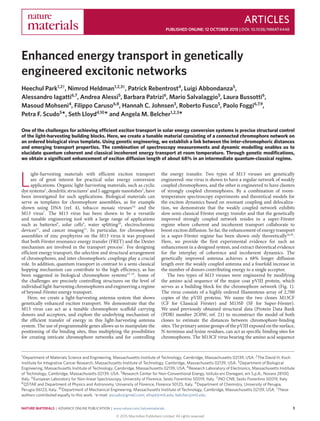
 26 Å 26 Å 26 Å 10 Å10 Å13 Å a ∼880 nm ∼7 nm 28 Å 28 Å 28 Å 33 Å 16 Å b c Figure 1 | Molecular models of the genetically engineered M13 viruses. a, A filamentous M13 virus is approximately 880 nm in length and ∼7 nm in diameter and composed of 2,700 copies of the major coat protein pVIII, which provides a scaffold for excitation energy transport over chromophores in a light-harvesting antenna system. b,c, Magnified M13 virus surfaces show the energy-transfer networks between chromophore-binding sites for M13CF and M13SF, respectively (blue colour, N-terminus; orange, newly added lysine residue; green, pre-existing lysine residue). Insets show schematic networks of energy transport in the M13CF and the M13SF, respectively. (Red gradient colour of ellipse indicates exciton delocalization. Blue arrows show incoherent exciton hopping. For the M13CF, pathways to any of the lysine binding sites are not shown because only 7% are occupied.) The M13CF has long inter-binding site distances of 16 Å and 33 Å within two pVIII proteins. In the M13SF, close inter-binding site distances of approximately 10 Å and 13 Å are formed. The pVIII coat protein assemblies were reconstructed from the structural model (PDB number 2C0W). of ADSPHTELPDPAK (ref. 9) at the N-terminal region of the pVIII was used because the distances between the two primary amines are longer than the corresponding distances in the wild type. The inter-binding site distance was estimated to be 33 Å on the pVIII (Fig. 1b). To create an entirely different network with closer distances, the M13SF virus was genetically engineered to have an additional third binding site on the pVIII. Among the possible candidates (data not shown), the M13SF carrying the shorter pVIII N-terminal sequence of AENKVEDPAK was identified as having inter-binding site distances of 10 Å on the pVIII (Fig. 1c). These distances are short enough to enhance the coupling strength of the bound chromophores, but long enough to avoid the complete quenching of fluorescence due to short-range Dexter exchange19,22 . The electronic coupling strength between donors for the 10 Å distance is about 460 cm−1 , calculated using the standard FRET exchange formula and the spectral overlap, and assuming parallel orientation of the dipoles. Various kinds of chromophores can be selected for coupling to the M13 virus biological scaffold through chemical modification of amine groups on the pVIII coat proteins. Alexa Fluor 488 and Alexa Fluor 594 were carefully selected as donor and acceptor chromophores, respectively (Supplementary Fig. 1). They show narrow and well-separated absorption bands and good spectral overlaps between donor emission and donor absorption, as well as between donor emission and acceptor absorption for energy transfer (Fig. 2a). Absorption peaks are at 491 nm for the donor and at 585 nm for the acceptor. Emission peaks are at 517 nm for the donor and at 611 nm for the acceptor. The acceptor emission peak is sufficiently far from the donor emission peak to resolve the signals of fluorescence when the donors are excited and the fluorescence intensities are detected from the acceptors. They also show high molar extinction coefficients (that is, strong dipole moments23 ) and relatively high quantum yields24 (Supplementary Section E.1.1). Moreover, a pre-activated carboxylic acid in each donor and acceptor allows specific conjugation to the primary amines on the pVIII by forming covalent amide bonds. A dense occupation of the sites comprising the energy-transfer network on the M13 virus surface is desired. The conjugation of the chromophores to the N-terminus and the lysine residues was confirmed by diverse control experiments (Fig. 2b (upper inset) and Supplementary Section E.3). On the M13CF (M13SF), on average 2,970 ± 180 (4,130 ± 70) donors occupy the 5,400 (8,100) possible binding sites per virus, estimated from the absorbance (Supplemen- tary Section E.2.1). For M13CF, 2,700 N-termini are fully occupied (∼100%) and only ∼200 lysine residues are occupied (∼7%). For M13SF, 2,700 N-termini are fully occupied (∼100%), ∼200 of the pre-existing lysine residues (∼7%), and ∼1,200 of the newly added lysines (∼44%) (Supplementary Section E.4). Thus, for M13SF, 1,200 out of the 2,700 pVIII copies have a donor pair separated by 10 Å. The donor absorbance shows peak shifts to 496 nm for the M13CF and 497 nm for the M13SF, which is evidence of an electronic coupling of the chromophores (Supplementary Fig. 6). From the occupation probabilities of the binding sites of the chromophore networks, the M13CF has closer to an ideal diamond grid of relatively well-separated N-terminus chromophores (Fig. 1b). Because the semi-classical Förster rates of transport depend on the distance r as 1/r6 (ref. 22), we can assume that each chromophore has four relevant nearest neighbours. The M13SF has the same basic diamond grid, but about 50% of the unit cells have additional vertices resulting from the lysines, which fill the empty space (Fig. 1c). The distance between nearest neighbours resides within the Förster radius between donors, which is estimated to be 37.5 Å on the M13CF, and 30.1 Å on the M13SF (Supplementary Section E.7). This guarantees connectivity of the network with at least more than 50% inter-chromophoric energy transport on both clones. For the M13SF, we expect that the stronger electronic couplings and the subsequent highly linked network lead to improved energy transport. 2 NATURE MATERIALS | ADVANCE ONLINE PUBLICATION | www.nature.com/naturematerials © 2015 Macmillan Publishers Limited. All rights reserved
  • 3. NATURE MATERIALS DOI: 10.1038/NMAT4448 ARTICLES a Normalizedintensity(a.u.) 0.0 0.2 0.4 0.6 0.8 1.0 Wavelength (nm) 400 450 500 550 600 650 700 750 800 DN absorption DN emission AC absorption AC emission Fluorescenceintensity(×107a.u.) 0.0 0.5 1.0 1.5 2.0 Wavelength (nm) 500 550 600 650 Free DN M13SF-400DN M13CF-DN M13SF-DN M13SF-DN Free DN b Figure 2 | Steady-state spectra and fluorescence quenching at room temperature. a, Absorption and emission spectra of free donor (DN) and free acceptor (AC). b, Fluorescence spectra of donor chromophores at an excitation wavelength of 495 nm. All samples have an absorbance of 0.3 at 495 nm with a 1 cm path length of light. M13SF-DN indicates the M13SF virus with ∼4,100 donors on the surface, and M13CF-DN indicates the M13CF virus with ∼2,900 donors. M13SF-400DN denotes a control sample of the M13SF virus with ∼400 donors. Significant quenching is observed in the M13SF-DN. The upper inset is a fluorescence microscope image of the M13SF-DN (excitation at 488 nm and detection at 528 nm). Scale bar, 5 µm. The bottom inset is a photograph taken under ambient light at the same absorbance of 0.5 with a 1 mm path length of light. The change in colour is apparent between the two samples. There exists an optimal distance between chromophores where the strong couplings lead to fast transport, but the increased exciton loss from concentration quenching does not dominate. The quenching of donor fluorescence is observed in both clones at a constant donor absorbance of 0.3 with a 1 cm path length to minimize the inner filter effects of the donors. M13SF shows more quenching than M13CF, which in turn shows more quenching than the free donors (Fig. 2b). Quantum yields for the samples were 1% for the M13SF and 5% for the M13CF, compared to a value of 92% for free donors (Supplementary Section E.6). This quenching can be explained by the close proximity between chromophores. To test this explanation, a control sample of more sparse chromophores, ∼400 donors on the M13SF, was prepared. This sample showed significantly less quenching than both previous samples (Fig. 2b), suggesting that the quenching effect of both clones comes largely from the inter-chromophoric interactions, rather than from the protein conjugation. Acceptor chromophores were scattered within the donor grid on the M13 virus to act as output sensors of exciton diffusion in Fig. 3a. When the donors on the M13 virus are excited, the excitation energy transfers between the donors until it reaches an acceptor. Once the acceptor receives the energy, then a part of the transferred energy is detected from the acceptors in the form of fluorescence intensities. To obtain quantitative insights into the exciton diffusion and the maximum number of donors contributing to a single acceptor, different ratios of the donors to acceptors were assembled along the viruses. Constant concentrations of donors with a varied concentration of acceptors were mixed into the solution of the virus. The total number of chromophores on the virus remained approximately constant (4,110 ± 250 for M13SF and 2,920 ± 240 for M13CF), whereas the number of acceptors bound to the virus surface varied. The samples had donor-to-acceptor ratios from 16:1 to 734:1 for M13SF and ratios from 19:1 to 394:1 for M13CF (Supplementary Tables 1 and 2), estimated by absorbance. All the samples were adjusted to the same donor absorbance at 495 nm at a level high enough to detect the fluorescence signal of the acceptors (Supplementary Figs 3 and 4). Steady-state fluorescence of the acceptors shows their sensi- tization by the donors. In the room-temperature measurements of steady-state fluorescence, all samples of the light-harvesting nanoantennae were excited at 495 nm, so that they absorb the same maximum number of incident photons. The emission near 610 nm is essentially ascribable to the acceptor as a consequence of energy transfer from the donor chromophores, whereas the 520 nm band is due to the donor emission. The overlapping background fluo- rescence of the donors was subtracted from the acceptor emission signal (Supplementary Section E.2.2). Each spectrum of fluores- cence was integrated over wavelength to obtain the total energy of fluorescence emitted from the acceptors (Fig. 3b). The integrated fluorescence of the M13CF increases linearly with increasing accep- tor concentration. On the other hand, the fluorescence of the M13SF increases at first and saturates for large acceptor concentrations. The saturation can be explained by a competition of the acceptors for the available excitation energy from the donors, as shown in Fig. 3a. The fluorescence data also enable us to elucidate the number of donors contributing to a single acceptor, as well as the excitonic diffusion length. The integrated fluorescence was normalized by the number of acceptors to obtain the total energy emitted from a single acceptor as a function of the donor-to-acceptor ratio. Then, the integrated fluorescence was normalized to have the maximum fluorescence of both clones be the same for a better comparison (Fig. 3c). The maximum fluorescence energy per acceptor for the M13SF was at a donor-to-acceptor ratio of 432:1 and for the M13CF at a ratio of 98:1. This already suggests a larger diffusion length for the M13SF clone. One can obtain an estimate of the exciton diffusion length solely from the Förster radius R0 and the average chromophoric distance RDD using a simple random walk model for excitons, leading to ldiff = R3 0/R2 DD. This estimate results in 5.8 nm for M13CF and 6.6 nm for M13SF (Supplementary Section T.4). This similar diffusion length does not explain the experimental results in Fig. 3c. We thus developed a phenomenological fitting method to extract a quantitative estimate of exciton diffusion length (Fig. 3c). This fitting method does not assume a classical or quantum nature of the underlying transport. The fitting function is based on the rationale that each acceptor harvests energy from a given area of donors on the virus. The acceptors compete for the available total area on the M13SF virus and, as their concentration increases, the harvesting areas of the acceptors overlap, which reduces the single acceptor trapping probability, exhibited by the saturation in Fig. 3a,b. The diffusion length is defined to be simply the radius of the harvested area around an acceptor and is obtained by the fitting to be 7.8 nm for M13CF, and 13.1 nm for M13SF, demonstrating an improvement of ∼68%. With the same theory, the number of donors contributing to the fluorescence of a single acceptor was estimated to be 29 for M13CF and 114 for M13SF, a fourfold increase. Thus, the experimental data show a significant improvement of energy transfer in M13SF relative to M13CF, which is not predicted by Förster theory (Supplementary Section T.2). NATURE MATERIALS | ADVANCE ONLINE PUBLICATION | www.nature.com/naturematerials 3 © 2015 Macmillan Publishers Limited. All rights reserved
  • 4. ARTICLES NATURE MATERIALS DOI: 10.1038/NMAT4448 Fluorescenceperacceptor(×108a.u.) 0 1 2 3 4 5 Donor-to-acceptor ratio 0 100 200 300 400 500 600 700 800 900 M13CF M13SF Integratedfluorescence(×108a.u.) 0.0 0.5 1.0 1.5 2.0 Acceptor concentration (nM) 0 50 100 150 200 250 300 M13CF M13SF 16:127:1 19:1 43:1 hv Donor Acceptora hv b c Figure 3 | Steady-state fluorescence data at room temperature showing exciton harvesting. a, Schematic representations where each acceptor harvests excitation energy from a given area of the donors on the viral surface (black circles with red centres, harvesting areas around acceptors; blue arrow, excitation of donors by incident light (hv); yellow arrow, excitation energy transport; orange arrow, fluorescence (hv) from an acceptor). From the left to the right panel as acceptor concentration increases on the M13SF virus, the harvesting areas overlap. b, Integrated fluorescence on donor excitation emitted from the acceptors as a function of acceptor concentration for both clones. Black text denotes donor-to-acceptor ratios. c, Fluorescence normalized by the number of acceptors as a function of donor-to-acceptor ratio. The M13CF fluorescence data were multiplied by a factor of 0.37 to have the same maximum as the M13SF data. Our fluorescence theory fits well with the M13CF and M13SF data. From the fitting curves of the fluorescence theory (solid lines), exciton diffusion lengths were obtained to be 7.8 nm for M13CF and 13.1 nm for M13SF. The donor-to-acceptor ratios are as follows: 19:1, 43:1, 98:1, 159:1 and 394:1 for the M13CF and 16:1, 27:1, 72:1, 118:1, 236:1, 432:1, 627:1 and 734:1 for the M13SF. Room-temperature measurements of transient-absorption (TA) spectroscopy reveal further details about the exciton dynamics. The purposes of these measurements are to quantify the exciton lifetime on the donors and to achieve an estimate of the trapping efficiency as a function of acceptor concentration. The lifetime is used for the numerical studies below. All the data were recorded at light intensities where singlet–singlet annihilation is not present (Supplementary Section E.8.2). The evolution-associated difference spectra (EADS) of the M13SF fully occupied with donors from a global analysis are shown in Fig. 4a. The normalized bleaching kinetic traces taken at 500 nm (pumped at 470 nm) show distinctive differences of excitonic behaviour between the donors on the M13 virus samples and free donors (Fig. 4b). The kinetics show a triple- exponential decay. The first time constant of the fitting arises from fast, internal and solvent relaxation contributions. Maximum entropy method (MEM) analysis showed that in the M13SF and M13CF the distributions of the second and the third time constants overlap and thus are averaged to obtain the exciton lifetimes (Supplementary Sections E.8.4 and E.8.5). The exciton lifetime of the donors is 2.7 times shorter in the densely clustered M13SF clone than in the M13CF clone, 155 ps and 422 ps, respectively. The control sample with only ∼400 donors exhibited a decay time of ∼2 ns, similar to free donors of ∼4 ns: the differences can be explained largely by chromophore–protein interactions. In addition, a measurement in the presence of acceptors bound to the virus shows further reduction of the donor excited state bleaching time (Supplementary Tables 3 and 4 and Supplementary Figs 14 and 17), consistent with donor-to-acceptor transfer processes. We set up a numerical simulation to predict our experimental results using three theories: a classical walk (CW), a decohered quantum walk (DQW) and a phenomenological Super-Förster theory (SFT). A classical walk describes the exciton motion as hopping between sites. The decohered quantum walk and the Super-Förster theory allow for strong inter-chromophore couplings and quantum coherence effects. We take into account the estimated detailed structural information, distance and network connectivity, and the probabilities of chromophore occupation at each binding site. Studying ensembles of virus–chromophores complexes enables us to require knowledge of only the averaged site occupation and also the averaged orientation of the chromophores. The chromophore couplings are derived from the Förster radii and the spectral overlap. The averaged relative dipole orientation is a free parameter, giving good results for a value corresponding to a non-isotropic averaged system (Supplementary Section T.5). As a result, the Förster CW numerical simulation predicted the fluorescence data of the M13CF (Supplementary Fig. 22), whereas it underestimated the performance of the M13SF (Fig. 5 and Supplementary Fig. 23). For the M13SF, we employed a decohered quantum walk model. It is based on a tight-binding Hamiltonian with the Haken–Strobl–Reinecker noise model25,26 and with estimates for the amount of site-energy static disorder and the dephasing rate. Although the model describes an infinite temperature pure-dephasing approximation, it includes exciton delocalization and strong coupling effects leading to clustering and allows us to simulate relatively large networks. It predicts the fluorescence data for the M13SF virus well (Fig. 5). Comparing the two calculations confirms that the exciton transfer of M13CF is well described by semi-classical Förster hopping, whereas the clustered network of the M13SF clone warrants a more careful treatment that takes into account strong couplings and quantum delocalization over clusters of donors. In addition, we developed the SFT to untangle the clustering effects of dipole moment and chromophore distance contained in the DQW model. It is based on the rationale that, owing to random disorder and strongly coupled dipole moments, in the transfer process excitons are delocalized on clusters of a few chromophores, while performing a classical hopping between adjacent clusters. First, these clusters can be considered as super-molecules sharing a collective transition dipole moment. Second, the spatial extent of the clusters in the M13SF reduces the effective distances between the clusters compared to the almost regular diamond grid of the M13CF. To get an idea of the clustering in the M13SF, the occupation probabilities of the binding sites predict that about 1.43 chromophores participate in a cluster on average. With these two Super-Förster corrections, the SFT predicts the fluorescence data of M13SF reasonably well (Fig. 5), with the distance correction being the dominating effect (Supplementary Section T.7). 4 NATURE MATERIALS | ADVANCE ONLINE PUBLICATION | www.nature.com/naturematerials © 2015 Macmillan Publishers Limited. All rights reserved
  • 5. NATURE MATERIALS DOI: 10.1038/NMAT4448 ARTICLES 0.00 0.02 0.04 0.06 Time (ps) 0 500 1,000 1,500 Free DN M13SF-400DN M13CF-DN M13SF-DNΔO.D. −ΔO.D. −0.06 −0.04 −0.02 0.00 0.02a Wavelength (nm) 350 400 450 500 550 600 650 700 750 2.0 ps 52 ps 560 ps b Figure 4 | Transient-absorption spectra at room temperature. a, Evolution-associated difference spectra (EADS) obtained from a global analysis of the transient-absorption data recorded for the M13SF-DN on excitation at 470 nm. b, Normalized bleaching kinetics of optical density change (− O.D.) probed at 500 nm as a function of delay time (dots). The samples were pumped at 470 nm. Kinetic curves were extracted at 500 nm from a global analysis (solid lines). Fluorescenceperacceptor(×108a.u.) 0 1 2 3 4 5 6 Donor-to-acceptor ratio 0 200 400 600 800 1,000 1,200 Experimental fit Decohered QW Super-Förster Classical Förster Figure 5 | Comparison of numerical simulations of classical and quantum transport theories for M13SF to the experiment. The classical walk description does not explain the M13SF. The newly developed Super-Förster theory and decohered quantum walk (QW) match the fluorescence per acceptor of the M13SF. For the experimental fit, the downward trend exhibited in Fig. 3c was corrected (Supplementary Section T.2). In this work, we have created a tunable light-harvesting material by using a genetically modifiable, self-assembled virus template. It enables us to probe chromophore network configurations along the spectrum between coherent and incoherent Förster exciton transport. We have created an intermediate configuration showing improved exciton transport compared to an incoherent transport configuration. This improvement arises from a regime of stronger coupling between the chromophores, where usually quantum coherence effects accompany the enhancement of transfer. We thus have provided evidence for an important energy-transfer design concept: beyond-Förster transport boosts the transfer efficiency. Theoretical work16 suggests that in this regime quantum coherence and incoherent mechanisms interact to enhance the exciton transfer. A similar design concept has been rationalized in photosynthetic energy transfer, with differences in the specific implementation details, such as vibrational environment or loss pathways. Vibronic couplings have attracted recent attention27–29 and are certainly demonstrated in our system in the form of non-radiative losses. Our system may prove especially useful for investigating higher- level energy transport mechanisms—for example, exciton relaxation effects and directing the exciton transport towards specific sites. In addition, we lay the fundamentals for a wide range of applications relying on efficient energy transfer, such as organic photovoltaics and light-driven catalysis. Methods Methods and any associated references are available in the online version of the paper. Received 25 December 2014; accepted 10 September 2015; published online 12 October 2015 References 1. Wasielewski, M. R. Self-assembly strategies for integrating light harvesting and charge separation in artificial photosynthetic systems. Acc. Chem. Res. 42, 1910–1921 (2009). 2. Goodson, T. G. Optical excitations in organic dendrimers investigated by time-resolved and nonlinear optical spectroscopy. Acc. Chem. Res. 38, 99–107 (2005). 3. Eisele, D. M. et al. Utilizing redox-chemistry to elucidate the nature of exciton transitions in supramolecular dye nanotubes. Nature Chem. 4, 655–662 (2012). 4. Woller, J. G., Hannestad, J. K. & Albinsson, B. Self-assembled nanoscale DNA-porphyrin complex for artificial light harvesting. J. Am. Chem. Soc. 135, 2759–2768 (2013). 5. Miller, R. A., Presley, A. D. & Francis, M. B. Self-assembling light-harvesting systems from synthetically modified tobacco mosaic virus coat proteins. J. Am. Chem. Soc. 129, 3104–3109 (2007). 6. Ma, Y. Z., Miller, R. A., Fleming, G. R. & Francis, M. B. Energy transfer dynamics in light-harvesting assemblies templated by the tobacco mosaic virus coat protein. J. Phys. Chem. B 112, 6887–6892 (2008). 7. Nam, Y. S. et al. Virus-templated assembly of porphyrins into light-harvesting nanoantennae. J. Am. Chem. Soc. 132, 1462–1463 (2010). 8. Lee, Y. J. et al. Fabricating genetically engineered high-power lithium-ion batteries using multiple virus genes. Science 324, 1051–1055 (2009). 9. Dang, X. N. et al. Virus-templated self-assembled single-walled carbon nanotubes for highly efficient electron collection in photovoltaic devices. Nature Nanotech. 6, 377–384 (2011). 10. Nam, Y. S. et al. Biologically templated photocatalytic nanostructures for sustained light-driven water oxidation. Nature Nanotech. 5, 340–344 (2010). 11. Nam, Y. S. et al. Virus-templated iridium oxide-gold hybrid nanowires for electrochromic application. Nanoscale 4, 3405–3409 (2012). 12. Ghosh, D. et al. M13-templated magnetic nanoparticles for targeted in vivo imaging of prostate cancer. Nature Nanotech. 7, 677–682 (2012). 13. Engel, G. S. et al. Evidence for wavelike energy transfer through quantum coherence in photosynthetic systems. Nature 446, 782–786 (2007). 14. Olaya-Castro, A., Lee, C. F., Fassioli Olsen, F. & Johnson, N. F. Efficiency of energy transfer in a light-harvesting system under quantum coherence. Phys. Rev. B 78, 085115 (2008). 15. Caruso, F., Chin, A. W., Datta, A., Huelga, S. F. & Plenio, M. B. Highly efficient energy excitation transfer in light-harvesting complexes: The fundamental role of noise-assisted transport. J. Chem. Phys. 131, 105106 (2009). NATURE MATERIALS | ADVANCE ONLINE PUBLICATION | www.nature.com/naturematerials 5 © 2015 Macmillan Publishers Limited. All rights reserved
  • 6. ARTICLES NATURE MATERIALS DOI: 10.1038/NMAT4448 16. Rebentrost, P., Mohseni, M., Kassal, I., Lloyd, S. & Aspuru-Guzik, A. Environment-assisted quantum transport. New J. Phys. 11, 033003 (2009). 17. Walschaers, M., Diaz, J. F., Mulet, R. & Buchleitner, A. Optimally designed quantum transport across disordered networks. Phys. Rev. Lett. 111, 180601 (2013). 18. Blankenship, R. E. Molecular Mechanisms of Photosynthesis (Blackwell Science, 2002). 19. Scholes, G. D., Fleming, G. R., Olaya-Castro, A. & van Grondelle, R. Lessons from nature about solar light harvesting. Nature Chem. 3, 763–774 (2011). 20. Panitchayangkoon, G. et al. Long-lived quantum coherence in photosynthetic complexes at physiological temperature. Proc. Natl Acad. Sci. USA 107, 12766–12770 (2010). 21. Marvin, D. A., Welsh, L. C., Symmons, M. F., Scott, W. R. P. & Straus, S. K. Molecular structure of fd (f1, M13) filamentous bacteriophage refined with respect to X-ray fibre diffraction and solid-state NMR data supports specific models of phage assembly at the bacterial membrane. J. Mol. Biol. 355, 294–309 (2006). 22. Lankiewicz, L., Malicka, J. & Wiczk, W. Fluorescence resonance energy transfer in studies of inter-chromophoric distances in biomolecules. Acta Biochim. Pol. 44, 477–489 (1997). 23. Turro, N. J., Ramamurthy, V. & Scaiano, J. C. Modern Molecular Photochemistry of Organic Molecules (Univ. Science Books, 2010). 24. Spence, M. T. Z. & Johnson, I.D. The Molecular Probes Handbook: A Guide to Fluorescent Probes and Labeling Technologies 11th edn (Live Technologies Corporation, 2010). 25. Haken, H. & Reineker, P. Coupled coherent and incoherent motion of excitons and its influence on line shape of optical-absorption. Z. Phys. 249, 253–268 (1972). 26. Haken, H. & Strobl, G. Exactly solvable model for coherent and incoherent exciton motion. Z. Phys. 262, 135–148 (1973). 27. Christensson, N., Kauffmann, H. F., Pullerits, T. & Mancal, T. Origin of long-lived coherences in light-harvesting complexes. J. Phys. Chem. B 116, 7449–7454 (2012). 28. Tiwari, V., Peters, W. K. & Jonas, D. M. Electronic resonance with anticorrelated pigment vibrations drives photosynthetic energy transfer outside the adiabatic framework. Proc. Natl Acad. Sci. USA 110, 1203–1208 (2013). 29. Halpin, A. et al. Two-dimensional spectroscopy of a molecular dimer unveils the effects of vibronic coupling on exciton coherences. Nature Chem. 6, 196–201 (2014). Acknowledgements This work was supported from Eni, S.p.A. (Italy) through the MIT Energy Initiative Program. H.P. thanks Kwanjeong Educational Foundation for its financial support, and G. W. Hwang for allowing us to use a fluorometer. F.C. has been supported by EU FP7 Marie-Curie Programme (Career Integration Grant) and by MIUR-FIRB grant (Project No. RBFR10M3SB). Author contributions H.P., N.H., P.F.S., M.M., R.F., S.L. and A.M.B. conceived the work. P.F.S., P.F., S.L. and A.M.B. supervised the overall work. H.P. and N.H. designed the experiments including protocols. H.P. prepared all the samples, performed the spectroscopic measurements, analysed the data, and interpreted the data with classical Förster theory. H.P. wrote a first version of the manuscript, based on which H.P., P.R. and N.H. developed the final version of the manuscript. H.P. and N.H. reconstructed the virus structure models, and performed the virus cloning. P.F.S. and R.F. made collaborations between MIT and Italy. P.R., P.F.S., L.A., M.M., R.F. and S.L. designed the theoretical work. P.R. and S.L. developed the fluorescence theory, and P.R. and H.P. applied it to the data. P.R. and S.L. developed the quantum transport theory, and P.R. and L.A. performed the quantum mechanical simulations. A.I., B.P., L.B. and P.F. performed the TA measurements and analysed the TA data. P.F., A.I. and H.P. interpreted the TA data. M.S. and R.F. helped H.P., N.H. and A.A. perform the QY measurements, and H.P. and A.A. analysed the QY data. F.C. collaborated in developing the computation model. H.C.J. helped H.P. with the virus preparations. H.P., N.H., P.R., P.F.S., S.L. and A.M.B. made major edits on the manuscript. All the authors gave helpful comments on the manuscript. Additional information Supplementary information is available in the online version of the paper. Reprints and permissions information is available online at www.nature.com/reprints. Correspondence and requests for materials should be addressed to P.F.S. or S.L. or A.M.B. Competing financial interests The authors declare no competing financial interests. 6 NATURE MATERIALS | ADVANCE ONLINE PUBLICATION | www.nature.com/naturematerials © 2015 Macmillan Publishers Limited. All rights reserved
  • 7. NATURE MATERIALS DOI: 10.1038/NMAT4448 ARTICLES Methods Construction of M13 virus clones. The M13CF clone with the N-terminal fusion sequence, DSPHTELP, was obtained through a panning and selection procedure9 . The M13SF clone with the N-terminal fusion sequence ENKVE was constructed as follows. Oligonucleotides encoding both strands of the fusion sequence were synthesized (Integrated DNA Technologies) with the following sequence: 5 (Phos)-GAGAATAAGGTTGAG-3 and 5 (Phos)- GATCCTCAACCTTATTCTCTGCA-3 . The two oligonucleotides were annealed in 10 mM Tris, 1 mM EDTA, and 100 mM NaCl by heating up to 95 ◦ C for 2 min and cooling down at a rate of 0.1◦ per second down to 25 ◦ C. The two annealed oligonucleotides were designed to have overhangs matching the cut sites of PstI and BamHI restriction enzymes. A genetically modified double-stranded M13KE viral DNA (ref. 30) was isolated from infected XL1-Blue (Agilent Technologies) Escherichia coli cells (QIAprep Spin Miniprep Kit, QIAGEN). The viral DNA was then digested with PstI and BamHI enzymes (New England Biolabs (NEB)). The resulting linear vector was dephosphorylated using alkaline phosphatase (NEB) to minimize re-ligation of the original DNA. The linear vector and the annealed oligonucleotides were ligated with T4 DNA ligase (NEB) at 16 ◦ C for 15 h. The resulting vector was transformed into XL1-Blue cells and plated on tetracycline/IPTG/XGAL plates to isolate single virus plaques. The next day plaques were grown for DNA isolation and sequencing. The positive clones having the ENKVE sequence were used for further amplification of larger virus yields. Sample preparation. Alexa Fluor 488 carboxylic acid succinimidyl ester and Alexa Fluor 594 carboxylic acid succinimidyl ester24 were obtained from Life Technologies as a donor and an acceptor chromophore, respectively. An excess amount of pre-activated chromophores, 20-fold over the number of binding sites was added to an M13 virus suspension (1010 viruses µl−1 ) in phosphate buffered saline (PBS, pH 7.4), and incubated overnight for complete conjugation. To remove unreacted chromophores, dialysis was performed for 24 h against PBS in the dark using 100 kD cutoff membrane (Spectrum Laboratories). During the dialysis, PBS was stirred magnetically at 400 r.p.m. and was frequently replaced for increasing purification efficiency. After the addition of PEG/NaCl, virus samples conjugated with chromophores were precipitated via centrifugation for 20 min at 14,000 r.p.m. at 4 ◦ C for further purification. The precipitated chromophore–virus samples were obtained from the centrifugation as shown in Supplementary Fig. 2a. Then, the precipitated samples were suspended with deionized water (RICCA Chemical Company). Fluorescence microscope. An inverted fluorescence microscope (Applied Precision, DeltaVision) consists of an epifluorescent illumination laser module, an oil immersion ×60 objective (Olympus) and a Photometrics CoolSNAP HQ camera. The virus antenna samples were deposited and dried on a glass coverslip. The samples were excited at 488 nm, detected at 528 nm, and imaged by using the fluorescence microscope. The uniform length of each fluorescent object in the upper inset of Fig. 2b indicates that the chromophores are assembled along the viral surface. Steady-state absorption and emission spectroscopy. Absorbance spectra were collected on a DU800 (Beckman Coulter) with a 1 cm path-length quartz cuvette. Fluorescence emission spectra were recorded on a FluoroLog-3 spectrofluorometer (Horiba Jobin Yvon) with a 1 cm path-length quartz cuvette. Transient-absorption spectroscopy. The transient-absorption (TA) spectroscopy has the output of an amplified Ti:sapphire laser system delivering ∼100 fs pulses to produce both the probe (white continuum) and the pump pulses. The 470 nm pump pulses (whose energy was set at 100 nJ pulse−1 ) were obtained by sum frequency generation (SFG) of the signal output of an optical parametric amplifier (TOPAS by Light Conversion) with a portion of the laser fundamental output at 800 nm. The repetition rate of the laser system was set at 100 Hz and the polarization angle between pump and probe beams was adjusted at 54.7◦ so as to exclude rotational contributions to the transient signal. The customized detection system was characterized by a double array (256 pixels) and was controlled by a front-end circuit. The data were acquired by means of a LabVIEW written computer program. The sample was contained in a 2 mm path-length quartz cell and was stirred continuously with a small magnetic bar to refresh the solution and avoid photo degradation. More detailed set-up information can be found in previous works31–33 . Fitting of fluorescence data. The fluorescence fitting function for Fig. 3 without the low-acceptor correction is given by FL(c) NA =α Ptrap NA =α c +1 Ntot 1− 1− πl2 diff Atot Ntot c+1 Here, the donor/acceptor concentration is c =ND/NA, where ND (NA) is the number of donors (acceptors). The total number of chromophores is Ntot =NA +ND. Ptrap is the probability of the initial excitation being inside at least one acceptor harvesting area, assumed to be circular shaped with radius ldiff (diffusion length). The total surface area on the phage is given by Atot. An experiment-specific linear scaling is given by α. The fit parameters are ldiff and α. Further details, including the low-acceptor correction, can be found in Supplementary Sections T.2 and T.3. Classical master equation. The classical master equation for the site occupation probabilities pm is given by34 d/dtpm =− n kmnpm + n knmpn −klosspm. The hopping rate from site m to site n is given by kmn. According to standard Förster theory35 , the rates between donors are given by kDD =kloss(RDD 0 /RDD)6 and between donor and acceptor by kDA =kloss(RDA 0 /RDA)6 . Here, R0 is the Förster radius for DD or DA transfer, respectively, kloss =kfl +knr is the exciton loss rate with radiative and non-radiative contribution, RDD and RDA and the distances between the two chromophores in question, taking into account the detailed geometry. In addition, zero acceptor to donor transfer is assumed, kAD =0. A uniform donor initial excitation pD(0)=1/ND is assumed. The probability of being trapped at the acceptors instead of lost at the donors is given by36 Ptrap =kloss ∞ 0 dt A pA(t), where the sum goes over all acceptors. More details can be found in Supplementary Section T.6. Quantum master equation. For the decohered quantum walk, a pure-dephasing model is employed based on the derivation by Haken, Strobl, and Reinecker25,26 under the Gauss–Markov approximation, leading to a master equation for the exciton density matrix (d/dt)ρex (t)=−(i/¯h)Hex ρex (t)+(i/¯h) ρex (t)Hex +L(ρex (t))+LDA(ρex (t)). The diagonals of the density matrix describe the site populations whereas the off-diagonals are inter-site quantum mechanical coherences. The Hamiltonian Hex =HS +Hloss includes the Hamiltonian for a single excitation in a lattice of sites34 , HS = m εm|m m|+ m<n Vmn(|m n|+|n m|), where the site energies are εm and the site–site couplings are Vmn. The site energies are assumed to be Gaussian distributed. To obtain the donor-to-donor couplings, the relation kDD =V2 DDWDD is used, where WDD is the normalized donor–donor spectral overlap. Thus, VDD =(RDD,3 0 /R3 DD) √ kloss/WDD. The single-exciton states |m denote the presence of the exciton at site m. The exciton loss is taken into account by the effective Hamiltonian14,16 Hloss =−(i/2) m kloss,m|m m|. The Lindblad37 operator describing the interaction of the exciton with the vibrational degrees of freedom is L(ρex (t))=γφ m(Amρex (t)A† m −(1/2)A† mAmρex (t) −(1/2)ρex (t)A† mAm). The Lindblad jump operators are Am =|m m| and the site-independent dephasing rate is γφ . In addition, irreversible donor-to-acceptor transfer is included by removing the coherent couplings VDA between donors and acceptors and introducing additional Lindblad jump operators in the master equation, LDA(ρex (t)). The initial state is taken as in the classical walk. The trapping probability at the acceptors can be computed by Ptrap =kloss ∞ 0 dt A A|ρex (t)|A , where the sum is over all acceptor sites. More details can be found in Supplementary Section T.8. The Super-Förster theory can be found in Supplementary Section T.7. References 30. Huang, Y. et al. Programmable assembly of nanoarchitectures using genetically engineered viruses. Nano Lett. 5, 1429–1434 (2005). 31. Lapini, A., Foggi, P., Bussotti, L., Righini, R. & Dei, A. Relaxation dynamics in three polypyridyl iron(II)-based complexes probed by nanosecond and sub-picosecond transient absorption spectroscopy. Inorg. Chim. Acta 361, 3937–3943 (2008). 32. Marcelli, A., Foggi, P., Moroni, L., Gellini, C. & Salvi, P. R. Excited-state absorption and ultrafast relaxation dynamics of porphyrin, diprotonated porphyrin, and tetraoxaporphyrin dication. J. Phys. Chem. A 112, 1864–1872 (2008). 33. Gentili, P. L., Bussotti, L., Ruzziconi, R., Spizzichino, S. & Foggi, P. Study of the photobehavior of a newly synthesized chiroptical molecule: (E)-(R-p,R-p)-1,2- Bis{4-methyl-[2]paracyclo[2](5,8)quinolinophan-2-yl}ethene. J. Phys. Chem. A 113, 14650–14656 (2009). 34. May, V. & Kühn, O. Charge and Energy Transfer Dynamics in Molecular Systems 2nd edn (Wiley-VCH, John Wiley, 2004). 35. Forster, T. ∗ Zwischenmolekulare Energiewanderung Und Fluoreszenz. Ann. Phys. 2, 55–75 (1948). 36. Sener, M. K. et al. Excitation migration in trimeric cyanobacterial photosystem I. J. Chem. Phys. 120, 11183–11195 (2004). 37. Breuer, H.-P. & Petruccione, F. The Theory of Open Quantum Systems (Oxford Univ. Press, 2002). NATURE MATERIALS | www.nature.com/naturematerials © 2015 Macmillan Publishers Limited. All rights reserved