![which is arguably a form of narrowed airway (the narrowing commonly occurring at
the velopharynx).2
CAUSES OF AIRWAY NARROWING AND AIRWAY STENOSIS
Some of the more common causes of airway narrowing and stenosis are listed in
Boxes 1–3. Some of these are genuine airway emergencies (eg, pediatric epiglot-
titis, obstructing foreign bodies, hematomas compressing the airway), whereas
some can be handled more electively (as with most cases of tracheal stenosis).
Note that patients presenting with stridor always require immediate clinical
attention.
Although in some of these cases tracheal intubation will be necessary to estab-
lish a definitive airway, in some other cases less invasive interventions may suffice.
For instance, patients with an edematous airway following prolonged airway instru-
mentation (eg, with a rigid bronchoscope) may have been treated with steroids (eg,
dexamethasone 8 mg intravenously [IV]) and topical dilute epinephrine (eg,
epinephrine 1:100,000 in 1–2 mL increments) but may need additional time for a
full therapeutic effect. In such cases heliox may be administered as an effective
temporizing measure. This is discussed further in Case Study 1. In some of these
patients noninvasive biphasic positive airway pressure ventilation would be another
temporizing measure.
It is worth emphasizing that excessive airway cuff pressures during tracheal intuba-
tion is a frequently unrecognized source of airway stenosis. Tracheal tube cuff
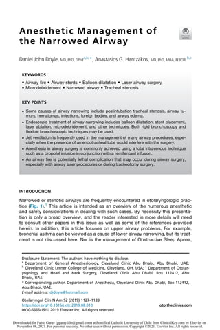
Fig. 1. (A) [Upper left] Patient with Grade 3 idiopathic subglottic stenosis, as seen with a
0
Hopkins laryngeal telescope. Suspension laryngoscopy and jet ventilation were used.
(B) [Lower left] Microdebridement of the same subglottic stenosis shown on Figure 1a. (C)
[Upper right] Endoscopic image of the same patient in Figure 1a and b after completion
of the microdebridement. (D) [Lower right] Treatment of subglottic stenosis with a flexible
CO2 laser system (Lumenis Duo Flexilase). The laser fiber is passed through the working
channel of a flexible endoscope.
Doyle Hantzakos
1128
Downloaded for Pablo Garay (pgaraylillo@gmail.com) at Pontifical Catholic University of Chile from ClinicalKey.com by Elsevier on
November 08, 2021. For personal use only. No other uses without permission. Copyright ©2021. Elsevier Inc. All rights reserved.](data:image/gif;base64,R0lGODlhAQABAIAAAAAAAP///yH5BAEAAAAALAAAAAABAAEAAAIBRAA7)
Recommended
More Related Content
What's hot
What's hot (20)
Similar to Anesthetic Management of the Narrowed Airway.pdf
Similar to Anesthetic Management of the Narrowed Airway.pdf (20)
Recently uploaded
Recently uploaded (20)
Anesthetic Management of the Narrowed Airway.pdf
- 1. Anesthetic Management of the Narrowed Airway Daniel John Doyle, MD, PhD, DPhil a,b, *, Anastasios G. Hantzakos, MD, PhD, MHA, FEBORL b,c INTRODUCTION Narrowed or stenotic airways are frequently encountered in otolaryngologic prac- tice (Fig. 1).1 This article is intended as an overview of the numerous anesthetic and safety considerations in dealing with such cases. By necessity this presenta- tion is only a broad overview, and the reader interested in more details will need to consult other papers in this issue as well as some of the references provided herein. In addition, this article focuses on upper airway problems. For example, bronchial asthma can be viewed as a cause of lower airway narrowing, but its treat- ment is not discussed here. Nor is the management of Obstructive Sleep Apnea, Disclosure Statement: The authors have nothing to disclose. a Department of General Anesthesiology, Cleveland Clinic Abu Dhabi, Abu Dhabi, UAE; b Cleveland Clinic Lerner College of Medicine, Cleveland, OH, USA; c Department of Otolar- yngology and Head and Neck Surgery, Cleveland Clinic Abu Dhabi, Box 112412, Abu Dhabi, UAE * Corresponding author. Department of Anesthesia, Cleveland Clinic Abu Dhabi, Box 112412, Abu Dhabi, UAE. E-mail address: djdoyle@hotmail.com KEYWORDS Airway fire Airway stents Balloon dilatation Laser airway surgery Microdebridement Narrowed airway Tracheal stenosis KEY POINTS Some causes of airway narrowing include postintubation tracheal stenosis, airway tu- mors, hematomas, infections, foreign bodies, and airway edema. Endoscopic treatment of airway narrowing includes balloon dilatation, stent placement, laser ablation, microdebridement, and other techniques. Both rigid bronchoscopy and flexible bronchoscopic techniques may be used. Jet ventilation is frequently used in the management of many airway procedures, espe- cially when the presence of an endotracheal tube would interfere with the surgery. Anesthesia in airway surgery is commonly achieved using a total intravenous technique such as a propofol infusion in conjunction with a remifentanil infusion. An airway fire is potentially lethal complication that may occur during airway surgery, especially with airway laser procedures or during tracheotomy surgery. Otolaryngol Clin N Am 52 (2019) 1127–1139 https://doi.org/10.1016/j.otc.2019.08.010 oto.theclinics.com 0030-6665/19/ª 2019 Elsevier Inc. All rights reserved. Downloaded for Pablo Garay (pgaraylillo@gmail.com) at Pontifical Catholic University of Chile from ClinicalKey.com by Elsevier on November 08, 2021. For personal use only. No other uses without permission. Copyright ©2021. Elsevier Inc. All rights reserved.
- 2. which is arguably a form of narrowed airway (the narrowing commonly occurring at the velopharynx).2 CAUSES OF AIRWAY NARROWING AND AIRWAY STENOSIS Some of the more common causes of airway narrowing and stenosis are listed in Boxes 1–3. Some of these are genuine airway emergencies (eg, pediatric epiglot- titis, obstructing foreign bodies, hematomas compressing the airway), whereas some can be handled more electively (as with most cases of tracheal stenosis). Note that patients presenting with stridor always require immediate clinical attention. Although in some of these cases tracheal intubation will be necessary to estab- lish a definitive airway, in some other cases less invasive interventions may suffice. For instance, patients with an edematous airway following prolonged airway instru- mentation (eg, with a rigid bronchoscope) may have been treated with steroids (eg, dexamethasone 8 mg intravenously [IV]) and topical dilute epinephrine (eg, epinephrine 1:100,000 in 1–2 mL increments) but may need additional time for a full therapeutic effect. In such cases heliox may be administered as an effective temporizing measure. This is discussed further in Case Study 1. In some of these patients noninvasive biphasic positive airway pressure ventilation would be another temporizing measure. It is worth emphasizing that excessive airway cuff pressures during tracheal intuba- tion is a frequently unrecognized source of airway stenosis. Tracheal tube cuff Fig. 1. (A) [Upper left] Patient with Grade 3 idiopathic subglottic stenosis, as seen with a 0 Hopkins laryngeal telescope. Suspension laryngoscopy and jet ventilation were used. (B) [Lower left] Microdebridement of the same subglottic stenosis shown on Figure 1a. (C) [Upper right] Endoscopic image of the same patient in Figure 1a and b after completion of the microdebridement. (D) [Lower right] Treatment of subglottic stenosis with a flexible CO2 laser system (Lumenis Duo Flexilase). The laser fiber is passed through the working channel of a flexible endoscope. Doyle Hantzakos 1128 Downloaded for Pablo Garay (pgaraylillo@gmail.com) at Pontifical Catholic University of Chile from ClinicalKey.com by Elsevier on November 08, 2021. For personal use only. No other uses without permission. Copyright ©2021. Elsevier Inc. All rights reserved.
- 3. pressures are easily measured with a cuff pressure manometer and should ordinarily be between 20 and 30 cm H2O. Excessive cuff pressures can result in tracheal steno- sis, tracheal necrosis, and even tracheal rupture as a result of diminished blood flow to tracheal mucosa. This hypothesized ischemic injury then produces healing fibrosis months or even years later.3 ENDOSCOPIC MANAGEMENT Airway endoscopy plays an important role in the diagnosis and treatment of airway tumors and especially to manage benign and malignant airway stenoses.4,5 More recently, endoscopic techniques have also been developed to treat end-stage emphy- sema, manage air leaks from bronchopleural fistulae, and treat intractable asthma via thermoablation.5–7 The frequency of central airway obstruction is increasing, paralleled with the increasing incidence of lung cancer. The development of endoscopic methods has allowed for less invasive management of these patients, with balloon dilation, laser therapy, microdebridement, and airway stenting as some of the options available to patients with central airway obstruction. Box 1 A list of some of the more common causes of airway narrowing and stenosis Tracheal stenosis (postintubation, GERD-related, post–lung transplantation, idiopathic) Tumors (intraluminal, extraluminal/extrinsic compression) Hematomas (eg, hematomas following thyroid surgery, carotid artery surgery, or trauma to the neck) Infections (eg, epiglottitis, Ludwig angina, retropharyngeal abscess) Foreign body aspiration (such items are frequently retrieved via rigid bronchoscopy) Airway edema (post–airway instrumentation, pregnancy, hereditary angioedema, smoke and hot gas inhalation) Abbreviation: GERD, gastroesophageal reflux disease. Box 2 Some common and uncommon airway management techniques in managing the narrow or stenotic airway Spontaneous ventilation in conjunction with an inhalation anesthetic induction Spontaneous ventilation in conjunction with an airway adjunct such as an OPA or NPA (following induction of anesthesia) Conventional positive pressure ventilation with an SGA (eg, i-gel) or ETT (wide bore preferred) or rigid bronchoscope Jet ventilation (see text) Positive/negative pressure ventilation via Ventrain system (see text) Extracorporeal membrane oxygenation (see text) Abbreviations: ETT, endotracheal tube; NPA, nasopharyngeal airway; OPA, oropharyngeal airway; SGA, supraglottic airway. Anesthetic Management of the Narrowed Airway 1129 Downloaded for Pablo Garay (pgaraylillo@gmail.com) at Pontifical Catholic University of Chile from ClinicalKey.com by Elsevier on November 08, 2021. For personal use only. No other uses without permission. Copyright ©2021. Elsevier Inc. All rights reserved.
- 4. BALLOON DILATATION Flexible bronchoscopic balloon dilatation is often used alone or in conjunction with other therapies to dilate narrowed airways.8–10 As an example, the CRE Pulmonary Balloon Dilatation Catheter series from Boston Scientific is designed to endoscopi- cally dilate narrowed airways via a 2.8 mm bronchoscope working channel. An excel- lent endoscopic video illustrating the technique is available at https://s3.amazonaws. com/prod.csurgeries.com/modify/csurgeries_20130923035501.mp4. LASER AIRWAY SURGERY Laser airway surgery can be done in 1 of 2 major ways: with a laser-safe endotra- cheal tube in place or using total intravenous anesthesia (TIVA) with jet ventilation. In the former situation volatile anesthetics may be used but conventional endotracheal tubes must be avoided, as they are flammable. (For a dramatic video showing an endotracheal tube [ETT] set on fire by a carbon dioxide [CO2] laser see https:// www.youtube.com/watch?v5zI8OAxpf9es). The lasers used in such settings are often CO2 lasers and Nd-YAG lasers, depending on the clinical situation. The CO2 laser offers precise cutting with a fine zone of coagulation that helps reduce surgical bleeding, whereas light emitted from an Nd-YAG laser can be transmitted through flexible quartz fibers that can be used in conjunction with a fiberoptic bronchoscope. Surgical lasers release great energy and can cause unintended tissue damage as well as operating room fires. This concern is discussed later in this article. An addi- tional concern with laser airway surgery is the smoke plume, which may be a vector for viral spread in some cases. As a result, continuous smoke evacuation is mandatory.11,12 Box 3 Helpful hints to bear in mind in dealing with patients with a narrow airway Patients presenting with stridor always command immediate attention. Although tracheal intubation is often required in such settings, less invasive interventions (eg, heliox administration) are sometimes appropriate as a temporizing measure. A joint review of available computed tomographic scans and airway videos by the anesthesiologist and surgeon together immediately before the procedure will help confirm the joint management plan. Although an inhalation induction is commonly recommended in pediatric patients with epiglottitis, this technique is usually unsatisfactory in adult patients with severe tracheal stenosis. (When an inhalational induction is attempted in such patients the induction is often slow and complicated by apneic episodes, hypoxemia, and a need to use bag-mask positive pressure ventilation to rescue the patient.) When using rigid bronchoscopy, avoid inhalational agents such as sevoflurane because they are not delivered reliably (scope going in and out) and they pollute the room terribly. Use a propofol infusion in conjunction with a remifentanil infusion instead. Muscle relaxation is usually needed. In addition, when using rigid bronchoscopy, it is frequently helpful to pack the oropharynx with saline soaked gauze to reduce the leak to manageable levels. (Leave a portion of each gauze outside the mouth as a reminder.) In cases where deep neuromuscular blockade is needed until the very end of the procedure, reversal with sugammadex is often preferable to using neostigmine and glycopyrrolate. Doyle Hantzakos 1130 Downloaded for Pablo Garay (pgaraylillo@gmail.com) at Pontifical Catholic University of Chile from ClinicalKey.com by Elsevier on November 08, 2021. For personal use only. No other uses without permission. Copyright ©2021. Elsevier Inc. All rights reserved.
- 5. MICRODEBRIDEMENT Microdebrider bronchoscopy is a new airway surgical technique that allows for precise removal of obstructing airway tissue. The technique is done under general anesthesia with either a rigid bronchoscope or (less commonly) a suspension laryngo- scope, with the microdebrider typically operating at 1000 to 3000 rpm.13,14 One impor- tant advantage of the technique is the avoidance of the complications of thermal techniques (eg, lasers or argon plasma coagulation) such as airway fires. STENTS Silicone and self-expandable metallic airway stents (SEMS) are the main stent types used in airway management.15,16 Although silicone stents are often the preferred mo- dality because they are easily repositioned, SEMS are frequently used in cases of airway wall malacia or stenosis, especially in the context of malignancy. SEMS have the advantage over silicone stents of a lower migration rate, thinner wall construction (allowing for greater cross-sectional airway diameter), and conformation to irregular airways. Although SEMS placement is ordinarily achieved endoscopically with relative ease, removal can be very difficult. Indications for SEMS removal include excessive or recurrent granulation, stent failure or fracture, infection, mucous plug formation, stent migration, or completion of successful treatment. Complications during stent removal may include pneumothorax; tracheal, bronchial, laryngeal, or pulmonary artery injury; and airway obstruction during or following stent removal. JET VENTILATION Jet ventilation is frequently used in the management of many otolaryngologic proced- ures, especially when the presence of an endotracheal tube would interfere with the surgery, such as with the treatment of some tracheal stenosis. Two techniques are available: (1) use of a simple hand-held device and (2) use of an automatic jet ventilator system. In both cases 100% oxygen delivery is possible but many automated jet ven- tilators have blenders to mix oxygen and air to reduce the FiO2. Although the Venturi effect causes entrainment of room air with the resulting gas mixture of less than 100%, it is prudent to use a gas mixture less than 100% and to ensure the FiO2 is less than 30% during laser surgery. In the case of hand-held devices, the operator starts operation with a driving pressure set to about around 20 psi (in adults) and provides manually delivered jets of a little less than 1 second duration delivered approximately 10 to 20 times a minute. In most cases the jet ventilation apparatus is attached to an anterior commissure laryngoscope or other type of otolaryngologic laryngoscope via a screw attachment to provide supraglottic ventilation. The main means of determining that ventilation is adequate is to watch the chest rise with each delivered jet pulse. For safety reasons the driving pressure should be adjusted down to the minimum needed to achieve adequate chest excursions. Note that obese individuals require higher driving pressures to achieve adequate chest excursions as compared with lean patients. In the case of an automatic jet ventilator system, a commercial product such as that provided by the Acutronic Company may be used to provide infraglottic ventilation. This is usually achieved in conjunction with an infraglottic ventilation catheter. Note that the ventilation rates for infraglottic automatic jet ventilation systems are usually much higher (in the range of 100 per minute and upwards) than for the supraglottic manual jet ventilation systems mentioned earlier (which typically operate in the range of 10–20 per minute). Anesthetic Management of the Narrowed Airway 1131 Downloaded for Pablo Garay (pgaraylillo@gmail.com) at Pontifical Catholic University of Chile from ClinicalKey.com by Elsevier on November 08, 2021. For personal use only. No other uses without permission. Copyright ©2021. Elsevier Inc. All rights reserved.
- 6. Most complications associated with the use of jet ventilation are due to use of a high pressure of the delivered gas, and barotrauma leading to pneumothorax, pneumomediastinum, or other problems can result from the high pressures used, which can run as high as 50 psi (which is equal to 3500 cm H2O pressure). This is particularly true if gas trapping due to an inadequate expiratory pathway takes place. ANESTHETIC TECHNIQUE The following anesthetic technique will serve as a rough guide to most cases involving conventional or bronchoscopic surgery used to manage a narrowed airway and is based on the clinical judgment of the surgeon and anesthesiologist who believe the narrowed airway is sufficient to permit anesthetic induction. If there is any doubt, an awake fiberoptic intubation with a small endotracheal tube or awake tracheostomy should be performed. After the induction of general anesthesia with propofol and muscle relaxation achieved with a paralytic such as rocuronium, anesthesia is commonly maintained using a TIVA technique such as a propofol infusion delivered at an initial rate of 100 mcg/kg/min in conjunction with a remifentanil infusion delivered at an initial rate of 0.1 mcg/kg/min. These infusions are then adjusted based on the factors such as the hemodynamics and the depth of anesthesia as guided by clinical findings and (commonly, but not always) by electroencephalographic analysis. In some centers it is common to add 1 mg of remifentanil to 500 mg (50 mL) of propofol and treat the combination as “superpropofol,” delivering it in a single syringe at an initial rate of 100 mcg/kg/min of propofol. When 100 mcg/kg/min of propofol is delivered in this scheme, the resulting rate of remifentanil infusion is then 0.2 mcg/kg/min. This arrangement has the advantage that only a single syringe pump is needed but has the disadvantage that the 2 infusions cannot be indepen- dently adjusted. Also if this scheme is used the pump should be correctly labeled to indicate the medications being infused. This should also be mentioned with any trans- fer of care that may occur during a procedure. PREVENTION AND MANAGEMENT OF AIRWAY FIRES An airway fire is potentially lethal complication that may occur during airway surgery, especially with airway laser procedures or during tracheotomy surgery. Case study 2 provides an example. For any fire to occur the triad of fuel (eg, ETT, drapes, sponges), oxidizer (O2 or N2O), and an ignition source (eg, laser or electrocautery) is needed. Consequently, in at-risk situations oxygen concentrations should be kept to a mini- mum, and nitrous oxide (which also supports combustion) should also be avoided. Further details are provided in the American Society of Anesthesiologists Operating Room Fire Algorithm (Fig. 2). In general, airway fires call for the immediate removal of the ETT, turning the oxygen/nitrous oxide sources off and flooding the field with saline from a prefilled 50 mL syringe. Although conventional wisdom holds that airway fires require the immediate removal of the ETT, it should be recognized that there are some situa- tions (eg, patients who were very difficult to intubate in the first place) where removal of the ETT would in all likelihood result in irreversible loss of the airway. Clinicians in such a setting face a particularly challenging choice: leave the tube in place and risk fire-related injury to the patient or remove the tube and risk loss of the airway.17 Doyle Hantzakos 1132 Downloaded for Pablo Garay (pgaraylillo@gmail.com) at Pontifical Catholic University of Chile from ClinicalKey.com by Elsevier on November 08, 2021. For personal use only. No other uses without permission. Copyright ©2021. Elsevier Inc. All rights reserved.
- 7. Fig. 2. American Society of Anesthesiologists Operating Room Fire Algorithm. a Ignition sources include but are not limited to electrosurgery or electrocautery units and lasers. b An oxidizer-enriched atmosphere occurs when there is any increase in oxygen concentra- tion above room air level and/or the presence of any concentration of nitrous oxide. c After minimizing delivered oxygen, wait a period of time (eg, 1–3 min) before using an ignition source. For oxygenation patients, reduce supplemental oxygen delivery to the minimum required to avoid hypoxia. Monitor oxygenation with pulse oximetry, and if feasible, inspired, exhaled, and/or delivered oxygen concentration. d After stopping the delivery of nitrous oxide, wait a period of the (eg, 1–3 min) before using an ignition source. e Unexpected flash, flame, smoke, or heat; unusual sounds (eg, a “pop,” “snap”, or “foomp”) or odors; unexpected movement of drapes; discoloration of drapes or breathing circuit; unexpected patient movement or complaint. f In this algorithm, airway fire refers to a fire in the airway or breathing circuit. g A CO2 fire extinguisher may be used on the patient if necessary. (From Apfelbaum JL, Caplan RA, Barker SJ, Connis RT, Cowles C, Ehrenwerth J, Nickinovich DG, Pritchard D, Roberson DW, Caplan RA, Barker SJ, Connis RT, Cowles C, de Richemond AL, Ehrenwerth J, Nickinovich DG, Pritchard D, Roberson DW, Wolf GL. Practice advisory for the prevention and management of operating room fires: an updated report by the American Society of Anesthesiologists Task Force on Operating Room Fires. Anesthesi- ology. 2013 Feb;118(2):271-90; with permission.) Anesthetic Management of the Narrowed Airway 1133 Downloaded for Pablo Garay (pgaraylillo@gmail.com) at Pontifical Catholic University of Chile from ClinicalKey.com by Elsevier on November 08, 2021. For personal use only. No other uses without permission. Copyright ©2021. Elsevier Inc. All rights reserved.
- 8. Lasers and Unique Considerations When lasers are in use it is prudent to remember that lasers used in surgery produce high amounts of optical energy. To warn everyone of this danger, one should place warning signs outside the operating room and opaque coverings should be placed on any operating room windows to prevent stray laser beams from exiting. Safety goggles should also be used; these are specific to each laser type. An even greater danger during laser airway surgery with an ETT in place is an airway fire, so laser-safe ETTs are necessary. For instance, the Mallinckrodt Laser-Flex ETT is a stainless-steel spiral wound tube with 2 PVC cuffs that are often filled with diluted methylene blue to allow early detection of a cuff leak. With 2 cuffs the distal cuff main- tains a seal if the proximal one is ruptured. Other popular tubes are the Sheridan LASER-TRACH and the Rusch Lasertubus, both which use an embossed copper foil covering a rubber tube. When used, the minimum acceptable FiO2 should be used and a 50-cc saline-filled syringe should be immediately available to use as an improvised fire extinguisher. Finally, note that the presence of high oxygen concentrations near the patient’s face can promote fires in this region, so delivering supplementary oxygen by face mask or nasal cannula requires special care. ANSI standard Z136.3 (Safe Use of Lasers in Health Care Facilities) provides additional relevant information. THE VENTRAIN SYSTEM Mechanical ventilation of patients is usually carried out using a conventional diam- eter (eg, 6–8 mm internal diameter [ID]) low-resistance cuffed tracheal tube inserted into the patient’s trachea. There are circumstances, however, where this arrange- ment is inappropriate. For instance, as noted earlier, with some forms of laryngeal surgery a narrow-diameter high-resistance catheter (eg, Hunsaker catheter) driven by a high-pressure gas source is used instead in order to offer the surgeon an un- impeded view of the glottis. Another situation is the “cannot intubate, cannot oxygenate/ventilate” (CICO/V) emergency, where percutaneous transtracheal jet ventilation via a very narrow bore high-resistance catheter is sometimes recommen- ded as a life-saving procedure. In all these cases expiration during ventilation occurs passively, but a recently introduced new technology now exists that sup- ports active expiration via a narrow-bore catheter. Known as the Ventrain system, the device allows for full ventilation via a mere 2 to 3 mm catheter, even in fully obstructed airways, using active expiration based on the Bernoulli Principle. The result is a system of ventilation that avoids extreme intrapulmonary pressures and the associated pulmonary (baro)trauma that sometimes plagues jet ventilation. As such the device is shown to be a lifesaver of babies and adults, providing full ventilation in CICO/V situations.18–21 As shown in Figs. 3 and 4, Ventrain is a compact, manually operated hand-held ventilator specifically designed for ventilation through narrow-bore (eg, 2–3 mm ID) catheters such as the Tritube narrow-bore cuffed endotracheal tube (Fig. 5). As noted earlier, what makes the Ventrain unique in comparison to other ventilation systems is its application of the Bernoulli principle to apply suction (as opposed to mere passive gas flow) during the expiratory phase of ventilation. This breakthrough in ventilator design allows for minute ventilation rates in excess of 7 L/min with relatively modest oxygen flows of 15 L/min and using catheters with internal diameters as narrow as 2 mm via any route of airway access.22 In CICO/V situations the Ventrain system has shown value during upper airway surgery by improving surgical exposure and avoiding the potential need for Doyle Hantzakos 1134 Downloaded for Pablo Garay (pgaraylillo@gmail.com) at Pontifical Catholic University of Chile from ClinicalKey.com by Elsevier on November 08, 2021. For personal use only. No other uses without permission. Copyright ©2021. Elsevier Inc. All rights reserved.
- 9. tracheostomy. Borg and colleagues23 were the first to describe the use of Ventrain during upper airway surgery, chronicling the use of the Ventrain during a laryngos- copy procedure in a stridorous patient suffering from an exophytic glottic tumor. A 2-mm ID transtracheal catheter was inserted into the cricothyroid membrane under local anesthesia and connected to the Ventrain using an oxygen flow of 15 L per minute. General anesthesia was then induced and the patient ventilated via the Ventrain at 15 breaths per minute (inspiration for 2 seconds, then expiration for 2 seconds). The procedure, which lasted 15 minutes, was uneventful and resulted in biopsy samples that allowed for the patient’s condition to be more reliably evaluated. Onwochei and colleagues24 described a 2-stage technique to manage airway obstruction and avoid a surgical tracheostomy in a 49-year-old woman who suffered from “postradiotherapy laryngeal fixation and transglottic stenosis” and had a pharyn- geal stricture in need of dilation. Their technique involved awake fiberoptic intubation, Fig. 3. The Ventrain device. This simple-to-operate manual ventilator is operated using one’s thumb and index finger. It features active expiration based on the Bernoulli principle, allow- ing ventilation through small-bore tubes. In addition to inspiratory (positive pressure) and expiratory (negative pressure) modes of operation, an equilibration (safety) mode is avail- able where no significant positive or negative pressure is present at the tip of the attached ventilation catheter. (Courtesy of Ventinova Medical, Eindhoven, The Netherlands; with permission.) Fig. 4. Ventrain used in conjunction with a small-bore airway catheter such as those used to exchange endotracheal tubes or those used to assist with tracheal extubation. In addition to providing a means to facilitate reintubation, such airway catheters can also be used with the Ventrain to maintain ventilation and oxygenation until a definitive airway is established. (Courtesy of Ventinova Medical, Eindhoven, The Netherlands; with permission.) Anesthetic Management of the Narrowed Airway 1135 Downloaded for Pablo Garay (pgaraylillo@gmail.com) at Pontifical Catholic University of Chile from ClinicalKey.com by Elsevier on November 08, 2021. For personal use only. No other uses without permission. Copyright ©2021. Elsevier Inc. All rights reserved.
- 10. followed by the transtracheal insertion of a Cricath flexible 2-mm needle cricothyrot- omy catheter, with ventilation using the Ventrain system. Similarly, Fearnley and col- leagues25 described the elective use of the Ventrain for upper airway obstruction in a patient with post-radiation fibrosis that had previously prevented passive expiration during attempted high-frequency jet ventilation. Kristensen and colleagues26 used the Ventrain in conjunction with a special 2.4-mm ID cuffed tracheal tube (Tritube) allowing intratracheal pressure monitoring in 7 ENT surgical patients. In all patients, adequate ventilation with intratracheal pressures between 5 and 20 cm H2O was performed. The investigators noted that the combina- tion of Ventrain with Tritube provided an “unprecedented view of the intubated airway during oral, pharyngeal, laryngeal or tracheal procedures” and even noted that the techniques has the “potential to replace temporary tracheostomy, jet-ventilation or extra-corporal membrane oxygenation in selected patients.” In their series, Kristensen and colleagues also mention that one patient was transported to the postanesthetic care unit (PACU) with a Tritube in place (with the cuff deflated) and was subsequently uneventfully extubated. Where a hand-operated technique is burdensome, clinicians may wish to use the new and Conformité Européenne-approved automatic ventilator Evone instead, allow- ing ventilation through the thin and cuffed Tritube. Product details are available at https://www.ventinovamedical.com/evone/. Barnes and Enk27 and Schmidt and col- leagues28 have recently executed useful studies awaiting publication dealing with the Evone that will interest those seeking further information. EXTRACORPOREAL MEMBRANE OXYGENATION IN THE MANAGEMENT OF THE CRITICAL AIRWAY In desperate cases, extracorporeal membrane oxygenation (ECMO) may occasionally be used in managing the critical airway. As an example, Holliday and Jackson29 re- ported on the use of ECMO to support a severely intoxicated patient with a life-threatening airway obstruction preventing adequate oxygenation and ventilation despite the presence of an ETT used to secure the airway. Fiberoptic bronchoscopy revealed a large tracheal food bolus situated beyond the ETT. With ECMO instituted, the obstructing food bolus was removed via rigid bronchoscopy under controlled conditions. A recent review by Hoetzenecker and colleagues30 provides a review of the published ECMO experience in relation to airway surgery in both in adult and pediatric patients. Fig. 5. The Tritube is a narrow-bore cuffed ETT with inner and outer diameters 2.4 mm and 4.4 mm, respectively. A Murphy eye is featured at the distal end. The ventilation lumen is attached to the Ventrain, whereas an inflatable cuff seals the airway. A pressure measure- ment lumen permits continuous intratracheal pressure measurements. This device may be useful in both elective and emergency airway settings. (Courtesy of Ventinova Medical, Eindhoven, The Netherlands; with permission.) Doyle Hantzakos 1136 Downloaded for Pablo Garay (pgaraylillo@gmail.com) at Pontifical Catholic University of Chile from ClinicalKey.com by Elsevier on November 08, 2021. For personal use only. No other uses without permission. Copyright ©2021. Elsevier Inc. All rights reserved.
- 11. CASE STUDY 1: USE OF HELIOX A 58-year-old female patient needed surgery for recurrent head and neck cancer. Because of her pathology, she was particularly difficult to intubate via a flexible bronchoscope. On extubation the patient became severely stridorous despite being wide awake and undergoing full reversal of neuromuscular blockade. Treatment included 2 IV doses of 8 mg dexamethasone (1 administered preextubation), 2 doses of nebulized racemic epinephrine (0.5 mL of 2.25% epinephrine added to 2.5 mL saline), and assisted mask ventilation with the patient sitting up at 60 , but despite this, the patient did not improve and was becoming exhausted. As reintubation would have been even more difficult than it had been earlier, a surgical airway was consid- ered the only option. The use of heliox, mixture of helium (70%) and oxygen (30%), delivered using a nonrebreathing face mask at 10 L/min saved the patient from needing a surgical airway. Approximately 5 to 10 breaths of heliox and the stridor vanished, and the patient’s work of breathing became much more manageable. The patient was then brought to the PACU with full monitoring and with a heliox tank in tow. She was then weaned off the heliox over several hours. Case Study 1 Discussion Airway obstructing conditions producing stridor may be modeled as breathing through an orifice (a situation where a tube length is smaller than its radius). Under such con- ditions, the approximate flow across the orifice varies inversely with the square root of the gas density. This is in contrast to the usual laminar flow situation during breathing, where gas flow varies inversely with gas viscosity. Note that although the viscosity values for helium and oxygen are similar, their densities are very different (density @ 20 C: air 1.293 g/L; nitrogen 1.250 g/L; oxygen 1.429 g/L; helium 0.178 g/L). In sum- mary, the low density of helium allows it to play an important role in the management of some forms of airway obstruction associated with gas turbulence, and heliox for delivery with a nonrebreathing face mask should be readily available in every operating room suite. CASE STUDY 2: AIRWAY FIRE The following case reported by Salaria and colleagues31 illustrates the complexity that may be involved in managing an airway fire. A 79-year-old man with severe aortic stenosis and multiple other comorbidities underwent an endoluminal minimally inva- sive mitral valve repair, aortic valve replacement, and MAZE procedure with a stormy postoperative course complicated by respiratory failure and necessitating an elective tracheostomy. One hundred percent oxygen was provided for preoxygenation before the tracheal incision. When the surgeon began to use monopolar electrocautery, “a 3-cm flame arose from the tracheal incision site.” In response, the field was flooded with saline, the ETT removed, the FiO2 decreased to room air, and the patent reintu- bated via the tracheal incision. Once the situation was stable, video laryngoscopy and fiberoptic visualization “revealed edematous, red, and inflamed mucosa in the right upper lobe and middle lobe bronchus” as well as “mild edema and red, inflamed mucosa” of the supraglottic structures. In the intensive care unit (ICU), a postoperative chest computed tomography “revealed bilateral patchy, nodular air space opacities and ground glass opacities with right perihilar bronchial thickening,” whereas “repeat fiberoptic bronchoscopy demonstrated blackened tracheal mucosa distal to the tracheal tube” with all 3 segments of the right upper lobe heavily charred. Despite fastidious care in the ICU that included “inhaled heparin every 8 hours, Anesthetic Management of the Narrowed Airway 1137 Downloaded for Pablo Garay (pgaraylillo@gmail.com) at Pontifical Catholic University of Chile from ClinicalKey.com by Elsevier on November 08, 2021. For personal use only. No other uses without permission. Copyright ©2021. Elsevier Inc. All rights reserved.
- 12. N-acetylcysteine, and albuterol” as well as daily bronchoscopy to clear mucous impaction, the patient ultimately died after developing multiorgan dysfunction. Case Study 2 Discussion Here the authors agree to what the investigators said about their case. “The cutting action of bipolar cautery generates lower temperatures, less tissue damage, and avoidance of sparks. In our case, the precipitating event was the use of monopolar diathermy to control bleeding in the presence of ventilation with 100% FiO2.” REFERENCES 1. Cooper JD. Tracheal injuries complicating prolonged intubation and tracheos- tomy. Thorac Surg Clin 2018;28(2):139–44. 2. Finkelstein Y, Wolf L, Nachmani A, et al. Velopharyngeal anatomy in patients with obstructive sleep apnea versus normal subjects. J Oral Maxillofac Surg 2014; 72(7):1350–72. 3. Feng TR, Ye Y, Doyle DJ. Critical importance of tracheal tube cuff pressure man- agement. World J Anesthesiol 2015;4(2):10–2. 4. Murgu SD, Egressy K, Laxmanan B, et al. Central airway obstruction: benign strictures, tracheobronchomalacia, and malignancy-related obstruction. Chest 2016;150(2):426–41. 5. Walters DM, Wood DE. Operative endoscopy of the airway. J Thorac Dis 2016; 8(Suppl 2):S130–9. 6. Paradis TJ, Dixon J, Tieu BH. The role of bronchoscopy in the diagnosis of airway disease. J Thorac Dis 2016;8(12):3826–37. 7. Ong PG, Debiane LG, Casal RF. Recent advances in diagnostic bronchoscopy. J Thorac Dis 2016;8(12):3808–17. 8. Wright CD. Nonoperative endoscopic management of benign tracheobronchial disorders. Thorac Surg Clin 2018;28(2):243–7. 9. Sharma SD, Gupta SL, Wyatt M, et al. Safe balloon sizing for endoscopic dilata- tion of subglottic stenosis in children. J Laryngol Otol 2017;131(3):268–72. 10. Ozturk K, Erdur O, Sofiyev F, et al. Noninvasive treatment of acquired subglottic stenosis. J Craniofac Surg 2016;27(5):e492–3. 11. Mowbray N, Ansell J, Warren N, et al. Is surgical smoke harmful to theater staff? a systematic review. Surg Endosc 2013;27(9):3100–7. 12. Ulmer BC. The hazards of surgical smoke. AORN J 2008;87(4):721–34 [quiz: 735]. 13. Lunn W, Garland R, Ashiku S, et al. Microdebrider bronchoscopy: a new tool for the interventional bronchoscopist. Ann Thorac Surg 2005;80(4):1485–8. 14. Casal RF, Iribarren J, Eapen G, et al. Safety and effectiveness of microdebrider bronchoscopy for the management of central airway obstruction. Respirology 2013;18(6):1011–5. 15. Cooper JD. Use of silicone tubes in the management of complex airway prob- lems. Thorac Surg Clin 2018;28(3):441–7. 16. Tjahjono R, Chin RY-K, Flynn P. Tracheobronchial stents in palliative care: a case series and literature review. BMJ Support Palliat Care 2018;8(3):335–9. 17. Chee WK, Benumof JL. Airway fire during tracheostomy: extubation may be contraindicated. Anesthesiology 1998;89(6):1576–8. Doyle Hantzakos 1138 Downloaded for Pablo Garay (pgaraylillo@gmail.com) at Pontifical Catholic University of Chile from ClinicalKey.com by Elsevier on November 08, 2021. For personal use only. No other uses without permission. Copyright ©2021. Elsevier Inc. All rights reserved.
- 13. 18. Willemsen MGA, Noppens R, Mulder ALM, et al. Ventilation with the Ventrain through a small lumen catheter in the failed paediatric airway: two case reports. Br J Anaesth 2014;112(5):946–7. 19. Escribá Alepuz FJ, Alonso Garcı́a J, Cuchillo Sastriques JV, et al. Emergency ventilation of infant subglottic stenosis through small-gauge lumen using the ventrain: a case report. A A Pract 2018;10(6):136–8. 20. Wahlen BM, Al-Thani H, El-Menyar A. Ventrain: from theory to practice. Bridging until re-tracheostomy. BMJ Case Rep 2017;2017. https://doi.org/10.1136/bcr- 2017-220403. 21. Heuveling DA, Mahieu HF, Jongsma-van Netten HG, et al. Transtracheal use of the cricath cannula in combination with the ventrain device for prevention of hyp- oxic arrest due to severe upper airway obstruction: a case report. A A Pract 2018; 11(12):344–7. 22. Hamaekers AEW, Borg PAJ, Enk D. Ventrain: an ejector ventilator for emergency use. Br J Anaesth 2012;108(6):1017–21. 23. Borg PAJ, Hamaekers AEW, Lacko M, et al. Ventrain for ventilation of the lungs. Br J Anaesth 2012;109(5):833–4. 24. Onwochei DN, El-Boghdadly K, Ahmad I. Two-stage technique used to manage severe upper airway obstruction and avoid surgical tracheostomy: a case report. A A Pract 2018;10(5):118–20. 25. Fearnley RA, Badiger S, Oakley RJ, et al. Elective use of the Ventrain for upper airway obstruction during high-frequency jet ventilation. J Clin Anesth 2016;33: 233–5. 26. Kristensen MS, de Wolf MWP, Rasmussen LS. Ventilation via the 2.4 mm internal diameter Tritube with cuff - new possibilities in airway management. Acta Anaesthesiol Scand 2017;61(6):580–9. 27. Barnes T, Enk D. Ventilation for low dissipated energy achieved using flow control during both inspiration and expiration. Trends in Anaesthesia and Critical Care 2019;24:5–12. 28. Schmidt J, Günther F, Weber J, et al. Flow-controlled ventilation (FCV) in the peri- operative setting – an observational two-centre first-in-human study. Eur J Anaesth 2019;36(5):327–34. 29. Holliday T, Jackson A. Emergency use of extracorporeal membrane oxygenation for a foreign body obstructing the airway. Crit Care Resusc 2010;12(4):273–5. 30. Hoetzenecker K, Klepetko W, Keshavjee S, et al. Extracorporeal support in airway surgery. J Thorac Dis 2017;9(7):2108–17. 31. Salaria ON, Suthar R, Abdelfattah S, et al. Perioperative management of an airway fire: a case report. A A Pract 2018;10(1):5–9. Anesthetic Management of the Narrowed Airway 1139 Downloaded for Pablo Garay (pgaraylillo@gmail.com) at Pontifical Catholic University of Chile from ClinicalKey.com by Elsevier on November 08, 2021. For personal use only. No other uses without permission. Copyright ©2021. Elsevier Inc. All rights reserved.