
Recommended
More Related Content
What's hot
What's hot (20)
Similar to Ridge augmentation seminar
Similar to Ridge augmentation seminar (20)
More from NikitaChhabariya
More from NikitaChhabariya (8)
Recently uploaded
Recently uploaded (20)
Ridge augmentation seminar
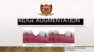
- 1. RIDGE AUGMENTATION PRESENTED BY- DR. NIKITA CHHABARIYA
- 2. CONTENT • Introduction • Classification • Ridge augmentation materials • Ridge augmentation procedures in • Mandible • Maxilla • Conclusion • References
- 3. INTRODUCTION Definition- Any procedure designed to enlarge or increase the size, extent, or quality of a deformed alveolar ridge.( GPT-9)
- 4. RIDGE AUGMENTATION • Onlay bone grafting – Autogenous / allogenic grafts • Interpositional / sandwich grafts • Sinus lift procedure Maxillary augmentation • Superior border augmentation (Iliac crest, rib graft, hydroxyapa tite) • Inferior border augmentation (Autogenous or allogenic freeze dried cadaveric mandible) • Interpositional / sandwich bone grafts • Visor osteotomy • Onlay grafting: Autogenous, allograft and alloplastic Mandibular augmentatio
- 5. CLASSIFICATION OF ALVEOLAR RIDGES AND TREATMENT PROTOCOL Class I -Alveolar ridge adequate in height but inadequate in width, usually with lateral deficiency or undercut areas: Patients received hydroxylapatite (HA) alone. Class II -Alveolar ridge deficient in both height and width presents a knife-edge appearance: Patients received HA alone. Class III- Alveolar ridge resorbed to level of the basilar bone, producing concave form on posterior areas of the mandible and a sharp, bony, ridge form with bulbous, mobile soft tissue in the maxilla: Patients received HA alone or HA mixed with autogenous, cancellous iliac bone. Class IV- Resorption of the basilar bone, producing pencil-thin, flat mandible or flat maxilla: Patients received HA mixed with autogenous, cancellous iliac bone Kent, J. N., Quinn, J. H., Zide, M. F., Guerra, L. R., & Boyne, P. J. (1983). Alveolar ridge augmentation using nonresorb hydroxylapatite with or without autogenous cancellous bone. Journal of Oral and Maxillofacial Surgery, 41(10), 629– 642. doi:10.1016/0278-2391(83)90016-2
- 6. • Loss of alveolar ridge height, width and decreased vestibular depth and denture bearing area. • Considerable basal bone resorption in the mandible, resulting in neurosensory disturbances. • Increased susceptibility to fracture of the atrophic jaws. Replacement of necessary supportive bone. • Progressive loss of denture stability and retention Indications for Ridge Augmentation Kent, J. N., Quinn, J. H., Zide, M. F., Guerra, L. R., & Boyne, P. J. (1983). Alveolar ridge augmentation using nonresor hydroxylapatite with or without autogenous cancellous bone. Journal of Oral and Maxillofacial Surgery,
- 7. Graft: Portion of a tissue or organ that after removal from its origin or donor site is positioned or inserted at a different place with the objective of reinforcing the existing tissues &/or correcting a structural defect.
- 8. Classification According to structure Cortical Cancellous Cortico- cancellous According to source Autograft Allograft Xenograft Alloplast According to embryologic origin Membranous Endochondral Fonseca, R.J. and Davis, W.H. Reconstructive Preprosthetic Oral and Maxillofacial Surgery. W.B. Saunders Co., Philadelphia,
- 10. SUPERIOR BORDER AUGMENTATION INDICATED • when severe resorption of the mandible results in inadequate height and contour and potential risk of fracture • when the treatment plan calls for placement of implants in areas of insufficient bone height or width. • mental foramen is situated in the superior border • Remaining bone < 10 mm Davis in the year 1970 Fonseca, R.J. and Davis, W.H. Reconstructive Preprosthetic Oral and Maxillofacial Surgery. W.B. Saunders Co., Philadelphia, 1986
- 11. PROCEDURE • Disadvantage 1. Morbidity of the donor site 2. Secondary surgical site 3. Necessity of the patient to withdraw denture till the surgical wound heals for period of 6-8 months autogenous bone graft is used. • Two segments of the rib, about 15 cm long, are obtained from the 5thand 9th ribs. • The rib is contoured by vertical scoring in the inner surface. • The second rib is made of corticocancellous paticles and mould around first rib. • Fixation is done by means of transosseous wiring or circumferential wiring. Fonseca, R.J. and Davis, W.H. Reconstructive Preprosthetic Oral and Maxillofacial Surgery. W.B.
- 12. INFERIOR BORDER AUGMENTATION OF MANDIBLE Indications: • severely atrophic edentulous mandible are a need for augmentation • prevention of pathologic fracture. • reduction and fixation (management) of nonunion or malunion in cases of fractures. Sanders and cox Sanders, B. (1982). Augmentation rib grafting to the inferior border of the mandible. Head & Neck
- 13. ADVANTAGES No dehiscence Less pain Better # stabilization 2o sulculoplasty not required Less bone resorption Indicated for pencil thin ridges Easier to perform skin graft vestibuloplasty DISADVANTAGES Scarring Presence of loose submandibular tissue Does not correct superior irregularities Sanders, B. (1982). Augmentation rib grafting to the inferior border of the mandible. Head & Neck
- 14. CASE REPORT- SANDERS, B. (1982). AUGMENTATION RIB GRAFTING TO THE INFERIOR BORDER OF THE MANDIBLE. HEAD & NECK SURGERY, • 31 patients have undergone inferior border rib grafting. Sanders, B. (1982). Augmentation rib grafting to the inferior border of the mandible. Head & Neck Article- Patient unable to wear satisfactory lower denture because of severely atrophic edentulous mandible. Preoperative appearance of alveolar ridge. lack of denture-bearing surface.
- 15. Alveolar ridge after skin-graft vestibuloplasty performed six months after rib grafting. Mandibular denture with extended flanges for excellent stability and retention. Results Two patients did not require vestibuloplasty. In one other patient, in whom the graft was used to support a mandible with a subperiosteal implant, no further treatment was deemed necessary. To date, all patients who had vestibuloplasty and those that did not require vestibuloplasty are wearing dentures satisfactorily Bone resorption rates to date have been minimal to moderate.
- 16. HORIZONTALOSTEOTOMY (DANIELSON AND NEMARICH)/SANDWICH TECHNIQUE Splitting of maxilla and mandible bone is grafted into osteotomy gap Can be used with osteointegrated implant Tucker, R.M. Ambulatory Preprosthetic Reconstructive Surgery
- 17. SEGMENTAL SANDWICH OSTEOTOMY AND TUNNEL TECHNIQUE FOR THREE-DIMENSIONAL RECONSTRUCTION OF THE JAW ATROPHY: A CASE REPORT • Case report - A 59-year-old woman with a severely atrophied right mandible was treated with the sandwich osteotomy technique filled with autologous bone graft harvested by a cortical bone collector from the ramus. International Journal of Implant Dentistry Mario Santagata et.al Article Clinical examination revealed that the mandible was edentulous bilaterally from the first molar to the second molar region. Radiographically, atrophy of the mandibular alveolar ridge in the same teeth site was observed- began to treat the right side.
- 18. • The entire bone fragment was displaced cranially, and the desirable position was obtained. • The gap was filled completely with autologous bone chips harvested from the mandibular ramus through a cortical bone collector. • No barrier membranes were used to protect the grafts. • The vertical incisions were closing with interruptive suturing of the flaps with a resorbable material.
- 19. VERTICAL OSTEOTOMY (HARLE,1975)/VISOR OSTEOTOMY 1. Was originated by Harle and modified by Peterson and Slade. 2. It is used where insufficient vertical mandibular bone height is present for the horizontal osteotomy technique but adequate bone width (approximately 10mm) is present. Tucker, R.M. Ambulatory Preprosthetic Reconstructive Surgery
- 20. 1.The mandible is split vertically and the lingual section is elevated to increase the mandibular height. 2. Cancellous bone or particulate bone and marrow is placed to correct the contours and fill in the gaps on the facial side of the elevated segment. 3.Transosteal wires hold the segments in place for a period of 3-4 months before vestibuloplasties are performed. The disadvantage is unavoidable nerve trauma and the resultant parasthesia. Tucker, R.M. Ambulatory Preprosthetic Reconstructive Surgery
- 21. COMBINED VERTICALAND HORIZONTAL OSTEOTOMY (KOOMEN ET AL) The combination of the ‘visor’ and ‘sandwich’ techniques was designed to over come the disadvantages in bone grafting. A modification of the visor osteotomy has been recommended for patients with at least 8 mm of bone height as measured at mental nerve region. the viability of the bone will be greater and the resorption decreased Stoelinga, P.J.W. Preprosthetic Reconstructive Surgery.
- 22. Procedure: • Vertical osteotomy cut is made in the posterior region to divide the segments buccolingually. • A horizontal osteotomy is performed in the anterior mandible to divide the anterior segment superiorly and inferiorly, and bone grafting was done into the osteotomised gap. • Two osteotomised segments are fixed with wires. Stoelinga, P.J.W. Preprosthetic Reconstructive Surgery.
- 23. ONLAY GRAFTING- • This procedure helps in increasing width of the ridge. Here graft material is placed on the buccal cortex either in putty form by mixing with saline/blood or in the form of blocks or split thickness rib/illiac crest graft. Tunnel is created via midline. Advantages: Improves width and to some extent height too. Can be used in anterior and posterior region. Stoelinga, P.J.W. Preprosthetic Reconstructive Surgery.
- 24. MAXILLARY AUGMENTATION Gross, B. D., James, R. B., & Fister, J. (1980). Use of pocket inlay grafts and tuberoplasty in maxillary prosthetic construction. The Journal of Prosthetic Dentistry, 43(6), 649–653. doi:10.1016/0022-3913(80)90381-9
- 25. • Advantages Development of increased height and form of the alveolar ridge and the palatal vault area. The anteroposterior position of the maxilla can be corrected. Disadvantages Need for a secondary donor site. Extensive post operative resorption. Postoperative secondary soft tissue procedures. Delay in wearing dentures for 6-8 months
- 26. Inter positional bone grafts Indications •In a bony deficient maxilla where there is adequate form to the palatal vault but insufficient ridge height, particularly in the zygomatic buttress and posterior tuberosity areas. Advantages •Stable and predictable results by changing maxillary position in the vertical, anteroposterior and transverse directions. Disadvantages •Need to harvest bone from the iliac bone crest •Possible secondary soft tissue surgery This technique effectively increases the ridge height from the lateral maxillary area to the crest of the ridge. lateral maxillary and lateral nasal walls and pterygoid maxillary suture area
- 27. Procedure • The lateral maxillary and lateral nasal walls and pterygoid maxillary suture area separated using surgical saws and osteotome and the maxilla is down fractured. • Bone grafts obtained from the iliac crest are shaped and wired in place in the lateral maxillary areas.
- 28. SINUS LIFT PROCEDURES - Riben C, Thor A. The Maxillary Sinus Membrane Elevation Procedure: Augmentation of Bone around Dental Implants without Grafts-A Review of a Surgical Technique. During long-term edentulism, resorption of the alveolar process occurs. Since the maxillary sinus also pneumatises during these circumstances the remaining bone volume can become very small and therefore clinicians and researchers have continuously developed techniques to overcome this problem. Increased bone volume in the maxillary sinus floor in order to enable installation of fixtures in the region
- 29. SUBANTRAL CLASSIFICATION AND TREATMENT OPTIONS Misch organized treatment options depending on available bone height Vlassis JM, Fugazzotto PA. A classification system for sinus membrane perforations during augmentation procedures with options for repair. J
- 30. INDIRECT SINUS AUGMENTATION TECHNIQUE sinus floor is lifted carefully by fracture of floor, separated from the Schneiderian membrane without damage to the membrane using a surgical mallet with controlled force. If required, autogenous graft material is inserted within the socket. The material is gradually displaced apically with the help of larger-diameter instruments. Riben C, Thor A. The Maxillary Sinus Membrane Elevation Procedure: Augmentation of Bone around Dental Implants without Grafts-A Review of a Surgical Technique. Int J Dent 2012;2012:105483.
- 31. DIRECT SINUS AUGMENTATION TECHNIQUE Autogenous bone grafts are harvested by shaving the mandibular bone from external oblique ridge area or parasymphysis region. Buccal window The implant can be placed on same sitting with the help of a stent for appropriate faciolingual and mesiodistal positioning. Vlassis JM, Fugazzotto PA. A classification system for sinus membrane perforations during augmentation procedures with options for repair. J Periodontol 1999;70:692-9
- 32. ONLAY AND INLAY AUGMENTATION • Indications: Severe resorption of the maxillary alveolar resulting in the absence of a clinical alveolar ridge and loss of adequate palatal vault form.
- 33. USE OF POCKET INLAY GRAFTS AND TUBEROPLASTY IN MAXILLARY PROSTHETIC CONSTRUCTION. THE JOURNAL OF PROSTHETIC DENTISTRY. • A white man, 59 years of age, was referred to the oral/maxillofacial surgery clinic for evaluation of his deficient maxilla. Gross, B. D., James, R. B., & Fister, J. (1980). Article Past medical history- had sustained a war injury to his face in 1944 which resulted in the avulsion of most of the maxillary alveolus and the entire hard palate Physical examination was unremarkable, except for the intraoral findings. Bilaterally, there was no clinically discernable alveolar ridge or tuberosity. Palpation revealed only a rim of bone in the area of the lateral wall of the maxillary sinus.
- 34. Pocket inlay skin grafts would be developed bilaterally in the region of the anterior lateral sinus walls, and bilateral tuberoplasties would be accomplished in the pterygoid region. PROCEDURE- Preoperatively, an overextended impression was made of the maxilla. The diagnostic cast was then marked to delineate the proposed surgical sites. With the patient present, the angulation and dimensions of the pockets and tuberoplasty were carved into the diagnostic cast. Acrylic resin was added to the patient’s dentures in these regions. Modeling compound was added at surgery for the final contour.
- 35. The original denture was modified using autopolymerizing acrylic resin and modeling compound to fit into the previously created pockets and the space created behind the tuberosity . The splitthickness skin graft was then draped over the prepared denture, and the denture was inserted into the prepared surgical sites. Stabilization was achieved with bilateral circumzygomatic wires. Ten’ days later the denture was removed, and the surgical sites were cleaned. At this time, a cast was made using the modified denture. The compound was replaced with acrylic resin. An impression tray was constructed from the cast. The denture was immediately reinserted,
- 37. TITANIUM MESH FOR BONE AUGMENTATION When the alveolar bone has severe vertical or horizontal bone defects, many clinical studies suggest that titanium mesh shows superior mechanical properties and great osteogenic performance during application Use of titanium mesh for staged 3D localized alveolar ridge augmentation Sajith 2015 int joral health The use of Ti-mesh allows the regeneration of sufficient bone volume for ideal implant placement. The clinical advantages related to this technique include the possibility of correcting severe vertical atrophies associated with considerable reductions in width and the lack of major complications if soft- tissue dehiscence and mesh exposures do occur.
- 38. CONCLUSION • Prosthetic-driven augmentation is recommended for a better outcome. If the clinician focuses only on ridge augmentation techniques to solve bone deficiency problems, he or she may overlook other treatment options that may have lower risks and less morbidity, such as using short, narrow, or tilted implants. After all, ridge augmentation is being performed for the ideal placement of dental implant.
- 39. REFERENCES • Peterson, L. J. Principles of Oral and Maxillofacial Surgery. Vol. II. J. B. Lippincott Co., Philadelphia, 1992. • Zarb, G. Prosthodontic View of Traditional and Contemporary Preprosthetic Surgery. Chapter 42, • Tucker, R.M. Ambulatory Preprosthetic Reconstructive Surgery. Chapter 43, • Stoelinga, P.J.W. Preprosthetic Reconstructive Surgery. • Fonseca, R.J. and Davis, W.H. Reconstructive Preprosthetic Oral and Maxillofacial Surgery. W.B. Saunders Co., Philadelphia, 1986. • Kent, J. N., Quinn, J. H., Zide, M. F., Guerra, L. R., & Boyne, P. J. (1983). Alveolar ridge augmentation using nonresorbable hydroxylapatite with or without autogenous cancellous bone. Journal of Oral and Maxillofacial Surgery, 41(10), 629–642. doi:10.1016/0278-2391(83)90016- 2 • Proussaefs, P., & Lozada, J. (2003). The use of resorbable collagen membrane in conjunction with autogenous bone graft and inorganic bovine mineral for buccal/labial alveolar ridge augmentation: A pilot study. The Journal of Prosthetic Dentistry, 90(6), 530– 538. doi:10.1016/s0022-3913(03)00521-3 • J Pharm Bioallied Sci. 2012 Aug; 4(Suppl 2): S414–S416. • dPre-prosthetic surgery: Mandible 10.4103/0975-7406.100312 PMCID: PMC3467894 jpd • Sandwich bone graft for vertical augmentation of the posterior maxillary region: a case report with 9-year follow-up Kenko Tanaka j.international implant • Riben, C., & Thor, A. (2012). The Maxillary Sinus Membrane Elevation Procedure: Augmentation of Bone around Dental Implants without Grafts—A Review of a Surgical Technique. International Journal of Dentistry, 2012, 1–9.
- 40. THANK YOU STAY SAFE AND HEALTHY