The pancrease.pptx
•Download as PPTX, PDF•
0 likes•106 views

The document summarizes the anatomy and histology of the pancreas. It describes the pancreas as having both exocrine and endocrine functions. The pancreas has five parts - the head, uncinate process, neck, body, and tail. It is supplied by branches of the splenic artery and drains into the splenic and portal veins. The pancreas contains acini that secrete enzymes into a duct system that empties into the duodenum. Islets of Langerhans contain endocrine cells that secrete hormones like insulin. Common clinical correlates of the pancreas include pancreatitis, which can be caused by factors like gallstones, alcohol use, and genetic mutations.

Recommended
More Related Content
What's hot
What's hot (20)
Similar to The pancrease.pptx
Similar to The pancrease.pptx (20)
More from Dr Ndayisaba Corneille
More from Dr Ndayisaba Corneille (20)
Recently uploaded
Recently uploaded (20)
The pancrease.pptx
- 1. Dr. NDAYISABA CORNEILLE CEO of CHG MBChB,DCM,BCSIT,CCNA Supported BY
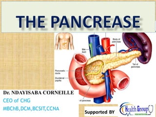
- 2. • The pancreas is an abdominal glandular organ with both digestive (exocrine) and hormonal (endocrine) functions. • The pancreas is an oblong-shaped organ positioned at the level of the transpyloric plane (L1). With the exception of the tail of the pancreas, it is a retroperitoneal organ, located deep within the upper abdomen in the epigastrium and left hypochondrium regions. • Within the abdomen, the pancreas has direct anatomical relations to several structures • Organs and vesselsVessels Dr Ndayisaba Corneille 2
- 3. • The pancreas lies near several major vessels and significant landmarks in vascular anatomy: • Stomach – Separated from the pancreas by the lesser sac, the stomach and pylorus lie anterior and to the pancreas. • Duodenum – The “C” shaped duodenum curves around and outlines the head of the pancreas. The first part of the duodenum lies anteriorly whereas • the second part of the duodenum including the ampulla of Vater lies laterally to the right of the pancreatic head Dr Ndayisaba Corneille 3
- 4. • Transverse mesocolon – Attaches to the anterior surface of the pancreas • Common bile duct – Descends behind the head of the pancreas before opening into the second part of the duodenum alongside the major pancreatic • duct through the major duodenal papilla • Spleen – located posteriorly and laterally. The lienorenal ligament is formed from peritoneum and connects the spleen to the tail of the pancreas. • The aorta and inferior vena cava pass posteriorly to the head of the pancreas. • The superior mesenteric artery lies behind the neck of the pancreas and anterior to the uncinate process. • Posterior to the neck of the pancreas, the splenic and superior mesenteric veins unite to form the hepatic portal vein. • As it journeys from its origin at the celiac plexus to the splenic hilum, the splenic artery traverses the superior border of the pancreas. Dr Ndayisaba Corneille 4
- 5. Embryology of the Pancreas • Derived from endodermal lining of the duodenum. • Formed from 2 buds (dorsal & ventral bud) • Initially dorsal bud is found in the dorsal mesogastrium & ventral bud lies close to bile duct but after rotation of the gut, ventral bud lies behind the dorsal bud, later their parenchyma & duct systems fuse. • Ventral bud forms the uncinate process&inferior part of the head of pancreas. Dr Ndayisaba Corneille 5
- 7. Cont’d • Main pancreatic duct of wirsung is formed from ventral bud &distal part of the dorsal pancreatic duct. • Proximal part of the duct either obliterates or persists as accessory pancreatic duct of Santorin. • Main duct & bile duct enter the duodenum at the main papilla. • Accessory duct if present enters at the minor papilla. Dr Ndayisaba Corneille 7
- 8. Cont’d • 10% of all cases the duct system fails to fuse &the double duct system persists. • Islets of langerhans develop in the 3rd month. • Insulin secretion begins by 5th month. • Glucagon & somato statin secreting cells also appear. Dr Ndayisaba Corneille 8
- 9. C L I N I C A L C O R R E L A T E S • Pancreatic Abnormalities • An annular pancreas is formed when the right portion of ventral bud migrates along the normal route but the left portion moves in the opposite direction hence duodenum gets surrounded by pancreatic tissue. This malformation constricts the duodenum and causes complete obstruction. Dr Ndayisaba Corneille 9
- 10. C L I N I C A L C O R R E L A T E S • Accessory pancreatic tissue; occur anywhere from the distal end of the esophagus to the tip of the primary intestinal loop. Commonly lies in the mucosa of the stomach and in Meckel’s diverticulum. Dr Ndayisaba Corneille 10
- 11. Anatomical Structure • The pancreas is typically divided into five parts: • Head – the widest part of the pancreas. It lies within the C-shaped curve created by the duodenum and is connected to it by connective tissue. • Uncinate process – a projection arising from the lower part of the head and extending medially to lie beneath the body of the pancreas. It lies posterior to the superior mesenteric vessels. • Neck – located between the head and the body of the pancreas. It overlies the superior mesenteric vessels which form a groove in its posterior aspect.Duct System • The exocrine pancreas is classified as a lobulated, serous gland which produces digestive enzyme precursors. It is composed of approximately one million ‘berry-like’ clusters of cells called acini, connected by short intercalated ducts. • The intercalated ducts unite with those draining adjacent lobules and drain into a network of intralobular collecting ducts, which in turn drain into the main pancreatic duct. Dr Ndayisaba Corneille 11
- 12. Dr Ndayisaba Corneille 12
- 13. Dr Ndayisaba Corneille 13
- 14. • The pancreatic duct runs the length of the pancreas and unites with the common bile duct, forming the hepatopancreatic ampulla of Vater. This structure then opens into the duodenum via the major duodenal papilla. • Secretions into the duodenum are controlled by a muscular valve – the sphincter of Oddi. It surrounds the ampulla of Vater, acting as a valve. • Body – centrally located, crossing the midline of the human body to lie behind the stomach and to the left of the superior mesenteric vessels. • Tail – the left end of the pancreas that lies within close proximity to the hilum of the spleen. It is contained within the splenorenal ligament with the splenic vessels. This is the only part of the pancreas that is intraperitoneal. Dr Ndayisaba Corneille 14
- 15. HISTOLOGY • The pancreas if invested by a thin loose collagenous capsule which extends its septa into the substance of the pancreatic tissue to form septa. • Septa form the boundaries of lobules. • Exocrine part consists of closely packed secretory acini which drain into a highly branched duct system. Dr Ndayisaba Corneille 15
- 16. Histology cont… • The different small ducts from the acini join to form interlobular ducts, which in turn join to form the main pancreatic duct. • The main pancreatic duct drains into the duodenum together with the bile duct. • A small accessory pancreatic duct drains into the duodenum proximally. Dr Ndayisaba Corneille 16
- 17. Histology • The endocrine tissue of the pancreas forms Islets of Langerhans which are of various sizes and scattered throughout the exocrine tissue. Dr Ndayisaba Corneille 17
- 18. Dr Ndayisaba Corneille 18
- 19. Dr Ndayisaba Corneille 19
- 20. Vasculature • The pancreas is supplied by the pancreatic branches of the splenic artery. The head is additionally supplied by the superior and inferior pancreaticoduodenal arteries which are branches of the gastroduodenal (from coeliac trunk) and superior mesenteric arteries, respectively. • Venous drainage of the head of the pancreas is into the superior mesenteric branches of the hepatic portal vein. The pancreatic veins draining the rest of the pancreas do so via the splenic vein. Dr Ndayisaba Corneille 20
- 21. • . Dr Ndayisaba Corneille 21
- 22. Lymphatics • The pancreas is drained by lymphatic vessels that follow the arterial supply. They empty into the pancreaticosplenal nodes and the pyloric nodes, which in turn drain into the superior mesenteric and coeliac lymph nodes Dr Ndayisaba Corneille 22
- 23. NERVE SUPPLY • parasympathetic supply through posterior vagal trunk and coeliac. • However, hormonal control of secretion is more important than neural. • Sympathetic vasoconstrictor impulses are from spinal cord T6 to T10 via splachnic nerve.. Dr Ndayisaba Corneille 23
- 24. Clinical Relevance: Pancreatitis • Pancreatitis refers to inflammation of the pancreas – this is can be acute or persist over an extended period (chronic pancreatitis). The causes of • pancreatitis can be remembered using the mnemonic – GET SMASHED: • Gall stones • Ethanol • Trauma • Steroids • Mumps • Autoimmune • Scorpion stings • Hypertriglyceridemia, hypercalcaemia and hyperparathyroidism • ERCP – endoscopic retrograde cholangiopancreatography • Drugs – such as sodium valproate, azathioprine and sulphonamide • Pancreatitis creates severe epigastric pain which often radiates to the back, nausea, vomiting and diarrhoea. • Treatment involves supportive measures such as IV fluids and analgesia. Antibiotics are rarely required, as most cases are not due to infection. • The underlying cause will then also need to be treated. Dr Ndayisaba Corneille 24
- 25. • Hepatopancratic ampulla blockage • Rapture of the pancrease. • Pancreatic cancer. Dr Ndayisaba Corneille 25
- 26. END Dr Ndayisaba Corneille THANKS FOR LISTENING By DR NDAYISABA CORNEILLE MBChB,DCM,BCSIT,CCNA Contact us: amentalhealths@gmail.com/ ndayicoll@gmail.com whatsaps :+256772497591 /+250788958241 26