
Recommended
More Related Content
What's hot
What's hot (20)
Similar to introduction of shalakya .pptx
Similar to introduction of shalakya .pptx (15)
introduction of shalakya .pptx
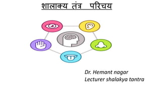
- 1. शालाक्य तंत्र परिचय Dr. Hemant nagar Lecturer shalakya tantra
- 2. निरुनि – शलाकया यत्कर्म क्रियते तत् शालाक्यं परिभाषा – शालाक्यं िामोर्ध्वजत्रुगतािां श्र्णियि्दिघ्राणानदसंनश्रतािां व्याधीिामुपशमिार्वम् (सु.सू.1) शालाक्यं तंत्र मे जत्रु अनथर् के ऊपि - श्र्ण (कणव) , िेत्र , मुख , िासा ,नशि आनद के आनश्रत िोगों तर्ा उिकी निनकत्सा का ्णवि आता है ।
- 3. संख्या सुश्रुत वाग्भट शार्मधर योर् रत्नाकर नेत्र रोर् 76 94 94 76 कर्म रोर् 28 25 18 28 नासा रोर् 31 18 18 34 र्ुख रोर् 65 75 74 67 क्रशरो रोर् 11 10 10 10
- 4. इनतहास - शालाक्य तंत्र उपदेष्टा – क्रवदेह राज क्रनर्ी - अष्टांर् आयुवेद र्े चरक और सुश्रुत क्रितीय तथा वार्भट चतुथम स्थान (ऊर्ध्वंर् क्रचक्रकत्सा) पर रखता है - वैक्रदक काल र्े शालाक्य तंत्र के कही वर्मन क्रर्लते है -ऋग्वेद र्े अक्रिनी कुर्ार ने दधीक्रच ऋक्रि के क्रशर का संधान क्रकया , नृपद पुत्र के बहरेपन को दूर क्रकया प्रावृक के अंधेपन को नाश क्रकया - चरक संक्रहता काय क्रचक्रकत्सा प्रधान है इसर्े कही कही शालाक्य का वर्मन प्राप्त ह होता है o चरक सूत्र – 17 व 18, चरक क्रचक्रकत्सा 9,10,12,26 चरक क्रसद्धी – 9 अर्ध्याय - सुश्रुत संक्रहता के उत्तर तंत्र र्े शालक्य तंत्र का क्रवस्तृत वर्मन क्रर्लता है o उत्तर तंत्र 1 से 19 अर्ध्याय- नेत्र रोर् क्रचक्रकत्सा o उत्तर तंत्र 20 से 21 अर्ध्याय – कर्म रोर् क्रचक्रकत्सा
- 5. o उत्तर तंत्र 22 से 24 अर्ध्याय – नासा रोर् क्रचक्रकत्सा o उत्तर तंत्र 25 से 26 अर्ध्याय – क्रशरो रोर् क्रचक्रकत्सा o क्रनदान स्थान – 16 अर्ध्याय – र्ुख रोर् क्रचक्रकत्सा - अष्टांर् हृदय ने उत्तर तंत्र के अर्ध्याय 8 से 24 तक शालाक्य तंत्र का वर्मन क्रकया है - अष्टांर् संग्रह ने क्रचक्रकत्सा स्थान के अर्ध्याय 9 से 28 तक शालाक्य का वर्मन क्रकया है - शार्मधर संक्रहता (13 वी सदी ) o पूवम खंड – नेत्र,कर्म, नासा, क्रशरो रोर् पररचय o र्र्ध्यर् खंड – औिध क्रनर्ामर् o उत्तर खंड – 8 अर्ध्याय – नस्य , 9 अर्ध्याय- धूर्पान ,10 अर्ध्याय- र्ंडूि कवल - भाव प्रकाश के उत्तर खंड र्े शालाक्य का वर्मन क्रर्लता है - र्ाधव क्रनदान के अर्ध्याय 56,57,58,59,60 र्े शालाक्य का वर्मन प्राप्त ह है - आधुक्रनक काल र्े शालाक्य तंत्र र्े क्रनम्न क्रवभार् है
- 6. o नेत्र क्रचक्रकत्सा - ophthalmology o कर्म नासा र्ल रोर् – ENT o र्ुख दांत रोर् क्रचक्रकत्सा – dentistry o क्रशरो रोर् क्रचक्रकत्सा – head and neurology
- 7. नेत्र रोग - ननदान पंचक सामान्य हेतु उष्णाभितप्तस्य जलप्रवेशाद्दूरेक्षणात्स्वप्नववपयययाच्च प्रसक्तसंरोदनकोपशोकक्लेशाभिघातादनतमैथुनाच्च || शुक्तारनालाम्लक ु लत्थमाषननषेवणाद्वेगववननग्रहाच्च | स्वेदादथो धूमननषेवणाच्च छदेववयघाताद्वमनानतयोगात्| बाष्पग्रहात्सूक्ष्मननरीक्षणाच्च नेत्रे ववकारान्जनयन्न्त दोषााः ||(सु.सू २६-२७) विहािाज निदाि – उष्ण से तप्त होकि अर्ाात् गर्मी से तप कि जल र्मे स्िाि आदद क े ललए जािा,दूि लगाताि देखिा, ददि र्मे सोिे या िात्री र्मे जागिण (स्िप्िविपयायाद्) धूर्म ग्रहण, छदी िेगधािण, िर्मि अनतयोग,िाष्पिेगधािण,सूक्ष्र्मिस्तुयों को लगाताि देखिे से आहिज निदाि – शुक्त,आििाल,अम्ल,क ु लत्र्,र्माष आदद क े सेिि से र्मािलसक निदाि – संिोदि,कोप,शोक,क्लेश,अनतर्मैर्ुि कििे से
- 8. सामान्य पूवयरूप तत्राववलं ससंरम्िमश्रुकण्डूपदेहवत्। गुरूषातोदरागाद्यैजुयष्टं चाव्यक्तलक्षणैाः।। सशूलं वत्मयकोषेषु शूकपूणायिमेव च । ववहन्यमानं रूपे वा क्रियास्वक्षक्ष यथा पुरा ।। (सु.सू २१-२३) िेत्र आविल,शोफ,अश्रु ि उपदेह (र्मलललप्त) होते है तर्ा गुरुता(कफ), तोद (िात), िाग(वपत्त) आदद त्रत्रदोष भाि अव्यक्त रूप र्मे होते है। ित्र्माकोष शुकपूणााभ ि शूलयुक्त होता है। रूप का िाश होिे लगता है ि अक्षि क्रियाये पहले की तिह िह ं िहती है ।
- 9. सामान्य संप्रान्प्त भसरानुसाररभि दोषै ववयगुणैरूर्धवयमागतैाः। जायन्ते नेत्रिागेषु रोगााः परमदारुणााः (सु.सू २०) निदाि सेिि िातआदद दोषप्रकोप लसिा से दोषगर्मि िेत्रभाग दोषस्स्र्त नेत्ररोग उत्पन
- 10. सामान्य चचक्रकत्सा सङ्क्क्षेपताः क्रियायोगो ननदानपररवजयनम्|वातादीनां प्रतीघाताः प्रोक्तो ववस्तरताः पुनाः(सु.सू २५) संिेप र्मे निदाि परििजाि ि विस्तित रूप र्मे िातआदद दोषिाशक चचक्रकत्सा कििा
- 11. • आकाि / size – द्द्ियङ्गुलबाहुल्यं – 2 अंगुल (स्ियं क े अंगूठे क े उदि् भाग से ) आयाम - द्व्यङ्गुलं सिातः साधं – 21 2 अंर्ुल = 5/2 अंर्ुल आकृ नत / Shape - सुिृत्तं गोस्तिाकािं संगठि / composition – सिाभूत / पञ्च र्महाभूत िेत्र शािीि न्द्याद्वयङ्गुलबाहुल्यं थ्ाङ्गुष्ठोदिसनममतम्| द्व्यङ्गुलं स्वतः साधं नभषङ्ियिबुद्बुदम् || सु्ृत्तं गोथतिाकािं स्वभूतगुणोद्भ्म् । (सु उ 1/10-11)
- 12. िेत्र बंधि द्रव्य नसिाणां कण्डिाणां ि मेदसः कालकथय ि । गुणाः कालात् पिः श्लेष्मा बन्धिेऽक्षणोः नसिायुतः। । - क्रसरा,कण्डिा ,र्मेद , कालकस्स्र् औि श्लेष्र्मा लर्मलकि अक्षि का बंधि किते है । िेत्र पञ्ि महाभूत संगठि पलं भु्ोऽननितो ििं ्ातात् कृ ष्णं नसतं जलात् आकाशादश्रुमागावच ज जायन्ते िेत्रबुद्बुदे । 1. भुवः (पृथ्िी द्िािा) - पलं (र्मांसंल भाग ) 2. अस्नि - िक्त भाग 3. जल – श्िेत भाग 4. वात – कृ ष्णं भाग 5. आकाश द्िािा - अश्रुर्मागा निर्मााण इस प्रकार पांचों र्हाभूत अक्रि के अलर् अलर् भार्ों का क्रनर्ामर् करते है ।
- 13. िेत्र मण्डल – 5 1. पक्ष्र् र्ण्डल 2. वत्र्म र्ण्डल 3. िेत र्ण्डल 4. कृष्र् र्ण्डल 5. दृक्रष्टर्ण्डल
- 14. पक्ष्म मण्डल - eye lash वत्मय मण्डल - eye lids श्वेत मण्डल- sclera कृ ष्ण मण्डल - cornea दृन्ष्टमण्डल - pupil , lens , retina
- 15. नेत्र संचध – 6 दो र्मण्डल जहां लर्मलते है िहां संचध बिाते है । क ु ल 6 संचधया होती है । 1. पक्ष्र्मित्र्मा संचध 2. ित्र्माशुक्ल संचध 3. शुक्लकृ ष्ण संचध 4. कृ ष्णदृस्ष्ि संचध 5. किीिक 6. अपांग संचध पक्ष्र्मित्र्मा संचध - lid margin ित्र्माशुक्ल – fornix शुक्लकृ ष्ण - limbus कृ ष्णदृस्ष्ि - pupillary margins किीिक संचध - inner canthus अपांग संचध को outer canthus
- 16. नेत्र पटल – 6 द्वे वत्मयपटले ववद्यात चत्वार् अन्यानन च अक्षक्षणण । क ु ल 6 पिल होते है - 2 ित्र्मा पिल + 4 अक्षि पिल 4 अक्षिपिल र्मे नतलर्मि व्याचध होती है । चाि अक्षि पिल इस प्रकाि है – तेजोजलाचश्रतं बाह्यं तेष्वन्यत् वपभशताचश्रतम्| मेदस्तृतीयं पटलमाचश्रतं त्वन्स्थ चापरम् (सु उ 1 ) 1. तेजोजल आचश्रत - बाह्य पिल/प्रर्र्म पिल 2. र्मांस आचश्रत – द्वितीय पिल 3. र्मेद आचश्रत - तृतीय पिल 4.अस्स्र् आचश्रत - अंत पिल/चतुर्ा पिल • प्रर्म पटल/बाह्य पटल – sclera and cornea • नवतीय पटल - uveal tissue • तृतीय पटल – Cortical part of lens • ितुर्व पटल – Nuclear part of lens
- 17. िेत्र के न्नभि भागों के आयाम सम्पूणा िेत्र का आयार्म – 2 ½ = 5/2 अंगुल कृ ष्णर्मण्डल आयार्म - िेत्र आयार्म का त्रत्रभागं - 5 2 * 1 3 = 5 6 अंगुल दृस्ष्िर्मण्डल आयार्म - कृ ष्णर्मण्डल का सप्तभागं - 5 6 * 1 7 = 5 42 अंगुल अक्षिपिल आयार्म - दृस्ष्िर्मण्डल का पञ्चभागं - 5 42 * 1 5 = 5 210 अंगुल
- 18. ANATOMY OF EYE The eye sits in a protective bony socket called the orbit. Six extraocular muscles in the orbit are attached to the eye. Dimensions of adult eyeball -Anteroposterior diameter – 24 mm -Horizontal DM – 23.5 mm -Vertical diameter – 23 mm -Circumference – 75 mm -Volume of eyeball – 6.5 ml -Weight of eyeball – 7 gm
- 19. Coats of Eyeball – 3 Outer fibrous coat, middle vascular coat, Inner nervous coat 1. Fibrous coat – 2 parts of fibrous coat -Anterior 1/6 part of is transparent -cornea -posterior 5/6 part is opaque -sclera -fibrous coat protect intraocular content 2. Vascular coat (uveal tissue) – 3 parts of uveal tissue from anterior to posterior – iris, ciliary body and choroid. -uveal tissue supplies nutrition of eyeball contents. 3. Nervous coat - Retina is part of nervous coat
- 20. Posterior segment -include structures posterior to lens viz. vitreous ,choroid, retina and optic disc. Anterior segment - include crystalline lens and structures anterior to it - Anterior segment divided into 2 chambers – anterior and posterior chamber. Anterior Chamber -bounded anteriorly by back of the cornea and posteriorly by iris and choroid’ -Deepness – 2.5mm -Content – 0.25 ml aqueous humour Posterior chamber - bounded anteriorly by posterior surface of iris and choroid and posteriorly by lens - Content – 0.06 ml aqueous humour. Segments and Chamber of eyeball
- 22. Layers of different structure of Eye Conjunctiva – 3 layers -epithelium, adenoid layer, fibrous layer Cornea – 6 layers – epithelium, bowman’s membrane, stroma, pre-Descemet’s membrane (dua’s layer), Descemet’s, endothelium. Sclera – 3 layers – Episcleral, sclera proper, lamina fusca. Iris – 4 layers – anterior limiting layer, iris stroma, anterior pigmented epithelial layer, posterior pigmented epithelial layer. Ciliary body – 5 layers – supraciliary lamina, stroma, layer of pigmented epithelium, layer of pigmented non-epithelium, internal limiting layer. Choroid – 3 layers – suprachoroidal lamina, stroma, basal lamina. Retina – 10 layers – pigmented epithelium, layer of rods and cones, external limiting membrane, outer nuclear layer, outer plexiform layer, inner nuclear layer, inner plexiform layer, ganglion layer, nerve fibre layer, internal limiting membrane. Arterial supply -Ophthalmic artery -central retinal artery Nerve supply - Trigeminal nerve Ophthalmic division -Oculomotor nerve - Trochlear nerve -Facial nerve
- 23. Physiological events of vision consists of following; Refraction of light entering the eye Focusing of image on the retina by accommodation of lens Convergence of image Photo-chemical activity in retina and conversion into neural impulse Processing in brain and perception PHYSIOLOGY OF VISION
- 24. Refraction of light entering the eye Light wave travels parallel to each other but they bend when passes from one medium to another. This phenomenon is called refraction. Before light reach retina it passes through cornea, aqueous humor, lens vitrous humor, so refraction takes place in every medium before it falls on retina. In normal eye, light wave focused on retina.
- 25. Accommodation is a reflex process to bring light rays from object into perfect focus on retina by adjusting the lens. For accommodation to view closer object, ciliary muscle contract and lens become thick which causes focus on closer object. Similarly, when distant object is viewed, ciliary muscles relaxes, so the tension of ligament become greater which pull lens and lens become thinner, due to which image forms on retina. The normal eye is able to accommodate light from object about 25 cm to infinity. Focusing of image on the retina by accommodation of lens
- 27. Convergence of image Human eye have binocular vision, it means although we have two eye, we perceive single image In binocular vision, two eye ball turns slightly inward to focus a close object so that both image falls on corresponding points on retina at same time. This phenomenon is called convergence.
- 28. Photochemical activity in rods: Photo-chemical activity in retina and conversion into neural impulse Each eye contains 125 million rods which contains light sensitive pigment-rhodopsin. The extra cellular fluids surrounding rod cells contains high concentration of Na+ ion and low concentration of K+ ions while concentration of Na+ is low and K+ is high inside rod cells. The concentration is maintained by Na-K pump Rhodopsin = scotopsin (protein) + retinal (carotenoid molecule) derivative of vitamin A. cis trans
- 29. In resting phase, K+ tends to move outside the rod cells creating slightly –ve charge inside. • light is falls on rod cell, it is absorbed by rhodopsin and it breaks into scotopsin and 11 cis- retinal. The process is known as bleaching. Light rod rhodopsin scotopsin+ 11 cis- retinal trans-retinal+ activate scotopsin • This reaction produces large amount of transducin which activates another enzyme phosphodiesterase.
- 30. • Phosphodiesterase causes to cease the flow of Na+ ion inside rod cell. This causes increased negative charge inside cell creating hyperpolarized state. • Hyperpolarized rod cells transmit the neural signal to bipolar cell. •Bipolar cell, amacrine cell and ganglion cell process the neural signal and generate action potential to transmit to brain via optic nerve.
- 31. Photochemical activity in cones: • Each eye contains 7 million cone cells. • The neural activity in cone cell is similar to that of rod cell but there are three different types of cone cells and each cone cell contains different photo-pigment and are sensitive to red, green and blue. •The final perceived color is combination of all three types of cone cell stimulated depending upon the level of stimulation. • cone cell iodopsin 11 cis-retinal + photopsin. •The perception of color depends upon which cone are stimulated.
- 32. Processing of image in brain and perception: •All visual information originates in retina due to stimulation of rods and cones are conveyed to brain. Processing of image in brain and perception