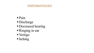
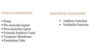
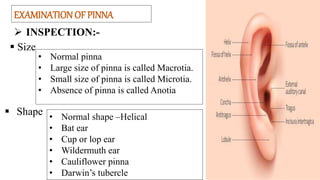
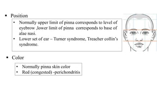
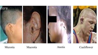
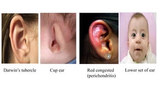
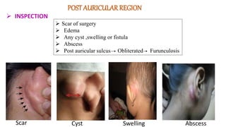
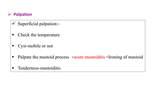
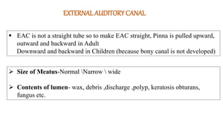
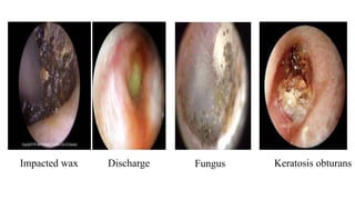
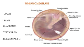
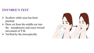
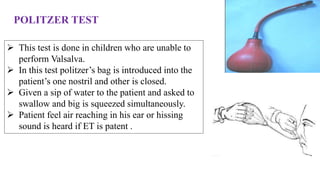
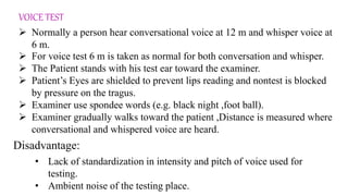
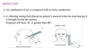
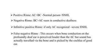
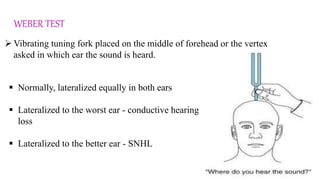
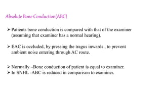
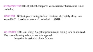
EXAMINATION OF EAR
Dr. Hemant Nagar
Dept of Shalakya Tantra
SYMTOMATOLOGY
 Pain
 Discharge
 Decreased hearing
 Ringing in ear
 Vertigo
 Itching
PHYSICAL EXAMINATION
 Pinna
 Pre-auricular region
 Post-auricular region
 External Auditory Canal
 Tympanic Membrane
 Eustachian Tube
FUNCTIONAL EXAMINATION
 Auditory Function
 Vestibular Function
EXAMINATION OF PINNA
 INSPECTION:-
 Size
 Shape • Normal shape –Helical
• Bat ear
• Cup or lop ear
• Wildermuth ear
• Cauliflower pinna
• Darwin’s tubercle
• Normal pinna
• Large size of pinna is called Macrotia.
• Small size of pinna is called Microtia.
• Absence of pinna is called Anotia
 Position
• Normally upper limit of pinna corresponds to level of
eyebrow ,lower limit of pinna corresponds to base of
alae nasi.
• Lower set of ear – Turner syndrome, Treacher collin’s
syndrome.
 Color
• Normally pinna skin color
• Red (congested) -perichondritis
Anotia
Microtia Macrotia Cauliflower
Darwin’s tubercle Cup ear Red congested
(perichondritis)
Lower set of ear
 PALPATION
 Digital Palpation -
 Cartilage and soft tissue -normal or any defect
 Mobility of skin -(It is lost in malignancy)
 Temperature –raised in inflammation
 Tragus sign- tenderness- furunculosis
Pre-Auricular region
Pre auricular sinus
Accessory tragus Cyst
Observe the presence or absence of
 Pre auricular sinus
 Accessory tragus
 Cyst
POST AURICULARREGION
 INSPECTION
Scar Cyst Abscess
 Scar of surgery
 Edema
 Any cyst ,swelling or fistula
 Abscess
 Post auricular sulcus Obliterated Furunculosis
Swelling
 Palpation
 Superficial palpation:-
 Check the temperature
 Cyst-mobile or not
 Palpate the mastoid process acute mastoiditis Ironing of mastoid
 Tenderness-mastoiditis
EXTERNAL AUDITORY CANAL
 EAC is not a straight tube so to make EAC straight, Pinna is pulled upward,
outward and backward in Adult
Downward and backward in Children (because bony canal is not developed)
 Size of Meatus-Normal Narrow  wide
 Contents of lumen- wax, debris ,discharge ,polyp, keratosis obturans,
fungus etc.
Discharge:-
• Consistency- Stringy discharge
-Non-stringy discharge
• Smell- Foul smelling discharge-indicate unsafe type of OM
-Non foul smelling discharge –indicate safe type of OM
• Quantity- profuse discharge
-moderate discharge
-scanty discharge
Impacted wax Discharge Fungus Keratosis obturans
TYMPANICMEMBRANE
COLOR
SHAPE
QUADRANTS
VERTICAL DM
HORIZONTAL DM
Quadrants of tympanic membrane:-
 Tympanic membrane divided into 4 parts by drawing an imaginary
line.
 One imaginary line extending downward from handle of malleus and
one more at the right angle to the first line at the umbo
1. Anterosuperior
2. Anteroinferior
3. Posteroinferior
4. posterosuperior
 Any pathology of TM should be mentioned
quadrant wise
COLOR OF T.M.
 Pearly white - Normal
 Red (congested) Drum - ASOM, Glomus tumor
 Blue Drum -Hemotympanum
 Pink Drum -Otospongiosis
 Chalky Drum-Tympanosclerosis
Red (Congested)ear drum Blue ear drum Chalky ear drum
RISING SUN SIGN:-
 Red vascular hue seen behind the intact
tympanic membrane
 Glomus tumour, high jugular bulb
SCHWARTZ SIGN – FLAMINGO PINK BLUSH:-
 In active phase of otosclerosis.
 It is seen because of increased
vascularity in sub-mucus layer of
promontory.
POSITION OF T.M.
Normal -Inclined downward and medially
Bulged TM -acute otitis media
-Hemotympanum
-OME with good ET function
Retracted TM -OME with poor ET function
-ET obstruction
Retraction Pocket – Usually in Attic region
annulus
PERFORATION
Central (in pars tensa)
-small size
-medium size
-subtotal
-total
Attic (in pars flaccida)
Marginal (at the periphery involving annulus)
SURFACE OF TM
hkjklhjghglhjhjjkg
gkhgkhkhkk
Small size central Medium size central Subtotal perforation
Total perforation Attic perforation Marginal perforation
Siegles speculum is used:-
 Normally tympanic membrane is mobile
 Reduced or immobility of T.M. -Any fluid in
middle ear
-tympanosclerosis
-perforation
LIGHT HOUSE SIGN:-
 small pin hole perforation of T.M. with pulsatile ear discharge
 seen in ASOM
MOBILITY OF T.M.
EUSTACHIAN TUBE EXAMINATION
Various method to know the patency of EUSTACHIAN TUBE
 Valsalva’s method
 Toynbee’s test
 ET Catheterization
 Politzer test
VALSALVA’S METHOD
Perform these steps in order:
 Pinch your nose.
 Close your mouth.
 Try to exhale, as if inflating a balloon.
 Air reach in middle ear through patent E.T.
 Tympanic membrane will move outward, which
can be verified by otoscopic examination
TOYNBEE’S TEST
 Swallow while nose has been
pinched
 Draw air from the middle ear into
the nasopharynx and cause inward
movement of T.M.
 Verified by the otoscopically
POLITZER TEST
 This test is done in children who are unable to
perform Valsalva.
 In this test politzer’s bag is introduced into the
patient’s one nostril and other is closed.
 Given a sip of water to the patient and asked to
swallow and big is squeezed simultaneously.
 Patient feel air reaching in his ear or hissing
sound is heard if ET is patent .
Auditory FUNCTIONAL EXAMINATION
Voice test
Tunic Fork test(256,512,1024 Hz)
-Rinne Test
-Weber Test
-Absolute Bone Conduction Test
CLINICAL TEST
VOICE TEST
 Normally a person hear conversational voice at 12 m and whisper voice at
6 m.
 For voice test 6 m is taken as normal for both conversation and whisper.
 The Patient stands with his test ear toward the examiner.
 Patient’s Eyes are shielded to prevent lips reading and nontest is blocked
by pressure on the tragus.
 Examiner use spondee words (e.g. black night ,foot ball).
 Examiner gradually walks toward the patient ,Distance is measured where
conversational and whispered voice are heard.
• Lack of standardization in intensity and pitch of voice used for
testing.
• Ambient noise of the testing place.
Disadvantage:
RINNE’S TEST
 Air conduction of ear is compared with its bone conduction .
 A vibrating tuning fork placed on patient’s mastoid when he stop hearing it
is brought beside the meatus.
If patient still hear, AC is greater than BC .
 Positive Rinne: AC>BC -Normal person /SNHL
 Negative Rinne: BC>AC-seen in conductive deafness
 Infinitive positive Rinne: if only AC recognized –severe SNHL
 False negative Rinne : This occurs when bone conduction on the
profoundly deaf ear is perceived louder than the AC but sound has
actually travelled via the bone and is picked by the cochlea of good
ear.
WEBER TEST
 Vibrating tuning fork placed on the middle of forehead or the vertex
asked in which ear the sound is heard.
 Normally, lateralized equally in both ears
 Lateralized to the worst ear - conductive hearing
loss
 Lateralized to the better ear - SNHL
Absolute Bone Conduction(ABC)
 Patients bone conduction is compared with that of the examiner
(assuming that examiner has a normal hearing).
 EAC is occluded, by pressing the tragus inwards , to prevent
ambient noise entering through AC route.
 Normally –Bone conduction of patient is equal to examiner.
 In SNHL -ABC is reduced in comparison to examiner.
SCHWABACH TEST: BC of patient compared with examiner but meatus is not
occluded.
BING’S TEST : BC test, place tuning fork on mastoid, alternately close and
open EAC Louder when canal occluded SNHL
GELLE’S TEST : BC test, using Siegel’s speculum and tuning fork on mastoid -
Decreased hearing when pressure is applied
Negative in ossicular chain fixation
ear examination.pptx

ear examination.pptx

  • 1.
    EXAMINATION OF EAR Dr.Hemant Nagar Dept of Shalakya Tantra
  • 2.
    SYMTOMATOLOGY  Pain  Discharge Decreased hearing  Ringing in ear  Vertigo  Itching
  • 3.
    PHYSICAL EXAMINATION  Pinna Pre-auricular region  Post-auricular region  External Auditory Canal  Tympanic Membrane  Eustachian Tube FUNCTIONAL EXAMINATION  Auditory Function  Vestibular Function
  • 4.
    EXAMINATION OF PINNA INSPECTION:-  Size  Shape • Normal shape –Helical • Bat ear • Cup or lop ear • Wildermuth ear • Cauliflower pinna • Darwin’s tubercle • Normal pinna • Large size of pinna is called Macrotia. • Small size of pinna is called Microtia. • Absence of pinna is called Anotia
  • 5.
     Position • Normallyupper limit of pinna corresponds to level of eyebrow ,lower limit of pinna corresponds to base of alae nasi. • Lower set of ear – Turner syndrome, Treacher collin’s syndrome.  Color • Normally pinna skin color • Red (congested) -perichondritis
  • 6.
  • 7.
    Darwin’s tubercle Cupear Red congested (perichondritis) Lower set of ear
  • 8.
     PALPATION  DigitalPalpation -  Cartilage and soft tissue -normal or any defect  Mobility of skin -(It is lost in malignancy)  Temperature –raised in inflammation  Tragus sign- tenderness- furunculosis
  • 9.
    Pre-Auricular region Pre auricularsinus Accessory tragus Cyst Observe the presence or absence of  Pre auricular sinus  Accessory tragus  Cyst
  • 10.
    POST AURICULARREGION  INSPECTION ScarCyst Abscess  Scar of surgery  Edema  Any cyst ,swelling or fistula  Abscess  Post auricular sulcus Obliterated Furunculosis Swelling
  • 11.
     Palpation  Superficialpalpation:-  Check the temperature  Cyst-mobile or not  Palpate the mastoid process acute mastoiditis Ironing of mastoid  Tenderness-mastoiditis
  • 12.
    EXTERNAL AUDITORY CANAL EAC is not a straight tube so to make EAC straight, Pinna is pulled upward, outward and backward in Adult Downward and backward in Children (because bony canal is not developed)  Size of Meatus-Normal Narrow wide  Contents of lumen- wax, debris ,discharge ,polyp, keratosis obturans, fungus etc.
  • 14.
    Discharge:- • Consistency- Stringydischarge -Non-stringy discharge • Smell- Foul smelling discharge-indicate unsafe type of OM -Non foul smelling discharge –indicate safe type of OM • Quantity- profuse discharge -moderate discharge -scanty discharge
  • 15.
    Impacted wax DischargeFungus Keratosis obturans
  • 16.
  • 17.
    Quadrants of tympanicmembrane:-  Tympanic membrane divided into 4 parts by drawing an imaginary line.  One imaginary line extending downward from handle of malleus and one more at the right angle to the first line at the umbo 1. Anterosuperior 2. Anteroinferior 3. Posteroinferior 4. posterosuperior  Any pathology of TM should be mentioned quadrant wise
  • 18.
    COLOR OF T.M. Pearly white - Normal  Red (congested) Drum - ASOM, Glomus tumor  Blue Drum -Hemotympanum  Pink Drum -Otospongiosis  Chalky Drum-Tympanosclerosis Red (Congested)ear drum Blue ear drum Chalky ear drum
  • 19.
    RISING SUN SIGN:- Red vascular hue seen behind the intact tympanic membrane  Glomus tumour, high jugular bulb SCHWARTZ SIGN – FLAMINGO PINK BLUSH:-  In active phase of otosclerosis.  It is seen because of increased vascularity in sub-mucus layer of promontory.
  • 20.
    POSITION OF T.M. Normal-Inclined downward and medially Bulged TM -acute otitis media -Hemotympanum -OME with good ET function Retracted TM -OME with poor ET function -ET obstruction Retraction Pocket – Usually in Attic region annulus
  • 21.
    PERFORATION Central (in parstensa) -small size -medium size -subtotal -total Attic (in pars flaccida) Marginal (at the periphery involving annulus) SURFACE OF TM
  • 22.
    hkjklhjghglhjhjjkg gkhgkhkhkk Small size centralMedium size central Subtotal perforation Total perforation Attic perforation Marginal perforation
  • 23.
    Siegles speculum isused:-  Normally tympanic membrane is mobile  Reduced or immobility of T.M. -Any fluid in middle ear -tympanosclerosis -perforation LIGHT HOUSE SIGN:-  small pin hole perforation of T.M. with pulsatile ear discharge  seen in ASOM MOBILITY OF T.M.
  • 24.
    EUSTACHIAN TUBE EXAMINATION Variousmethod to know the patency of EUSTACHIAN TUBE  Valsalva’s method  Toynbee’s test  ET Catheterization  Politzer test
  • 25.
    VALSALVA’S METHOD Perform thesesteps in order:  Pinch your nose.  Close your mouth.  Try to exhale, as if inflating a balloon.  Air reach in middle ear through patent E.T.  Tympanic membrane will move outward, which can be verified by otoscopic examination
  • 26.
    TOYNBEE’S TEST  Swallowwhile nose has been pinched  Draw air from the middle ear into the nasopharynx and cause inward movement of T.M.  Verified by the otoscopically
  • 27.
    POLITZER TEST  Thistest is done in children who are unable to perform Valsalva.  In this test politzer’s bag is introduced into the patient’s one nostril and other is closed.  Given a sip of water to the patient and asked to swallow and big is squeezed simultaneously.  Patient feel air reaching in his ear or hissing sound is heard if ET is patent .
  • 28.
    Auditory FUNCTIONAL EXAMINATION Voicetest Tunic Fork test(256,512,1024 Hz) -Rinne Test -Weber Test -Absolute Bone Conduction Test CLINICAL TEST
  • 29.
    VOICE TEST  Normallya person hear conversational voice at 12 m and whisper voice at 6 m.  For voice test 6 m is taken as normal for both conversation and whisper.  The Patient stands with his test ear toward the examiner.  Patient’s Eyes are shielded to prevent lips reading and nontest is blocked by pressure on the tragus.  Examiner use spondee words (e.g. black night ,foot ball).  Examiner gradually walks toward the patient ,Distance is measured where conversational and whispered voice are heard. • Lack of standardization in intensity and pitch of voice used for testing. • Ambient noise of the testing place. Disadvantage:
  • 30.
    RINNE’S TEST  Airconduction of ear is compared with its bone conduction .  A vibrating tuning fork placed on patient’s mastoid when he stop hearing it is brought beside the meatus. If patient still hear, AC is greater than BC .
  • 31.
     Positive Rinne:AC>BC -Normal person /SNHL  Negative Rinne: BC>AC-seen in conductive deafness  Infinitive positive Rinne: if only AC recognized –severe SNHL  False negative Rinne : This occurs when bone conduction on the profoundly deaf ear is perceived louder than the AC but sound has actually travelled via the bone and is picked by the cochlea of good ear.
  • 32.
    WEBER TEST  Vibratingtuning fork placed on the middle of forehead or the vertex asked in which ear the sound is heard.  Normally, lateralized equally in both ears  Lateralized to the worst ear - conductive hearing loss  Lateralized to the better ear - SNHL
  • 33.
    Absolute Bone Conduction(ABC) Patients bone conduction is compared with that of the examiner (assuming that examiner has a normal hearing).  EAC is occluded, by pressing the tragus inwards , to prevent ambient noise entering through AC route.  Normally –Bone conduction of patient is equal to examiner.  In SNHL -ABC is reduced in comparison to examiner.
  • 34.
    SCHWABACH TEST: BCof patient compared with examiner but meatus is not occluded. BING’S TEST : BC test, place tuning fork on mastoid, alternately close and open EAC Louder when canal occluded SNHL GELLE’S TEST : BC test, using Siegel’s speculum and tuning fork on mastoid - Decreased hearing when pressure is applied Negative in ossicular chain fixation