Nanobiotechnology in medical diagnostics
•
1 like•644 views

Leveraging nanotechnology and biology for medical diagnostics. Including novel techniques such as immuno-PCR and using phages as reporters, as well as using Izon's qNano to detect DNA hybridization and potential uses in point-of-care applications.

Recommended
Recommended
More Related Content
What's hot
What's hot (20)
Viewers also liked
Viewers also liked (15)
Similar to Nanobiotechnology in medical diagnostics
Similar to Nanobiotechnology in medical diagnostics (20)
Recently uploaded
Recently uploaded (20)
Nanobiotechnology in medical diagnostics
- 1. Revista Mexicana de Ingeniería Química CONTENIDO Volumen 8, número 3, 2009 / Volume 8, number 3, 2009 213 Derivation and application of the Stefan-Maxwell equations (Desarrollo y aplicación de las ecuaciones de Stefan-Maxwell) Stephen Whitaker Biotecnología / Biotechnology 245 Modelado de la biodegradación en biorreactores de lodos de hidrocarburos totales del petróleo intemperizados en suelos y sedimentos (Biodegradation modeling of sludge bioreactors of total petroleum hydrocarbons weathering in soil and sediments) S.A. Medina-Moreno, S. Huerta-Ochoa, C.A. Lucho-Constantino, L. Aguilera-Vázquez, A. Jiménez- González y M. Gutiérrez-Rojas 259 Crecimiento, sobrevivencia y adaptación de Bifidobacterium infantis a condiciones ácidas (Growth, survival and adaptation of Bifidobacterium infantis to acidic conditions) L. Mayorga-Reyes, P. Bustamante-Camilo, A. Gutiérrez-Nava, E. Barranco-Florido y A. Azaola- Espinosa 265 Statistical approach to optimization of ethanol fermentation by Saccharomyces cerevisiae in the presence of Valfor® zeolite NaA (Optimización estadística de la fermentación etanólica de Saccharomyces cerevisiae en presencia de zeolita Valfor® zeolite NaA) G. Inei-Shizukawa, H. A. Velasco-Bedrán, G. F. Gutiérrez-López and H. Hernández-Sánchez Ingeniería de procesos / Process engineering 271 Localización de una planta industrial: Revisión crítica y adecuación de los criterios empleados en esta decisión (Plant site selection: Critical review and adequation criteria used in this decision) J.R. Medina, R.L. Romero y G.A. Pérez Revista Mexicana de Ingenier´ıa Qu´ımica Academia Mexicana de Investigaci´on y Docencia en Ingenier´ıa Qu´ımica, A.C. 1 Volumen 13, N´umero 1, Abril 2013 ISSN 1665-2738 1 Vol. 13, No. 1 (2014) 9-18 NANOBIOTECHNOLOGY FOR MEDICAL DIAGNOSTICS NANOBIOTECNOLOGI´A PARA EL DIAGNO´ STICO ME´DICO K. Kourentzi1 and R. C. Willson1;2;3;4 1Department of Chemical Biomolecular Engineering, University of Houston, Houston, TX 77204, USA. 2Department of Biology Biochemistry, University of Houston, Houston, TX 77004, USA 3Houston Methodist Research Institute, Houston, TX, 77030, USA 4Centro de Biotecnolog´ıa FEMSA, Departamento de Biotecnolog´ıa e Ingenier´ıa de Alimentos, Tecnol´ogico de Monterrey, Monterrey, NL 64849, Mexico Received June 14, 2013; Accepted December 31, 2013 Abstract Traditional core areas of chemical engineering education are being extended by new expertise in science and engineering at the molecular and nanometer scale. Chemical engineers have been pursuing a dynamic role in the design and development of new generations of diagnostic platforms exploiting dierent nanomaterials and “are the forefront of this rapidly developing field, with the potential to propel discoveries from the bench to bedside” (Ruan et al., 2012). Nanobiotechnology leverages existing expertise from engineering and biology, promotes interdisciplinary discoveries and addresses key elements of next-generation clinical applications. In the present review we attempt to give an overview of the latest technologies that in our opinion hold great promise as the basis of powerful biodiagnostic tools. Keywords: bioassays, ELISA, Immuno-PCR, phage, nanofabrication. Resumen La ´areas tradicionales de la educaci´on en ingenier´ıa qu´ımica est´an siendo extendidas por nuevas experiencias en ciencia e ingenier´ıa a escalas molecular y nanom´etrica. Los ingenieros qu´ımicos has estado persiguiendo jugar un rol din´amico en el dise˜no y desarrollo de nuevas generaciones de plataformas de diagn´ostico explotando diferentes nanomateriales y “son el frente de este campo que se encuentra r´apidamente en desarrollo, con el potencial de impulsar descubrimientos que vayan desde el laboratorio hasta el tratamiento de pacientes.” (Ruan et al., 2012). La nanobiotecnolog´ıa sirve como palanca en la experiencia que se tiene en ingenier´ıa y biolog´ıa, promueve descubrimientos interdisciplinarios y atiende elementos clave de la siguiente generaci´on de aplicaciones cl´ınicas. En la presente revisi´on tratamos de proporcionar un visi´on general de las ´ultimas tecnolog´ıas que en nuestra opini´on constituyen una gran promesa como base para herramientas poderosas de diagn´ostico. Palabras clave: bioensayos, ELISA, Inmuno-PCR, fago, nanofabricaci´on. 1 The state of the art-ELISA and PCR We begin with a brief discussion of the two most widely-used technologies, which are being advanced by the integration of nanoscale elements and to which new approaches are inevitably compared. For 40 years, the gold standard for detecting protein molecules has been ELISA (enzyme -linked immunosorbent assay) in which a surface-captured analyte is detected by binding an antibody conjugated to a signal-generating enzyme reporter. Enzymes can generate absorbance, fluorescence, chemiluminescence or luminescence from appropriate substrates, and each of these is commonly used with proper instrumentation. Technical innovations such as miniaturization, integration with microfluidics (e.g. GyroLab) and au- Corresponding author. E-mail: willson@uh.edu +1-713-743-4308 Publicado por la Academia Mexicana de Investigaci´on y Docencia en Ingenier´ıa Qu´ımica A.C. 9
- 2. Kourentzi and Willson/ Revista Mexicana de Ingenier´ıa Qu´ımica Vol. 13, No. 1 (2014) 9-18 Figure 1. Detection of protein biomolecules; adapted with permission (Giljohann {it et al}., Manuscrito sometido a la Revista Mexicana de Ingeniería Química 12 2009). Fig. 1. Detection of protein biomolecules; adapted with permission (Giljohann et al., 2009). tomation, along with engineered reporters that carry multiple enzymes (e.g. 10 nm gold nanoparticles (Jia et al., 2009)) and development of sensitive substrates have taken the classic assay to a new level, but sensitivity (Figure 1) and narrow linear dynamic range remain still an issue. Using PCR, detection of nucleic acids achieves remarkable sensitivity, down to a few molecules, by exploiting the natural mechanisms of DNA replication during cell division. PCR, or “polymerase chain reaction”, the Nobel-recognized 1983 discovery of Dr. Kary Mullis, is used to amplify a specific region of a given DNA molecule bounded by two complementary DNA primers using a heat-stable DNA-copying polymerase. Heating denatures the double-stranded target into two single strands, each of which is made double-stranded by polymerase extension of the complementary primer, so that the target sequence is doubled in concentration. The reaction is repeated multiple times and leads to exponential amplification of the DNA fragment. After tens of cycles, million-fold amplification of the DNA target region makes detection relatively easy, but at the cost of time and complex temperature-cycling PCR apparatus. Note that most of the emerging nano-bio- diagnostic methods depend upon molecular recognition, in which a molecule such as an antibody or DNA probe, binds or hybridizes to its target. Biochemistry and physiology depend on molecular recognition in every aspect of their functioning, and the “nano” side of a bio-nano collaboration often is most impressed by the ability of the “bio” side to obtain specific, high-anity recognition tools, which bind the analyte of interest. The essential following element of a complete diagnostic is the transduction of this recognition and binding into a human-readable output signal, and it is in this transduction step that nanostructured elements usually make their contribution. This review is organized according to signal transduction methods used for detection. 2 Optical readout Metal nanoparticles (typically gold and silver particles with diameters ranging from 10-150 nm) support surface plasmons (oscillations of the electrons at the nanoparticle surface) that result in extraordinary optical properties that are not exhibited by any other class of material (Saha et al., 2012; Weintraub, 2013). By changing their size, shape, and surface coating, the colors of nanoparticles can be tuned across the visible and near-infrared region of the electromagnetic spectrum. Solutions of spherical gold nanoparticles are ruby red in color due to the strong scattering and absorption in the green region of the spectrum. Solutions of silver nanoparticles are yellow due to the plasmon resonance in the blue region of the spectrum (red and green light are unaected). Sensors utilizing plasmonic nanoparticles allow for a rather simple detection, even by optical means. Mirkin and co-workers were the first to utilize metal nanoparticles for the plasmonic-based detection of nucleic acids (Mirkin et al., 1996; Elghanian et al., 1997). The analyte molecules cause the bridging of DNA-functionalized metal nanoparticles (gold or silver) generating aggregates with a concomitant change of solution color from red to blue as a consequence of interacting particle surface plasmons and aggregate scattering properties. More recently, de Rica and Stevens reported a plasmonic ELISA (de la Rica et al., 2012) for the ultrasensitive detection of proteins with the naked eye. Their significant observation was that nanoparticles can be generated by the reduction of gold ions in the presence of hydrogen peroxide. However, the concentration of hydrogen 10 www.rmiq.org
- 3. Kourentzi and Willson/ Revista Mexicana de Ingenier´ıa Qu´ımica Vol. 13, No. 1 (2014) 9-18 peroxide directly aects the reaction and in the presence of high concentration of hydrogen peroxide, a red colored solution of non-aggregated, spherical gold particles is formed. Then they adapted this process as a signal generation mechanism for ELISA by utilizing the very-active catalase enzyme that when bound by the analyte decreases the concentration of hydrogen peroxide and favors the generation of blue-colored aggregates of nanoparticles. The authors demonstrated the detection of HIV-1 capsid antigen p24 at ag/ml level in serum by visual scoring and thus they opened the road for the adoption of classical ELISA in settings, e.g., in developing countries, that lack sophisticated laboratory instrumentation. Another approach pioneered by Halas, West, and co-workers is based on the rational design of nanoshells (core-shell spherical particles consisting of a dielectric core with a thin, metallic shell) that attenuate light strongly in the near-infrared region where blood does not. Using nanoshells, they were able to demonstrate an immunoassay performed in whole blood without the need for purification/separation steps (Hirsch et al., 2005). Ultimate sensitivity is at the level of single molecules. Counting single molecules comes with practical challenges but oers distinct advantages over ensemble measurements (Walt 2013). Building on their fiber optic microarray technology, Walt group at Tufts have developed a single-molecule digital ELISA (Rissin et al., 2010) where single immunocomplexes captured on beads are detected in arrays of femtoliter size wells using fluorescence imaging. They reported the detection of PSA in serum in femtomolar level using the same reagents as in a classic ELISA. The technology (Single Molecule Array, SiMoA) has been commercialized and a pilot study to quantitatively measure biomarkers of inflammation from patients with Crohn’s disease has been reported (Song et al., 2011). Quantum dots (Q dots), semiconductor nanocrystals (2-8 nm), exhibit size-dependent optical and electrical properties (Alivisatos 1996) and show great promise as multiplexable fluorescent reporters in diagnostic assays (reviewed in (Samir et al., 2012)). 3 Immuno-PCR The combination of antibody-like protein molecular recognition with PCR’s enormous DNA amplification sensitivity is an intuitively attractive concept which has been visited repeatedly since Sano et al. (1992) coined the term Immuno-PCR in 1992. In Immuno-PCR, a chimeric molecule is used consisting of an antibody (which recognizes the target) linked to a sensitively-detectable amplifiable DNA. Immuno-PCR generally achieves a 100-10,000- fold improvement in the detection limit compared to standard ELISAs, but has still failed ubiquitously to establish itself in the analytical laboratory, mainly due to the complicated preparation of immuno- PCR reagents, non-specific binding, and lack of reproducibility (Burbulis et al., 2007: Adler et al., 2008; Malou et al., 2011). The pioneering work of Mirkin et al. pushed the limits of protein detection to low femtomolar levels. Ultrasensitive detection is achieved by the introduction of 15 nm gold nanoparticles co-loaded with analyte-specific antibodies and many copies of DNA reporters (extensively reviewed previously (Rosi et al., 2005; Giljohann et al., 2009)). The DNA reporters are finally detected by hybridization onto a microarray by a conventional flatbed scanner. After significant fine-tuning of the assay format (Bao et al., 2006) this technology showed improved dose response and has even become an analytically useful assay (Verigene system; Nanosphere). Alternative immuno-PCR formats based on nanostructures have been reported. For example, Mason et al. (2006) developed a liposome-based PCR detection construct where the DNA reporters were encapsulated inside the lipid bilayer of a 115 nm liposome into which ganglioside receptors (known to bind biological toxins, including cholera toxin) were incorporated and reported sub-attomolar detection sensitivities for cholera toxin in human urine (Mason et al., 2006). More recently, “generic” immuno-liposome constructs were reported that can accommodate any biotinylated recognition molecule (e.g. antibodies (He et al., 2012)). 4 Phage as reporters Going beyond traditional phage display library screening, viruses have taken up new roles as building blocks for generation of highly sophisticated structures useful in diverse applications such as drug delivery and diagnostics (Douglas et al., 2006). Phage nanoparticles present monodisperse but versatile scaolds that can accommodate a large number of recognition (antibodies, aptamers, lectins, etc) and reporter (enzymes) elements leading to ultrasensitive, modular, bio-detection reporters www.rmiq.org 11
- 4. Kourentzi and Willson/ Revista Mexicana de In genier´ıa Qu´ımica Vol. 13, No. 1 (2014) 9-18 510 511 512 Figure 2. 513 Viruses used in bionanotechnology. (a) Tobacco mosaic virus, TMV 514 (b) Bacteriophage M13. (c) Cowpea chlorotic mottle virus (d) Cowpea mosaic virus 515 (CPMV); adapted with permission (Soto {it et al}., 2010). 516 517 518 519 520 521 522 523 524 525 Fig. 2. Viruses used in bionanotechnology. (a) Tobacco mosaic virus, TMV (b) Bacteriophage M13. (c) Cowpea chlorotic mottle virus (d) Cowpea mosaic virus (CPMV); adapted with permission (Soto et al., 2010). (Soto et al., 2010) (Figure 2). For example, M13 bacteriophage displaying short peptides that recognize small molecular weight compounds (e.g. 3-phenoxybenzoic acid or brominated diphenyl ether 47) was used as the anity element in an ELISA and detected by an anti-phage antibody conjugated to horseradish peroxidase enzyme (HRP) (Kim et al., 2009, Kim et al., 2010). Cowpea mosaic virus (CPMV) decorated with Cy5 fluorescent dye significantly increased assay sensitivity in a microarray-based genotyping of Vibrio cholera O139 (Soto et al., 2006). A highly sensitive and selective diagnostic assay for troponin I has been reported that utilizes HBV virus nanoparticles. The virus particles display antibody-binding protein A that is used for the oriented immobilization of the anti-analyte antibody as well as a hexahistidine sequence so that the chimeric nanoparticles would have a strong anity for nickel (Park et al., 2009). The analyte-chimeric virus complex is captured on three-dimensional nickel nanostructures, sandwiched by an anti-analyte monoclonal antibody and finally detected by a secondary antibody labeled with quantum dots. The sensitivity was surprisingly boosted to attomolar level which represents six to seven orders of magnitude greater sensitivity than current ELISA assays. Utilizing PCR-based detection of the phage DNA as the signal generating mechanism also promises ultrasensitive detection of small molecules (Kim et al., 2011) and proteins. For example T7 phage modified with antibodies was used for the detection of human HbsAg using real-time PCR as the output (Zhang et al., 2013). 5 Mass/size detection Translating biomolecular recognition into nanomechanical signals oers a label-free approach of detecting the presence of an analyte (Fritz et al., 2000, Majumdar 2002). Specific biomolecular reactions confined to one surface of a microfabricated diving-board shaped microcantilever beam induce surface stress and cause the mechanical bending of the cantilever. However, until recently micromechanical cantilevers have been limited to detection of purified targets in high concentrations. The Manalis group at MIT has been developing vacuum-enclosed silicon microcantilivers with embedded microchannels of picoliter volume (suspended microchannel resonators, SMR) whose resonance frequency quantifies the mass of the cantilever (Burg et al., 2007, Churana et al., 2007). These cantilivers when protected with nonfouling surface coatings enable the sensitive detection of protein molecules in undiluted serum (von Muhlen et al., 2010). A commercial instrument that encompasses the suspended microchannel resonators (Archimedes; Anity Systems) is now available. Particles can also be detected on smaller size scales by their reduction of the electrical conductance of small orifices. When particles suspended in an electrolyte traverse an aperture they cause a change in resistance or a blockade of the ionic current that is proportional to the particle volume. The idea of resistive pulse sensing has been successfully commercialized as the Coulter counter in the 1950’s for characterization of larger colloidal and cellular suspensions, especially blood. Driven by the advances in nanofabrication techniques, making 12 www.rmiq.org Manuscrito sometido a la Revista Mexicana de Ingeniería Química 13
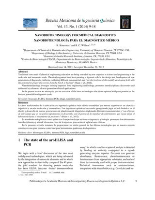
- 5. Kourentzi and Willson/ Revista Mexicana de Ingenier´ıa Qu´ımica Vol. 13, No. 1 (2014) 9-18 526 527 Figure 3. 528 Nanopore set-up in the IZON qNano instrument, (B) transient current drops created by 529 particles passing through the nanopore, with a scheme indicating calculations for baseline 530 duration, blockade full width at half maximum (FWHM) and magnitude (dI) and (C) 531 DNA hybridization on DNA-functionalized dextran particles; adapted with permission 532 (Booth {it et al}., 2013). 533 534 Fig. 3. Nanopore set-up in the IZON qNano instrument, (B) transient current drops created by particles passing through the nanopore, with a scheme indicating calculations for baseline duration, blockade full width at half maximum (FWHM) and magnitude (dI) and (C) DNA hybridization on DNA-functionalized dextran particles; adapted with permission (Booth et al., 2013). artificial nanoscale pores has been an interesting analyte-induced particle aggregation is detected by the goal (reviewed in (Kozak et al., 2011)). However, increased magnitude of the blockade events (Platt et only recently have dynamically-adjustable (tunable) al., 2012). elastomeric nanopores become available (Garza- Licudine et al., 2010, Roberts et al., 2010) as part of the qNano particle analyzer (IZON Science Ltd) 6 Paramagnetic microparticles as that allows the characterization of nanoparticles by monitoring the magnitude, duration and frequency labels of the blockade events as the particles traverse the nanopore (Drescher et al., 2012). The magnitude of the generated blockade depends on the particle-to- pore volume ratio and thus for a given aperture the measured change in resistance is proportional to the particle volume; blockade duration correlates with electrophoretic mobility and surface charge. In the qNano, the elastomeric membrane allows for real-time tuning of the pore size by applying a macroscopic stretch to the membrane and enables the real-time tuning of the sensitivity of the measurement. Beyond particle characterization the device appears to be an attractive platform to develop point-of-care diagnostics. Recently detection of DNA hybridization in the qNano has been reported; upon hybridization to complementary DNA, the surface charge of DNA functionalized particles changed (Booth et al., 2013) (Figure 3). Platt and coworkers have demonstrated a proof of concept protein detection assay in which We and others have been integrating paramagnetic microparticles, traditionally used in o-line sample capture, cleanup and concentration (van Reenen et al., 2013) with optical biosensors for the ultrasensitive detection of proteins and pathogens (Mani et al., 2011). We have been developing microfabricated retroreflectors as bio-sensing surfaces. Retroreflectors return light directly to its source and are readily detectable with inexpensive optics. Suspended corner cube retroreflectors (Figure 4), 5 m in size, consisting of a transparent epoxy core and three surfaces coated with gold are promising ultra-bright labels for use in a rapid, low-labor diagnostic platform (Sherlock et al., 2011). On the other hand linear arrays of retroreflectors combined with micron-sized magnetic particles acting as light-blocking labels provide a low-cost platform for multiplex detection of Manuscrito sometido a la Revista Mexicana de Ingeniería Química 14 www.rmiq.org 13
- 6. Kourentzi and Willson/ Revista Mexicana de Ingenier´ıa Qu´ımica Vol. 13, No. 1 (2014) 9-18 535 536 537 Figure 4. 538 (Left) Scanning electron micrographs of corner cube retroreflectors. (Right) 539 Schematic of the fabrication sequence for corner cube retroreflectors; pending 540 permission (Sherlock {it et al}., 2011). 541 542 Fig. 4. (Left) Scanning electron micrographs of corner cube retroreflectors. (Right) Schematic of the fabrication sequence for corner cube retroreflectors; pending permission (Sherlock et al., 2011) pathogens (Les reviewed 2013). We have been also investigating the use of an implantable micron-sized retroreflector-based platform and Optical Coherence Tomography (OCT) as a non-invasive and depth-resolved imaging technique for reflectance measurements of micro-retroreflectors in the subcutaneous tissue (Ivers et al., 2010). Diraction-based biosensors rely on optical scattering and have been explored as sensitive protein detection platforms. Signal enhancement techniques include micro fabricating solid diraction gratings or by inducing, in the presence of analyte, the assembly of microbeads into diraction patterns. Lee and co-workers have demonstrated an aptamer-based assay to detect platelet-derived growth actor B on a microprinted gold-coated glass slide where the assembled bead patterns allow visual analysis using a bright-field microscope (Lee et al., 2010). Recently, a microfluidic chip-based magnetic bead surface coverage assay has been reported in which large magnetic beads that have captured analytes from a serum sample ‘roll’ over a pattern of small beads functionalized with anti-analyte antibodies, to which they can bind selectively, achieving attomolar detection sensitivity using optical microscopy (Tekin et al., 2013). Leveraging existing technology utilized in magnetic data storage hard drives has enabled dramatic progress to be achieved in the magnetic biosensors arena in recent years. Micrometer-sized magnetic particles are promising reporters since even the most complex biological samples lack detectable magnetic background and thus do not interfere with the detection mechanism of the magnetic labels. In 1998, Baselt and co-workers were the first to demonstrate a prototype GMR-based biosensor (Baselt et al., 1998). Giant magnetoresistive (GMR) spin-valve sensors detect the presence of magnetic particles by measuring the change of resistance of the conductive layer due to the presence of the magnetic particles. Most recently two research groups have been developing GMR biosensing devices. The Wang group at Stanford has demonstrated multiplex protein detection using 50 nm magnetic nanotags at subpicomolar levels with a dynamic range of more than four orders of magnitude in clinically-relevant serum samples without the need for any washing protocol (Osterfeld et al., 2008; Gaster et 14 www.rmiq.org Manuscrito sometido a la Revista Mexicana de Ingeniería Química 15
- 7. Kourentzi and Willson/ Revista Mexicana de Ingenier´ıa Qu´ımica Vol. 13, No. 1 (2014) 9-18 al., 2009). The J.-P. Wang group at University of Minnesota demonstrated the applicability of sub 13 nm high-moment magnetic nanoparticles in a novel GMR biosensor that achieved detection of as few as 600 molecules ( one zeptomole) of streptavidin (Srinivasan et al., 2009) and the possibility to rapidly quantify femtomolar concentrations of a biomarker in human serum in 5 min (Li et al., 2010). 7 Conductivity Inorganic nanostructures exhibit unique tunable electrical properties and are exploited as signal transduction elements for ultrasensitive, rapid, real-time sensing (Rosi et al., 2005; Kierny et al., 2012). The tunable conducting properties of semiconductor nanowires allow for label-free electrical detection of analytes through changes in conductance induced by binding events occurring on the nanowire surface and thus provide an attractive diagnostics platform. Since the first report of electrical detection of picomolar concentrations of streptavidin in solution in 2001 (Cui et al., 2001) the Lieber group has pioneered bottom-up strategies to fabricate silicon nanowires and they have demonstrated the ultrasensitive detection of various targets including proteins, nucleic acids, and viruses (Patolsky et al., 2006). Developments in nanowire sensors for multiplexed detection of biomolecules have been recently reviewed (He et al., 2008). The Lieber group has also demonstrated a multiplex assay for three cancer markers with a detection of 0.9 pg/mL in desalted but undiluted serum samples (Zheng et al., 2005). Carbon has been showing great potential in its newly-popular forms, carbon nanotubes (CNTs) and graphene. Nonspecific binding is always an issue with these materials, however. Graphene, a two-dimensional hexagonal network of carbon atoms only one atom thick, has been extensively investigated for various applications due to its prominent structural and electrical properties, and can be used as the basis of extremely powerful biosensor systems and integrated assays with high sensitivity. A number of studies reported the use of graphene in biosensors, and the electrical detection of biomolecules using ultrathin 2D graphene sheets can potentially achieve high sensitivity. Carbon nanotubes (CNTs) are hollow cylindrical nanostructures composed of single or multiple sheets of graphene containing carbon atoms in a honeycomb arrangement. Single-wall CNTs (SWNT) arrayed vertically are electrically conductive and allow for the construction of high-density sensors with high sensitivity. SWNT arrays combined with multiwall carbon nanotubes decorated with multiple copies of antibodies and enzyme reporters have achieved highly sensitive detection of a cancer biomarker in serum and in tissue lysates (Yu et al., 2006). More recently, Cai et al. (2010), fabricated arrays of carbon nanotube tips with molecular-imprinted polymer coating for ultrasensive sensing of proteins. Concluding remarks Nanoscience, nanotechnology, and advanced fabrication methods have made access to previously-impossible structures almost routine. Chemical engineers are taking advantage of these new tools, integrating them into a wide range of promising new technologies. The future of this synergy looks very promising. Acknowledgments RCW acknowledges the Robert A. Welch Foundation for support under grant E-1264, and the Hungton- Woestemeyer Professorship. References Adler, M., Wacker, R. and Niemeyer, C. M. (2008). Sensitivity by combination: immuno-PCR and related technologies. Analyst 133, 702-718. Alivisatos, A. P. (1996). Semiconductor clusters, nanocrystals, and quantum dots. Science 271, 933-937. Bao, Y. P., Wei, T. F., Lefebvre, P. A., An, H., He, L., Kunkel, G. T. and Muller, U. R. (2006). Detection of protein analytes via nanoparticle-based bio bar code technology. Analytical Chemistry 78, 2055-2059. Baselt, D. R., Lee, G. U., Natesan, M., Metzger, S. W., Sheehan, P. E. and Colton, R. J. (1998). A biosensor based on magnetoresistance technology. Biosensors and Bioelectronics 13, 731-739. Booth, M. A., Vogel, R., Curran, J. M., Harbison, S. and Travas-Sejdic, J. (2013). Detection www.rmiq.org 15
- 8. Kourentzi and Willson/ Revista Mexicana de Ingenier´ıa Qu´ımica Vol. 13, No. 1 (2014) 9-18 of target-probe oligonucleotide hybridization using synthetic nanopore resistive pulse sensing. Biosensors and Bioelectronics 45, 136-140. Burbulis, I., Yamaguchi, K., Yu, R., Resnekov, O. and Brent, R. (2007). Quantifying small numbers of antibodies with a ‘near-universal’ protein-DNA chimera. Nature Methods 4, 1011- 1013. Burg, T. P., Godin, M., Knudsen, S. M., Shen, W., Carlson, G., Foster, J. S., Babcock, K. and Manalis, S. R. (2007). Weighing of biomolecules, single cells and single nanoparticles in fluid. Nature 446, 1066-1069. Cai, D., Ren, L., Zhao, H., Xu, C., Zhang, L., Yu, Y., Wang, H., Lan, Y., Roberts, M.F., Chuang, J.H., Naughton, M.J., Ren, Z. and Chiles, T.C. (2010). A molecular -imprint nanosensor for ultrasensitive detection of proteins. Nature Nanotechnology 5, 597-601. Churana, R., Godin, M., Knudsen, S. and Manalis, S. (2007). Mass-based readout for agglutination assays. Applied Physics Letters 91, e193902. Cui, Y., Wei, Q., Park, H. and Lieber, C. M. (2001). Nanowire nanosensors for highly sensitive and selective detection of biological and chemical species. Science 293, 1289-1292. de la Rica, R. and Stevens, M. M. (2012). Plasmonic ELISA for the ultrasensitive detection of disease biomarkers with the naked eye. Nature Nanotechnology 7, 821-824. Douglas, T. and Young, M. (2006). Viruses: making friends with old foes. Science 312, 873-875. Drescher, D., Giesen, C., Traub, H., Panne, U., Kneipp, J. and Jakubowski, N. (2012). Quantitative imaging of gold and silver nanoparticles in single eukaryotic cells by laser ablation ICP-MS. Analytical Chemistry 84, 9684-9688. Elghanian, R., Storho, J. J., Mucic, R. C., Letsinger, R. L. and Mirkin, C. A. (1997). Selective colorimetric detection of polynucleotides based on the distance-dependent optical properties of gold nanoparticles. Science 277, 1078-1081. Fritz, J., Baller, M. K., Lang, H. P., Rothuizen, H., Vettiger, P., Meyer, E., Guntherodt, H., Gerber, C. and Gimzewski, J. K. (2000). Translating biomolecular recognition into nanomechanics. Science 288, 316-318. Garza-Licudine, E., Deo, D., Yu, S., Uz-Zaman, A. and Dunbar,W. B. (2010). Portable nanoparticle quantization using a resizable nanopore instrument - the IZON qNano. Conference proceedings: Annual International Conference of the IEEE Engineering in Medicine and Biology Society 2010, 5736-5739. Gaster, R. S., Hall, D. A., Nielsen, C. H., Osterfeld, S. J., Yu, H., Mach, K. E., Wilson, R. J., Murmann, B., Liao, J. C., Gambhir, S. S. and Wang, S. X. (2009). Matrix-insensitive protein assays push the limits of biosensors in medicine. Nature Medicine 15, 1327-1332. Giljohann, D. A. and Mirkin, C. A. (2009). Drivers of biodiagnostic development. Nature 462, 461- 464. He, B., Morrow, T. J. and Keating, C. D. (2008). Nanowire sensors for multiplexed detection of biomolecules. Current Opinion in Chemical Biology 12, 522-528. He, J., Evers, D. L., O’Leary, T. J. and Mason, J. T. (2012). Immunoliposome-PCR: a generic ultrasensitive quantitative antigen detection system. Journal of Nanobiotechnology 10, 26. Hirsch, L. R., Halas, N. J. and West, J. L. (2005). Whole-blood immunoassay facilitated by gold nanoshell-conjugate antibodies. Methods in Molecular Biology 303, 101-111. Ivers, S. N., Baranov, S. A., Sherlock, T., Kourentzi, K., Ruchhoeft, P., Willson, R. and Larin, K. V. (2010). Depth-resolved imaging and detection of micro-retroreflectors within biological tissue using Optical Coherence Tomography. Biomedical Optics Express 1, 367- 377. Jia, C. P., Zhong, X. Q., Hua, B., Liu, M. Y., Jing, F. X., Lou, X. H., Yao, S. H., Xiang, J. Q., Jin, Q. H. and Zhao, J. L. (2009). Nano-ELISA for highly sensitive protein detection. Biosensors and Bioelectronics 24, 2836-2841. Kierny, M. R., Cunningham, T. D. and Kay, B. K. (2012). Detection of biomarkers using recombinant antibodies coupled to nanostructured platforms. Nano Reviews 3. 16 www.rmiq.org
- 9. Kourentzi and Willson/ Revista Mexicana de Ingenier´ıa Qu´ımica Vol. 13, No. 1 (2014) 9-18 Kim, H. J., Ahn, K. C., Gonzalez-Techera, A., Gonzalez-Sapienza, G. G., Gee, S. J. and Hammock, B. D. (2009). Magnetic bead-based phage anti-immunocomplex assay (PHAIA) for the detection of the urinary biomarker 3-phenoxybenzoic acid to assess human exposure to pyrethroid insecticides. Analytical Biochemistry 386, 45-52. Kim, H. J., McCoy, M., Gee, S. J., Gonzalez- Sapienza, G. G. and Hammock, B. D. (2011). Noncompetitive phage anti-immunocomplex real-time polymerase chain reaction for sensitive detection of small molecules. Analytical Chemistry 83, 246-253. Kim, H. J., Rossotti, M. A., Ahn, K. C., Gonzalez- Sapienza, G. G., Gee, S. J., Musker, R. and Hammock, B. D. (2010). Development of a noncompetitive phage anti-immunocomplex assay for brominated diphenyl ether 47. Analytical Biochemistry 401, 38-46. Kozak, D., Anderson, W., Vogel, R. and Trau, M. (2011). Advances in Resistive Pulse Sensors: Devices bridging the void between molecular and microscopic detection. Nano Today 6, 531- 545. Lee, J., Icoz, K., Roberts, A., Ellington, A. D. and Savran, C. A. (2010). Diractometric detection of proteins using microbead-based rolling circle amplification. Analytical Chemistry 82, 197- 202. Les, C. B. (2013). From bike reflector to virus detector. Photonics Spectra. Li, Y., Srinivasan, B., Jing, Y., Yao, X., Hugger, M. A., Wang, J. P. and Xing, C. (2010). Nanomagnetic competition assay for low-abundance protein biomarker quantification in unprocessed human sera. Journal of the American Chemical Society 132, 4388-4392. Majumdar, A. (2002). Bioassays based on molecular nanomechanics. Disease Markers 18, 167-174. Malou, N. and Raoult, D. (2011). Immuno-PCR: a promising ultrasensitive diagnostic method to detect antigens and antibodies. Trends in Microbiology 19, 295-302. Mani, V., Chikkaveeraiah, B. V. and Rusling, J. F. (2011). Magnetic particles in ultrasensitive biomarker protein measurements for cancer detection and monitoring. Expert Opinion on Medical Diagnostics 5, 381-391. Mason, J. T., Xu, L., Sheng, Z. M., He, J. and O’Leary, T. J. (2006). Liposome polymerase chain reaction assay for the sub-attomolar detection of cholera toxin and botulinum neurotoxin type A. Nature Protocols 1, 2003- 2011. Mirkin, C. A., Letsinger, R. L., Mucic, R. C. and Storho, J. J. (1996). A DNA-based method for rationally assembling nanoparticles into macroscopic materials. Nature 382, 607-609. Osterfeld, S. J., Yu, H., Gaster, R. S., Caramuta, S., Xu, L., Han, S. J., Hall, D. A., Wilson, R. J., Sun, S. H., White, R. L., Davis, R. W., Pourmand, N. and Wang, S. X. (2008). Multiplex protein assays based on real-time magnetic nanotag sensing. Proceedings of the National Academy of Sciences of the United States of America 105, 20637-20640. Park, J. S., Cho, M. K., Lee, E. J., Ahn, K. Y., Lee, K. E., Jung, J. H., Cho, Y., Han, S. S., Kim, Y. K. and Lee, J. (2009). A highly sensitive and selective diagnostic assay based on virus nanoparticles. Nature Nanotechnology 4, 259- 264. Patolsky, F., Zheng, G. and Lieber, C. M. (2006). Nanowire sensors for medicine and the life sciences. Nanomedicine 1, 51-65. Platt, M., Willmott, G. R. and Lee, G. U. (2012). Resistive pulse sensing of analyte-induced multicomponent rod aggregation using tunable pores. Small 8, 2436-2444. Rissin, D. M., Kan, C. W., Campbell, T. G., Howes, S. C., Fournier, D. R., Song, L., Piech, T., Patel, P. P., Chang, L., Rivnak, A. J., Ferrell, E. P., Randall, J. D., Provuncher, G. Walt, D. R. and Duy, D. C. (2010). Single-molecule enzyme-linked immunosorbent assay detects serum proteins at subfemtomolar concentrations. Nature Biotechnology 28, 595-599. Roberts, G. S., Kozak, D., Anderson, W., Broom, M. F., Vogel, R. and Trau, M. (2010). Tunable nano/micropores for particle detection and discrimination: scanning ion occlusion spectroscopy. Small 6, 2653-2658. www.rmiq.org 17
- 10. Kourentzi and Willson/ Revista Mexicana de Ingenier´ıa Qu´ımica Vol. 13, No. 1 (2014) 9-18 Rosi, N. L. and Mirkin, C. A. (2005). Nanostructures in biodiagnostics. Chemical Reviews 105, 1547- 1562. Ruan, G. and Winter, J. (2012). Chemical Engineering at the Intersection of Nanotechnology and Biology. Chemical Engineering Progress, 41-51. Saha, K., Agasti, S. S., Kim, C., Li, X. and Rotello, V. M. (2012). Gold nanoparticles in chemical and biological sensing. Chemical Reviews 112, 2739-2779. Samir, T. M., Mansour, M. M., Kazmierczak, S. C. and Azzazy, H. M. (2012). Quantum dots: heralding a brighter future for clinical diagnostics. Nanomedicine 7, 1755-1769. Sano, T., Smith, C. L. and Cantor, C. R. (1992). Immuno-PCR: very sensitive antigen detection by means of specific antibody-DNA conjugates. Science 258, 120-122. Sherlock, T., Nasrullah, A., Litvinov, J., Cacao, E., Knoop, J., Kemper, S., Kourentzi, K., Kar, A., Ruchhoeft, P. and Willson, R. (2011). Suspended, micron-scale corner cube retroreflectors as ultra-bright optical labels. Journal of Vacuum Science Technology B 29, V. Song, L., Hanlon, D. W., Chang, L., Provuncher, G. K., Kan, C. W., Campbell, T. G., Fournier, D. R., Ferrell, E. P., Rivnak, A. J., Pink, B. A., Minnehan, K. A., Patel, P. P., Wilson, D. H., Till, M. A., Faubion, W. A. and Duy, D. C. (2011). Single molecule measurements of tumor necrosis factor alpha and interleukin-6 in the plasma of patients with Crohn’s disease. Journal of Immunological Methods 372, 177-186. Soto, C. M., Blum, A. S., Vora, G. J., Lebedev, N., Meador, C. E., Won, A. P., Chatterji, A., Johnson, J. E. and Ratna, B. R. (2006). Fluorescent signal amplification of carbocyanine dyes using engineered viral nanoparticles. Journal of the American Chemical Society 128, 5184-5189. Soto, C. M. and Ratna, B. R. (2010). Virus hybrids as nanomaterials for biotechnology. Current Opinion in Biotechnology 21, 426-438. Srinivasan, B., Li, Y., Jing, Y., Xu, Y., Yao, X., Xing, C. and Wang, J. P. (2009). A detection system based on giant magnetoresistive sensors and high-moment magnetic nanoparticles demonstrates zeptomole sensitivity: potential for personalized medicine. Angewandte Chemie International Edition in English 48, 2764-2767. Tekin, H. C., Cornaglia, M. and Gijs, M. A. (2013). Attomolar protein detection using a magnetic bead surface coverage assay. Lab on a Chip 13, 1053-1059. van Reenen, A., de Jong, A. M. and Prins, M. W. (2013). Accelerated particle-based target capture–the roles of volume transport and near-surface alignment. The Journal of Physical Chemistry B 117, 1210-1218. von Muhlen, M. G., Brault, N. D., Knudsen, S. M., Jiang, S. and Manalis, S. R. (2010). Label-free biomarker sensing in undiluted serum with suspended microchannel resonators. Analytical Chemistry 82, 1905-1910. Walt, D. R. (2013). Optical methods for single molecule detection and analysis. Analytical Chemistry 85, 1258-1263. Weintraub, K. (2013). Biomedicine: The new gold standard. Nature 495, S14-16. Yu, X., Munge, B., Patel, V., Jensen, G., Bhirde, A., Gong, J. D., Kim, S. N., Gillespie, J., Gutkind, J. S., Papadimitrakopoulos, F. and Rusling, J. F. (2006). Carbon nanotube amplification strategies for highly sensitive immunodetection of cancer biomarkers. Journal of the American Chemical Society 128, 11199-11205. Zhang, H., Xu, Y., Huang, Q., Yi, C., Xiao, T. and Li, Q. (2013). Natural phage nanoparticle-mediated real-time immuno-PCR for ultrasensitive detection of protein marker. Chemical Communications 49, 3778-3780. Zheng, G., Patolsky, F., Cui, Y., Wang, W. U. and Lieber, C. M. (2005). Multiplexed electrical detection of cancer markers with nanowire sensor arrays. Nature Biotechnology 23, 1294- 1301. 18 www.rmiq.org