
Recommended
More Related Content
What's hot
What's hot (20)
Similar to Sole Of Foot Final.pptx
Similar to Sole Of Foot Final.pptx (20)
More from MeetVaghasiya20
More from MeetVaghasiya20 (8)
Recently uploaded
Recently uploaded (20)
Sole Of Foot Final.pptx
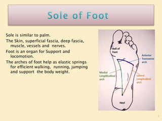
- 1. 1 Sole is similar to palm. The Skin, superficial fascia, deep fascia, muscle, vessels and nerves. Foot is an organ for Support and locomotion. The arches of foot help as elastic springs for efficient walking, running, jumping and support the body weight.
- 2. 2 The skin of the sole, like that of the palm is: 1. Thick for protection 2. Firmly adherent to the underlying plantar aponeurosis and 3. Creased. These features increase the efficiency of the grip of the sole on the ground.
- 3. 3 The skin of the sole, like that of the palm is: The skin is mainly supplied by three cutaneous nerves The nerves are: a) Medial calcanean branches of the tibial nerve, to the posterior and medial portions. b) Branches from the medial plantar nerve to the larger, anteromedial portion including the medial 3½ digits. c) Branches from the lateral plantar nerve to the smaller anterolateral portion including the lateral 1½ digits.d. Small areas on medial and lateral sides are innervated by saphenous and sural nerves
- 4. 4 The superficial fascia of the sole is fibrous and dense. Fibrous bands bind the skin to the deep fascia or plantar aponeurosis, and divide the subcutaneous fat into small tight compartments which serve as water-cushions and reinforce the spring-effect of the arches of the foot during walking, running and jumping. The fascia is very thick and dense over the weight-bearing points. It contains cutaneous nerves and vessels
- 6. 1. Plantar Aponeurosis in the sole. 2. Deep transverse metatarsal ligament between Metatarsophalangeal joints. 3. The fibrous flexor sheaths in the toe. 6
- 7. THICKENED CENTRAL BAND OF THE DEEP FASCIA IN THE SOLE OF THE FOOT. 7 The aponeurosis is triangular in shape. The apex is proximal. It is attached to the medial tubercle of the calcaneum, proximal to the attachment of the flexor digitorum brevis. The base is distal. It divides into five processes near the heads of the metatarsal bones. The digital nerves and vessels pass through the intervals between the processes PLANTAR APONEUROSIS
- 9. THICKENED CENTRAL BAND OF THE DEEP FASCIA IN THE SOLE OF THE FOOT. 9 Each process splits, opposite the metatarso- phalangeal joints, into a superficial and a deep slip. The superficial slip is attached to dermis of skin. The deep slip divides into two parts which embrace the flexor tendons, and blend with the fibrous flexor sheaths and with the deep transverse metatarsal ligaments. PLANTAR APONEUROSIS
- 10. THICKENED CENTRAL BAND OF THE DEEP FASCIA IN THE SOLE OF THE FOOT. 10 Function 1. It fixes the skin of sole. 2. It protects deeper structure. 3. It help to maintaining longitudinal arches of foot. 4. It gives origin to muscles of the first layer of sole. PLANTAR APONEUROSIS
- 11. THE MUSCLE OF THE SOLE IS ARRANGED IN FOUR (4) LAYERS. 11 1. FIRST LAYER (SUPERFICIAL) a) FLEXOR DIGITORUM BRVIS b) ABDUCTOR HALLUCIS c) ABDUCTOR DIGITI MINIMI
- 13. 13 Origin : Medial Tubercle of calcaneum and plantar aponeurosis. Insertion : Middle phalanges of digits 2-5 Nerve supply : Medial planter nerve Action : Flexion on proximal interpharangeal and Metatarsopharlyngeal joint.
- 14. 14 Origin : Medial tubercle of calcaneum, flexor retinaculum, medial intermuscular septum. Insertion : Medial Plantar portion of proximal phalanx of great toe. Nerve supply : Medial Plantar Nerve Actions : Abducts great toe at Metatarsopharlyngeal joint and flexes great toe at Metatarsopharlyngeal joint.
- 15. 15 Origin : Medial and lateral processes of posterior calcaneal tuberosity Insertion : Lateral side of base of proximal phalanx of 5th toe and 5th metatarsal Nerve supply : Lateral plantar (S2,3) Action : Flexes and abducts 5th toe. Support lateral longitudinal arch
- 16. 16 1. FLEXOR DIGITORUM LONGUS 2. FLEXOR DIGITORUM ACCESSERIOUS (QUADRATUS PLANTAE) 3. FLEXOR HALLUCIS LONGUS 4. LUMBRICALS
- 17. 17 Origin: From upper two thirds of the medial part of the posterior surface below the soleal line. Insertion: The muscle divides into four tendons. Each is inserted into the plantar surface of distal phalanx of second to fifth digit. Nerve Supply: Tibial nerve (S2, S3) Action : Plantar flexion of lateral four toes and of ankle. Maintains medial longitudinal arch.
- 19. Origin : It arises by two heads: a. Medial head is large and fleshy; it arises from the medial concave surface of the calcaneum b. Lateral head is smaller and tendinous; it arises from the calcaneum in front of the lateral tubercle.The two heads unite at an acute angle. Insertion : The muscle fibres are inserted into the lateral side of the flexor digitorum longus 19
- 21. 21 Nerve supply : Main trunk of lateral plantar nerve. Action : Straightens the pull of the long flexor muscles. Flexes the toes through the long tendons.
- 22. Origin: Lower three-fourths of the posterior surface of fibula except lowest 2.5 cm and adjoining interosseous membrane. Insertion: plantar surface of the base of distal phalanx of the great toe. Nerve Supply: Tibial nerve Function: Plantar flexor of the big toe, plantar flexor of ankle joint, maintains medial longitudinal arch. 22
- 24. Origin: They arise from the tendons of theflexor digitorum longus. The first lumbrical is unipennate, and theothers are bipennate First lumbrical arises from medial side of 1st tendon of flexor digitorum longus. Second lumbrical arises from adjacent sides of 1st and 2nd tendons of flexor digitorum longus. Third lumbrical arises from adjacent sides of 2nd and 3rd tendons of flexor digitorum longus. Fourth lumbrical arises from adjacent sides of 3rd and 4th tendons of flexor digitorum longus. 24
- 25. 25
- 26. Insertion : Their tendons pass forwards on the medial sides of the metatarsophalangeal joints of the lateral four toes, and then dorsally for insertion into the extensor expansion. Nerve supply : The first muscle by the medial plantar nerve; and the other three by the deep branch of lateral plantar nerve Action : They maintain extension of the digits at the inter- phalangeal joints so that in walking and running the toes do not buckle under 26
- 27. 27
- 29. Origin: It arises by a Y-shaped tendon:a. The lateral limb, from the medial part of the plantar surface of the cuboid bone, behind the groove for the peroneus longus and from the adjacent side of the lateral cuneiform boneb. The medial limb is a direct continuation of the tendon of tibialis posterior into the foot. 29
- 30. 30 Insertion : The muscle splits into medial and lateral parts, each of which ends in a tendon. Each tendon is inserted into the corresponding side of the base of the proximal phalanx of the great toe. Nerve supply : Medial Plantar Nerve Actions : Flexes the proximal phalanx at the metatarsophalangeal joint of the great toe.
- 31. Origin : It arises by two heads: a. The oblique head is large, and arises from the bases of the second, third, and fourth metatarsals, from the sheath of the tendon of the peroneus longus b. The transverse head is small, and arises from the deep metatarsal ligament, and the plantar ligaments of the metatarsophalangeal joints of the third, fourth and fifth toes (transverse head has no bony origin) 31
- 33. 33 Insertion : On the lateral side of the base of the proximal phalanx of the big toe, in common with the lateral tendon of the flexor hallucis brevis. Nerve supply : Deep Branch of lateral plantar nerve, which terminates in this muscle. Actions : Adductor of great toe towards second toe. Maintains transverse arches of foot.
- 34. Origin : a. Base of the fifth metatarsal bone b. Sheath of the tendon of the peroneus longus Insertion : Into the lateral side of the base of the proximal phalanx of the little toe. 34
- 36. 36 Nerve supply : Superficial branch of lateral plantar nerve Action : Flexes the proximal phalanx at the metatarsophalangeal joint of the little toe
- 37. 37 1.Plantar interossei 2.Dorsal interossei 3.Tibialis Posterior 4.Peroneus Longus
- 39. 39 Origin : Bases and medial sides of third, fourth and fifth metatarsals. Insertion : Medial sides of bases of proximal phalanges and dorsal digital/extensor expansions of 3rd, 4th and5th toes Nerve supply : First and second by lateral plantar (deep branch). Third by lateral plantar (superficial branch) Function : Adductors of third, fourth and fifth toes toward the axis. Flexor of metatarsophalangeal and extensor of interphalangeal joints of third, fourth and fifth toes
- 40. 40 Origin : Adjacent sides of metatarsal bones Insertion : Bases of proximal phalanges and dorsal digital expansion of toes; first on medial side of 2nd toe; second on lateral side of 2nd toe; third on lateral side of 3rd toe and fourth on lateral side of 4th toe.
- 41. 41 Nerve supply : First, second, third by lateral plantar (deep branch), fourth dorsal interosseous by superficial branch of lateral plantar. Function : Abductors of toes from axis of second toe. First and second cause medial and lateral abduction of second toe. Third and fourth for abduction of 3rd and 4th toes
- 42. Origin : Posterior surfaces of leg bones Insertion : Tuberosity of navicular Nerve supply : Tibial Nerve Function : Plantar Flexion of ankle 42 Tibialis Posterior
- 43. Origin: Upper part of lateral surface of fibula Insertion : Base of 1st metatarsal Nerve Supply : Superficial peroneal nerve Function : Evertor of foot 43
- 44. origin and course :. Largest terminal branch of Tibial nerve. It passes forwards between Abductor hallucis and flexor digitorum brevis and divides into its branches. Root Value : L4, L5, S1 Branches: The Muscular branches supply the Four muscle. 1. Abductor hallucis 2. Flexor digitorum brevis 3. Flexor hallucis brevis 4. First lumbrical muscle 44
- 45. Branches 45 Its muscular branches supply four muscles as follows. 1. The abductor hallucis. 2. The flexor digitorum brevis. 3. The flexor hallucis brevis receives a branch from the first digital nerve. 4. The first lumbrical muscle receives a branch from the second digital nerve.
- 46. Branches 46
- 47. Branches 47 Cutaneous branches supply the skin of the medial part of the sole, and of the medial 3½ toes through four digital branches. The first digital nerve supplies the medial side of the great toe. The second nerve supplies the adjacent sides of the first and second toes. The third nerve supplies the adjacent sides of the second and third toes. The fourth nerve supplies the adjacent sides of the third and fourth toes. Each digital nerve gives off a dorsal branch which supplies structures around the nail of the digit concerned. Articular branches supply joints of the tarsus and metatarsus.
- 48. 48 Origin: Small terminal branch of Tibial nerve. Passes laterally and forwards till base of fifth Metatarsal, where it divides Superficial and Deep branches. Its Root value is Ventral Primary Rami of S2,S3, its supply 14 muscles of the sole.
- 49. Branches : The main trunk supplies two muscles-the flexor digitorum accessorius and the abductor digiti minimi, and the skin of the sole. The main trunk ends by dividing into superficial and deep branches. The superficial branch divides into two branches- lateral and medial. The lateral branch supplies three muscles-flexor digiti minimi brevis, the third plantar and fourth dorsal interossei, and the skin on the lateral side of the little toe.
- 50. Branches : The medial branch communicates with the medial plantar nerve, and supplies the skin lining the fourth interdigital cleft. The deep branch supplies nine muscles, including the second, third and fourth lumbricals; first, second and third dorsal interossei; first and second plantar interossei and adductor hallucis.
- 51. 51 Beginning, Course and Termination : Medial plantar artery is a smaller terminal branch of the posterior tibial artery. It lies along the medial border of foot and divides into branches.
- 52. 52 Branches It gives off cutaneous, muscular branches to the overlying skin and to the adjoining muscles, and three small superficial digital branches that end by joining the first, second and third plantar metatarsal arteries which are branches of the plantar arch
- 54. 54 Beginning, Course and Termination : Lateral plantar artery is the larger terminal branch of the posterior tibial artery. At the base of the fifth metatarsal bone, it gives a superficial branch and then continues as the plantar arch
- 56. 56 Branches : Muscular branches supply the adjoining muscles. Cutaneous branches supply the skin and fasciae of the lateral part of the sole. Anastomotic branches reach the lateral border of the foot and anastomose with arteries on the dorsum of the foot. A calcanean branch is occasionally given off to the skin of the heel.
- 57. 57 Beginning, Course and Termination : Plantar arch is formed by the direct continuation of the lateral plantar artery after it has given off the superficial branch and is completed medially by the dorsalis pedis artery. It extends from the base of the fifth metatarsal bone to the proximal part of the first intermetatarsal space, and lies between the third and fourth layers of the sole. It is accompanied by venae comitantes. The deep branch of the lateral plantar nerve lies in the concavity of the plantar arch
- 58. 58 Branches of the Plantar Arch : 1. Four plantar metatarsal arteries run distally, one in each intermetatarsal space. Each artery ends by dividing into two plantar digital branches for adjacent sides of two digits. The first artery also gives off a branch to the medial side of the great toe. The lateral side of the little toe gets a direct branch from the lateral plantar artery.
- 60. 60 Branches of the plantar arch : 2. The plantar arch gives off three proximal perforating arteries that pass through the second, third and fourth intermetatarsal spaces and communicate with the dorsal metatarsal arteries which are the branches of the arcuate artery. The distal end of each plantar metatarsal artery gives off a distal perforating artery which joins the distal part of the corresponding dorsal metatarsal artery
- 61. Plantar fasciitis occurs in policemen due to stretching of the plantar aponeurosis. This results in pain in the heel region, especially during standing.
- 62. A neuroma may be formed on the branch of medial plantar nerve between 3rd and 4th metatarsal bones. It is called Morton's neuroma This causes pain between third and fourth metatarsals. It may be also due to pressure on digital nerve between 3rd and 4th metatarsals. Any of the digital nerves, especially the one in the third interdigital cleft may develop neuroma. This is a painful condition
- 63. Fracture of shaft of 2nd/3rd/4th/metatarsal bones is called 'march fracture. It is seen in army personnel, policemen as they have to march a lot. It occurs due to decalcification and vascular necrosis. Toes may be spread out or splayed. Longitudinal arches are exaggerated leading to pes cavus
- 64. Normal architecture of foot is subjected to insults due to 'high heels'. Females apparently look taller, smarter but may suffer from sprains and dislocations of the ankle joint
- 65. If medial border of foot is raised, person walks on lateral border of foot. The condition is called 'talipes varus’ If lateral border of foot is raised, person walks on medial border of foot. The condition is called 'talipes valgus' .
- 66. If foot is dorsiflexed, person walks on the hee lcondition is called 'talipes calcaneus’ . If foot is plantar flexed, person walks on toes. The condition is called 'talipes equinus' .
- 67. Most common is talipes equinovarus in which theheel is medial, the foot is plantar flexed and invertedwith high medial longitudinal arch