SlideShare a Scribd company logo
1 of 404
IT’S ALIVE !!!! 
1.1 Understand that living organisms share the following characteristics: they require nutrition; they respire; they excrete their waste; they respond to their 
surroundings; they move; they control their internal conditions; they reproduce; they grow and develop. 
M Movement All living things move, even plants 
R Respiration Getting energy from food 
S Sensitivity Detecting changes in the surroundings 
G Growth All living things grow and develop 
R Reproduction Making more living things of the same 
type (species). 
E Excretion Getting rid of waste 
N Nutrition Taking in and using food and other 
nutrients 
In addition, all living organisms contain nucleic acids (DNA/RNA) and 
have the ability to control their internal conditions (homeostasis). 
Finally, all living organisms can die.
ALIVE OR NOT ALIVE!! 
ALIVE OR NOT ALIVE 
1.1 Understand that living organisms share the following characteristics: they require nutrition; they respire; they excrete their waste; they respond to their 
surroundings; they move; they control their internal conditions; they reproduce; they grow and develop.
GROUPS OF LIVING ORGANISMS 
1.2 describe the common features shared by organisms within the following main groups: plants, animals, fungi, bacteria, protoctists and viruses, and for each 
group describe examples and their features as follows (details of life cycle and economic importance are not required)
PLANTS 
Plants: These are multicellular organisms; their cells contain chloroplasts and are able to carry out photosynthesis; their cells have cellulose cell walls; they 
store carbohydrates as starch or sucrose 
Plants all have the following in common: 
1) Multicellular organisms (made of lots of cells) 
2) Cells contain chloroplasts and are able to carry out photosynthesis. 
Photosynthesis takes simple inorganic molecules and turns them into 
simple sugar (glucose). Glucose can be turned into complex organic 
molecules such as carbohydrates.
PLANTS 
Plants: These are multicellular organisms; their cells contain chloroplasts and are able to carry out photosynthesis; their cells have cellulose cell walls; they 
store carbohydrates as starch or sucrose 
3) Cells have cellulose cell walls (cellulose is a 
carbohydrate)
PLANTS 
Plants: These are multicellular organisms; their cells contain chloroplasts and are able to carry out photosynthesis; their cells have cellulose cell walls; they 
store carbohydrates as starch or sucrose 
4) They store carbohydrates as starch. 
Carbohydrates are polysaccharides (many sugars/glucose linked 
together) 
or sucrose. 
Sucrose is a disaccharide (two sugars linked together).
Examples you need to know: 
1) flowering plants, such as a cereals 
A) Maize 
PLANTS 
Examples include flowering plants, such as a cereal (for example maize), and a herbaceous legume (for example peas or beans) 
B) Wheat
PLANTS 
Examples include flowering plants, such as a cereal (for example maize), and a herbaceous legume (for example peas or beans) 
2) Herbaceous legume (e.g. peas or beans). 
Root nodules
ANIMALS 
Animals: These are multicellular organisms; their cells do not contain chloroplasts and are not able to carry out photosynthesis; they have no cell walls; they 
usually have nervous coordination and are able to move from one place to another; they often store carbohydrate as glycogen 
1) Animals include: 
sponges, molluscs, worms, starfish, insects, crustaceans, 
fish, amphibians, reptiles, birds, mammals 
Animals without a backbone (vertebral column) are invertebrates. 
Animals with a vertebral column are called vertebrates. 
2) Animals are Multicellular organisms
ANIMALS 
Animals: These are multicellular organisms; their cells do not contain chloroplasts and are not able to carry out photosynthesis; they have no cell walls; they 
usually have nervous coordination and are able to move from one place to another; they often store carbohydrate as glycogen 
3) They have a nervous system 
4) They often store carbohydrate as glycogen in the 
liver and muscles.
ANIMALS 
Animals: These are multicellular organisms; their cells do not contain chloroplasts and are not able to carry out photosynthesis; they have no cell walls; they 
usually have nervous coordination and are able to move from one place to another; they often store carbohydrate as glycogen 
5) Animal cells do not contain chloroplasts and are 
not able to carry out photosynthesis 
6) Animal cells have no cell walls
ANIMALS 
Animals: These are multicellular organisms; their cells do not contain chloroplasts and are not able to carry out photosynthesis; they have no cell walls; they 
usually have nervous coordination and are able to move from one place to another; they often store carbohydrate as glycogen
ANIMALS 
Examples include mammals (for example humans) and insects (for example housefly and mosquito) 
Examples you need to know: 
1) Human (mammal)
2) Insects 
Examples include mammals (for example humans) and insects (for example housefly and mosquito) 
a) Housefly 
ANIMALS 
Why the housefly? 
- They regurgitate stomach enzymes onto their food 
- but also because other flies are interesting 
A) Tsetse fly 
B) Screw fly 
C) Drosophila
ANIMALS 
Examples include mammals (for example humans) and insects (for example housefly and mosquito) 
Lastly….. the use of flies ended the belief in the 
spontaneous generation of life
Examples include mammals (for example humans) and insects (for example housefly and mosquito) 
b) Mosquito 
ANIMALS 
Video 
Bite 
So why the Mosquito? (it is really just another fly!) 
- Complex life cycle 
- Can be a vector for malaria, yellow fever, abrovirus 
- Control measures have damaged ecosystems
FUNGI 
Fungi: These are organisms that are not able to carry out photosynthesis; their body is usually organised into a mycelium made from thread-like structures 
called hyphae, which contain many nuclei; some examples are single-celled; their cells have walls made of chitin; they feed by extracellular secretion of 
digestive enzymes onto food material and absorption of the organic products; this is known as saprotrophic nutrition; they may store 
carbohydrate as glycogen 
Characteristics of Fungi: 
1) They feed by saprophytic nutrition (feeds on dead 
organic material & digestion takes place outside the 
organism. 
2) Fungi feed by excreting extracellular secretions of 
digestive enzymes onto food and absorbing the digested 
products. 
3) Cells are joined together to form threads, called hyphae. 
Hyphae contain many nuclei, because they are made 
from many cells. 
4) Hyphae join together to make a network of threads called 
mycelium
FUNGI ARE NOT PLANTS 
5) Cells do not contain chloroplasts and are not able 
to carry out photosynthesis 
6) Cell walls are made from a protein called chitin 
(not cellulose) 
7) They store carbohydrates as glycogen 
(not starch) 
FUNGI 
Fungi: These are organisms that are not able to carry out photosynthesis; their body is usually organised into a mycelium made from thread-like structures 
called hyphae, which contain many nuclei; some examples are single-celled; their cells have walls made of chitin; they feed by extracellular secretion of 
digestive enzymes onto food material and absorption of the organic products; this is known as saprotrophic nutrition; they may store 
carbohydrate as glycogen
FUNGI 
Fungi Examples include Mucor , which has the typical fungal hyphal structure, and yeast, which is single-celled 
Examples you must know: 
1) Mucor (bread mold) 
Why do you need to know about Mucor? 
- Easy to experiment on in the lab 
(temperature, moisture) 
- Mucor must use energy to absorb nutrients in it’s hyphae 
- Was used to produce penicillin, the first antibiotic.
FUNGI 
Fungi Examples include Mucor , which has the typical fungal hyphal structure, and yeast, which is single-celled 
2) Yeast (single celled fungi) 
Why do you need to know about Yeast? 
- Easy to experiment on in the lab 
(temperature & sugar concentration) 
- Yeast is used in the production of beer & bread
BACTERIA 
Bacteria: These are microscopic single-celled organisms; they have a cell wall, cell membrane, cytoplasm and plasmids; they lack a nucleus but contain a 
circular chromosome of DNA; some bacteria can carry out photosynthesis but most feed off other living or dead organisms 
Bacteria Characteristics: 
1) Made from single cells 
2) Cells do not contain a nucleus. Bacteria cells have a small piece of circular DNA instead of a 
nucleus (a bacterial chromosome/nucleoid). 
2) Bacteria also have DNA in the form of circular plasmids 
3) most gain their nutrients by saprophytic nutrition (feed off dead organisms) or by parasitic 
nutrition (feed off living organisms). Some bacteria can carry out photosynthesis. 
You need to know their structure: 
TED
BACTERIA 
Bacteria: These are microscopic single-celled organisms; they have a cell wall, cell membrane, cytoplasm and plasmids; they lack a nucleus but contain a 
circular chromosome of DNA; some bacteria can carry out photosynthesis but most feed off other living or dead organisms 
Some pretty pictures of bacteria
BACTERIA 
Examples include Lactobacillus bulgaricus , a rod-shaped bacterium used in the production of yoghurt from milk, and Pneumococcus , a spherical bacterium 
that acts as the pathogen causing pneumonia 
Example you need to know: 
1) Lactobacillus bulgaricus 
- Used to make yogurt 
- Produces lactic acid (excretes) 
- Rod shaped 
- Not dangerous to humans 
Why Lactobacillus bulgaricus? 
- It is used in how human food production 
- lowers the pH of milk to inhibit harmful 
to human bacterial growth
Examples include Lactobacillus bulgaricus , a rod-shaped bacterium used in the production of yoghurt from milk, and Pneumococcus , a spherical bacterium 
2) Pneumococcus 
BACTERIA 
that acts as the pathogen causing pneumonia 
- pathogen causes pneumonia (inflammation of one or both lungs) 
- killed with antibiotics 
- spherical bacteria shape 
Why Pneumococcus? 
- reduces surface area of alveoli 
- use of antibiotics in treatment can lead to resistant 
strains
PROTOCTISTS 
Protoctists: These are microscopic single-celled organisms. 
When you don’t fit in scientists drop you into the 
Protoctists group! 
Protoctists Characteristics: 
- most are single celled (exception is seaweeds) 
- Some have animal-like features - protozoa 
- Some have plant-like features – algae 
swim
PROTOCTISTS 
Some, like Amoeba, that live in pond water, have features like an animal cell, while others, like Chlorella, have chloroplasts and are more like plants. A 
pathogenic example is Plasmodium , responsible for causing malaria 
Examples you need to know: 
1) Amoeba 
- protozoa 
- live in pond water 
- Pathogen amoeba can cause Amoebic dysentery 
(kills 70,000 people every year) 
Why Amoeba? 
- Highlights the need for clean drinking water supplies 
- Uses amoebiasis to feed (similair to phagocytosis) 
eating
Some, like Amoeba, that live in pond water, have features like an animal cell, while others, like Chlorella, have chloroplasts and are more like plants. A 
2) Chlorella 
PROTOCTISTS 
pathogenic example is Plasmodium , responsible for causing malaria 
- ‘more’ like plants than animals 
- have chloroplasts 
Why Chlorlla? 
- both animal and plant characteristics 
- Possible food source (1940’s solution to world hunger) 
view
3) Plasmodium 
- Protozoa 
- pathogen that causes malaria 
- uses a mosquito as a vector (transfer) 
- not killed by antibiotics 
Why plasmodium? 
- it is the greatest pathogen killer! 
TED 
PROTOCTISTS 
Some, like Amoeba, that live in pond water, have features like an animal cell, while others, like Chlorella, have chloroplasts and are more like plants. A 
pathogenic example is Plasmodium , responsible for causing malaria
VIRUS 
Viruses: These are small particles, smaller than bacteria; they are parasitic and can reproduce only inside living cells; they infect every type of living 
organism. They have a wide variety of shapes and sizes; they have no cellular structure but have a protein coat and contain one type of nucleic acid, either 
DNA or RNA 
Viruses Characteristics: 
1) Much smaller than bacteria. 
1) They are not made from cells (no cellular structures) 
3) Totally parasitic and reproduce inside host cells. 
3) They infect every type of living cell 
3) Have either DNA or RNA as genetic material 
3) Have a protein coat
VIRUS 
Viruses: These are small particles, smaller than bacteria; they are parasitic and can reproduce only inside living cells; they infect every type of living 
organism. They have a wide variety of shapes and sizes; they have no cellular structure but have a protein coat and contain one type of nucleic acid, either 
DNA or RNA 
You just have to recognize a virus, but there are several shapes
VIRUS 
Viruses: These are small particles, smaller than bacteria; they are parasitic and can reproduce only inside living cells; they infect every type of living 
organism. They have a wide variety of shapes and sizes; they have no cellular structure but have a protein coat and contain one type of nucleic acid, either 
DNA or RNA 
REVIEW FROM YEAR 8
VIRUS 
Examples include the tobacco mosaic virus that causes discolouring of the leaves of tobacco plants by preventing the formation of chloroplasts, the influenza 
virus that causes gfluc and the HIV virus that causes AIDS 
Examples you need to know: 
1) Tobacco Mosaic Virus 
- infects crop plant and prevents formation of chloroplasts 
- symptoms are discoloration of leaves 
Why Tobacco Mosaic Virus? 
- first identification of a virus as a pathogen 
(1898, Martinus W. Beijerinck) 
- Genetic engineering
VIRUS 
Examples include the tobacco mosaic virus that causes discolouring of the leaves of tobacco plants by preventing the formation of chloroplasts, the influenza 
virus that causes gfluc and the HIV virus that causes AIDS 
2) Influenza Virus 
- Viral pathogen causes the flu 
- Transmitted through air/ contact 
Why the Flu? 
- Pandemics (spanish flu) 
- Mutations leading to new strains 
- Vaccinations 
vaccine 
Flu 
History
Just thought you would like to know.
3) HIV (human immunodeficiency virus) 
- Pathogen causes AIDS (acquired immunodeficiency syndrome) 
- Virus attacks white blood cells (immune system) 
- Host killed by secondary disease 
- Transmitted by blood to blood contact 
Why HIV? 
- Immune system 
- Mutations 
VIRUS 
Examples include the tobacco mosaic virus that causes discolouring of the leaves of tobacco plants by preventing the formation of chloroplasts, the influenza 
virus that causes gfluc and the HIV virus that causes AIDS
PATHOGEN 
1.3 recall the term gpathogenc and know that pathogens may be fungi, bacteria, protoctists or viruses. 
Pathogen: 
An agent that causes infection or disease, especially a 
microorganism, such as a bacterium or protozoan, or 
a virus.
LEVELS OF ORGANIZATION 
2.1 describe the levels of organisation within organisms: organelles, cells, tissues, organs and systems. 
Organisms are made from organizations of smaller structures. You 
need to know the following hierarchy of structures. 
Organelles - intracellular structures that carry out specific functions within a cell 
Nucleus Chloroplast Mitochondria Ribosome Vacuole 
Cells - the basic structural and functional unit from which all biological organisms are made 
Neurone Skin cell Muscle 
cell 
Tissues - a group of specialized cells, which are adapted to carry out a specific function 
Organs - a collection of two or more tissues, which carries out a specific function or functions 
Organ Systems - a group of two or more organs 
Phagocyte Red Blood Cell 
Muscle Nerves Blood Bone Adipose (Fat) 
Heart Skin Brain Artery Kidney 
Pulmonary Cardiac Nervous Endocrine Skeletal
LEVELS OF ORGANIZATION 
2.1 describe the levels of organisation within organisms: organelles, cells, tissues, organs and systems.
CELL STRUCTURE (PLANT & ANIMAL) 
2.2 describe cell structures, including the nucleus, cytoplasm, cell membrane, cell wall, chloroplast and vacuole 
You need to know the differences between plant and animal cells, the functions of the 
organelles and be able to recognize them in a microscope picture or drawing. 
Mircro 
scope
CELL STRUCTURE (PLANT & ANIMAL) 
2.2 describe cell structures, including the nucleus, cytoplasm, cell membrane, cell wall, chloroplast and vacuole
CELL STRUCTURE (PLANT & ANIMAL) 
2.3 describe the functions of the nucleus, cytoplasm, cell membrane, cell wall, chloroplast and vacuole 
Functions of the Organelles 
(These are the basic definitions you must know) 
Cytoplasm - site of chemical reactions in the cell 
Cell Membrane - controls what enters / leaves the cell (selectively permeable) 
Nucleus - contains nucleic acids, which code for the synthesis of specific proteins. These 
proteins control all activity in the cell 
Mitochondrion - site of respiration 
Chloroplast - site of photosynthesis (contains chlorophyll) 
Cell Wall - made from cellulose. Strengthens the cell and allows it to be turgid 
Sap Vacuole - contains the cell sap. Acts as a store of water, or of sugars or, in some cases, of 
waste products the cell needs to excrete. Helps keep plant cell turgid.
PLANTS VS ANIMALS 
2.4 compare the structures of plant and animal cells. 
IF YOU ARE EVER ASKED TO DRAW AND LABLE A CELL IT MUST NOT BE A GENERAL CELL, 
BUT A SPECIFIC CELL 
Cell 
theory
CELL STRUCTURE (PLANT & ANIMAL) 
2.2 describe cell structures, including the nucleus, cytoplasm, cell membrane, cell wall, chloroplast and vacuole 
SOME SAMPLE CELL DIAGRAMS: 
White blood cell 
SPERM CELL 
Root hair cell
CHEMICAL ELEMENTS OF ORGANIC MOLECULES 
2.5 identify the chemical elements present in carbohydrates, proteins and lipids(fats and oils) 
To be a basic organic molecule you must have: 
Some have: or even 
CARBOHYDRATES PROTIENS LIPIDS 
Carbon Carbon Carbon 
Hydrogen Hydrogen Hydrogen 
Oxygen Oxygen Oxygen 
Nitrogen & Sulphur
CHEMICAL ELEMENTS OF ORGANIC MOLECULES 
2.5 identify the chemical elements present in carbohydrates, proteins and lipids(fats and oils)
Making Complex Organic Structures (molecules) 
2.6 describe the structure of carbohydrates, proteins and lipids as large molecules made up from smaller basic units: starch and glycogen from simple sugar; 
protein from amino acids; lipid from fatty acids and glycerol 
Components of the main Food Groups: 
The main food groups are: 
1) Carbohydrate 
2) Lipids (fats) 
3) Proteins 
DEFINITIONS: 
Monomer: Single unit 
Polymer: Two or more monomers 
chemically combined together 
All three groups are polymers made from smaller molecules known as monomers. 
1) Carbohydrates are large polymer molecules made from one or more monomer 
sugars. 
Two carbohydrates you need to know are Starch and Glycogen. Both have glucose as 
their monomer. 
2) Proteins are polymers of Amino Acids (there are 20 amino acids) 
3) Lipid polymers are made from one glycerol molecule and three fatty acid molecules 
joined together. So lipids are made of two different types of monomers.
Making Complex Organic Structures (molecules) 
2.6 describe the structure of carbohydrates, proteins and lipids as large molecules made up from smaller basic units: starch and glycogen from simple sugar; 
protein from amino acids; lipid from fatty acids and glycerol 
CARBOHYDRATE BONDS ARE CALLED: 
Glycosidic Bonds
Making Complex Organic Structures (molecules) 
2.6 describe the structure of carbohydrates, proteins and lipids as large molecules made up from smaller basic units: starch and glycogen from simple sugar; 
protein from amino acids; lipid from fatty acids and glycerol 
BONDS IN PROTEINS ARE CALLED: 
Peptide Bonds
Making Complex Organic Structures (molecules) 
2.6 describe the structure of carbohydrates, proteins and lipids as large molecules made up from smaller basic units: starch and glycogen from simple sugar; 
protein from amino acids; lipid from fatty acids and glycerol 
BONDS IN FATTY ACIDS AND GLYCEROL ARE CALLED: 
Ester Bond
REVIEW: Glucose is a Monomer of several 
Polymers 
2.6 describe the structure of carbohydrates, proteins and lipids as largemolecules made up from smaller basic units: starch and glycogen from simple sugar; protein from 
amino acids; lipid from fatty acids and glycerol 
Carbohydrate that is the 
chief form of stored 
energy in plants 
Carbohydrate that is the 
main component of the 
cell walls of most plants 
Carbohydrate is stored in 
the liver and muscles in 
man and animals
Making Cellulose 
(not required in your syllabus) 
2.6 describe the structure of carbohydrates, proteins and lipids as large molecules made up from smaller basic units: starch and glycogen from 
simple sugar; protein from amino acids; lipid from fatty acids and glycerol
Test for Glucose 
2.7 describe the tests for glucose and starch 
NEGATIVE TEST: Blue Solution (No change) 
POSITIVE TEST: Colour Precipitate (Change) 
Benedict’s Test: 
- In test tube with 2 ml of Benedict's reagent. 
- add 5-6 drops of the test carbohydrate 
solution and mix well. 
- Place the test tube in a boiling water bath for 
5 minutes. 
- Observe any change in color or precipitate 
formation. 
- Cool the solution. 
- Observe the colour change from blue to 
green, yellow, orange or red depending upon 
the amount of reducing sugar present in the 
test sample. 
0.5% 1% 2%<x
Test for Starch 
2.7 describe the tests for glucose and starch 
NEGATIVE TEST: orange/brown Solution (No change) 
POSITIVE TEST: Black Solution (Change) 
Iodine Test: 
- Add 2 drops of iodine solution to about 2 mL of 
the carbohydrate containing test solution. 
- A blue-black colour is observed which is indicative 
of presence of starch.
Enzymes AKA Organic Catalysts 
2.8 understand the role of enzymes as biological catalysts in metabolic reactions 
1) Enzymes are large molecules that speed up the chemical reactions inside cells. 
2) Enzymes have a specific job (break/make substances) 
3) Enzymes are specific to a particular substrate (protein, carbohydrate, lipid) 
4) Enzymes are a type of protein, and like all proteins, they are made from long chains of 
different amino acids. 
5) Enzymes are not used up in the reactions they catalyze (speed up) 
6) Enzymes are affected by temperature and pH 
Enzymes are BIOLOGICAL CATALYSTS
So What is an difference between an Inorganic Catalyst and 
an Enzyme 
2.8 understand the role of enzymes as biological catalysts in metabolic reactions 
Hydrogen peroxide breaks down to water and oxygen 
hydrogen peroxide 
water + oxygen 
manganese oxide 
2H2O2 2H2O O2 + 
The escaping oxygen causes the foaming
So What is an difference between an Inorganic Catalyst and 
an Enzyme 
2.8 understand the role of enzymes as biological catalysts in metabolic reactions 
• They occur inside cells or are secreted by the cells. 
• Catalase is the enzyme that catalyses the break 
down of hydrogen peroxide. 
Catalase
Naming Enzymes 
2.8 understand the role of enzymes as biological catalysts in metabolic reactions 
To name an enzyme in most cases just add ‘-ase’ to 
the ending of the substrate. 
SUBSTRATE ENZYME 
Protein Protease 
Lipid (fats) Lipase 
Maltose (disaccharide) Maltase 
Carbohydrate Amylase (it used to be called 
Other special cases are: 
Carbohydrase) 
Specific proteases are Pepsin and Tripsin 
Catalase increase the rate of H2O2 H20 + O2
Enzymes AKA Organic Catalysts 
2.8 understand the role of enzymes as biological catalysts in metabolic reactions 
Enzymes are soluble protein molecules that can speed up chemical reactions in cells. These 
reactions include : 
• Respiration 
• Photosynthesis 
• Making new proteins 
For this reason enzymes are called biological catalysts.
Enzymes AKA Organic Catalysts 
2.8 understand the role of enzymes as biological catalysts in metabolic reactions 
Each enzyme will only speed up one type of reaction as the shape of the enzyme molecule 
needs to match the shape of the molecule it reacts with (the substrate molecule). This is called 
the lock and key model. 
The part of the enzyme molecule that matches the substrate is called the active site.
Rates of enzyme reactions can be measured by recording the time for a 
substrate to disappear or a product to appear. 
trypsin 
Rates of Enzymes 
2.8 understand the role of enzymes as biological catalysts in metabolic reactions 
protein polypeptides 
white clear 
Controlled variables: 
•Volume and concentration of substrate (milk) 
•Volume and concentration of enzyme (trypsin) 
•pH (controlled by buffers) 
•Temperature 
WHAT KIND OF UNITS 
WILL RATES OF 
REACTION HAVE?
Temperature’s effect on Enzyme activity 
2.9 understand how the functioning of enzymes can be affected by changes in temperature, including changes due to change in active site 
At low temperatures, enzyme reactions are slow. They speed up as the 
temperature rises until an optimum temperature is reached. After this 
point the reaction will slow down and eventually stop. 
The enzyme activity increases as 
temperature increases because: 
1) More collisions between 
substrate and enzymes 
2) More kinetic energy in each 
collision between substrate 
and enzymes 
3) More successful collisions 
because of 1 & 2. 
The enzyme activity decreasing 
as temperature increases after a 
point because: 
1) Enzyme’s active site starts to 
change shape (denature) 
Enzyme Activity against Temperature 
Rate 
Of 
Reaction 
Optimum 
temperature 
0 10 20 30 40 50 60 70 
Temperature/oC 
Enzyme is 
Molecules gain denaturing 
kinetic energy
Temperature’s effect on Enzyme activity 
2.9 understand how the functioning of enzymes can be affected by changes in temperature, including changes due to change in active site 
If the shape of the enzyme changes, its active site may no longer work. We say the enzyme has 
been denatured. They can be denatured by high temperatures or extremes of pH. Note that it is 
wrong to say the enzyme has been killed. Although enzymes are made by living things, they are 
proteins, and not alive. 
You can investigate the effect of temperature on the enzyme amylase using starch and iodine, 
putting the mixture in water baths at different temperatures.
pH’s effect on Enzyme activity 
2.10 understand how the functioning of enzymes can be affected by changes in active site caused by changes in pH (TA) 
Enzymes and pH 
Most enzymes work fastest in neutral conditions. Making the solution more acid or alkaline will 
slow the reaction down. At extremes of pH the reaction will stop altogether. 
Some enzymes, such as those used in digestion, are adapted to work faster in unusual pH 
conditions and may have an optimum pH of 2 (very acidic) if they act in the stomach.
pH’s effect on Enzyme activity 
2.10 understand how the functioning of enzymes can be affected by changes in active site caused by changes in pH (TA) 
Raising and lowering the pH can: 
• Make more hydrogen bonds or 
• Break hydrogen bonds 
These hydrogen bonds hold the protein’s 
active site in the correct shape
Experiments on Enzymes: TEMPERATURE 
2.11 describe experiments to investigate how enzyme activity can be affected by changes in temperature. 
Amylase Iodine and Starch solution 
In different temperatures. 
Measuring: time for iodine test to be 
negative 
Yeast and glucose solution vs Temperature. 
Measuring: CO2 produced (ml) 
Saliva and 
Starch solution 
Vs temperature 
Measuring: 
time for iodine 
test to become 
negative
POSSIBLE CORMMS QUESTIONS TOPICS 
2.11 describe experiments to investigate how enzyme activity can be affected by changes in temperature. 
Enzymes are used in biological washing powders 
• Proteases break down the coloured, insoluble proteins that 
cause stains to smaller, colourless soluble polypeptides. 
• Can wash at lower temperatures 
Enzymes are used in the food industry 
• Pectinase break down substances in apple cell 
walls and enable greater juice extraction. 
• Lactase breaks down lactose in milk into 
glucose and galactose. 
This makes milk drinkable for lactose 
intolerant people.
POSSIBLE CORMMS QUESTIONS TOPICS 
2.11 describe experiments to investigate how enzyme activity can be affected by changes in temperature. 
Enzymes are used in seed germination 
starch 
embryo plant 
amylase 
secreted 
maltose
Key words 
catalyst catalyse protein 
catalase amylase 
pectinase trypsin pepsin 
substrate active site product 
temperature denature 
enzyme 
pH 
optimum 
lactase 
protease
Movement into and out of a cell 
DEFINITIONS 
2.12 understand definitions of diffusion, osmosis and active transport 
Diffusion: The net movement of the particles of a gas or a 
solute from an area of high concentration to an area of low 
concentration down a concentration gradient. 
Osmosis: The net movement of water down a concentration 
gradient from an area of high concentration of water 
molecules to an area of low concentration of water molecules 
across a partially permeable membrane. 
Active transport: The movement of substances against a 
concentration gradient and/or across a cell membrane, using 
energy.
Movement into and out of a cell 
Diffusion 
2.12 understand definitions of diffusion, osmosis and active transport 
Diffusion: The net movement of the particles of a gas or a solute from an area of high 
concentration to an area of low concentration down a concentration gradient. 
link 
link
Movement into and out of a cell 
Diffusion 
2.12 understand definitions of diffusion, osmosis and active transport 
In Diffusion experiments you must only change 
one variable (IV), all other variables must be 
controlled. Examples are: 
- Temperature (increases Kinetic energy) 
- Stirring (increases Kinetic energy) 
- Surface area of the boundary region 
- Thickness / distance molecules have to diffuse 
- The size of the concentration gradient 
- The surface area to volume ratio
Movement into and out of a cell 
Osmosis 
2.12 understand definitions of diffusion, osmosis and active transport 
Osmosis: The net movement of water down a concentration gradient from an area of high 
concentration of water molecules to an area of low concentration of water molecules across a 
partially permeable membrane. 
Link
Movement into and out of a cell 
Active Transport 
2.12 understand definitions of diffusion, osmosis and active transport 
Active transport: The movement of substances against a concentration gradient and/or across a 
cell membrane, using energy. This also requires a carrier protein in the cell membrane. 
link 
Chemical energy is 
called ATP.
Movement into and out of a cell 
Review 
2.13 understand that movement of substances into and out of cells can be by diffusion, osmosis and active transport 
In cells molecules can move through the cell 
membrane by: 
Diffusion: 
Small molecules move directly through the cell membrane from high concentration to 
low concentration. (NO ENERGY REQUIRED) 
Large molecules move through facilitated diffusion using protein channels from high 
concentration to low concentration. (NO ENERGY REQUIRED) 
Osmosis: 
Water moves from high concentration to low concentration directly through the cell 
membrane. (NO ENERGY REQUIRED) 
Active transport: 
Moves molecules and Ions through the cell membrane from low concentration to high 
concentration. (ENERGY REQUIRED, CARRIER PROTEIN REQUIRED)
TURGID CELLS 
2.14 understand the importance in plants of turgid cells as a means of support (TA) 
EXAMINATION POINTS (step by step) 
(4 Mark Question) 
1) Plant cells are normally turgid (swollen full of water). 
2) This is important because it provides strength to plants (rigidity). 
3) Plant cells have a cell wall to stop them bursting when turgid. 
4) When plant cells start to lose water they become flaccid. 
5) Flaccid plants lose their strength and start to wilt. 
6) Eventually, flaccid cells become plasmolysed as the cell membrane begins to peel 
away from the cell wall. 
7) This kills the cell.
RBC Example (not in syllabus) 
2.14 understand the importance in plants of turgid cells as a means of support (TA)
Variables affecting movement into and out of cells 
2.15 understand the factors that affect the rate of movement of substances into and out of cells, to include the effects of surface area to volume ratio, 
temperature and concentration gradient 
VARIABLES THAT AFFECT MOVEMENT RATE OF SUBSTANCES INTO AND 
OUT OF CELLS: 
1) Temperature 
• As temperature increases movement increases 
• Eventually increased temperature ruptures the plasma membrane & 
denatures the enzymes 
• killing the cell. 
2) Concentration Gradient 
• The higher the concentration gradient of a substance the faster the 
rate of diffusion 
• This is only if the substance can cross the plasma membrane 
(osmosis/water) 
3) Surface area/Volume ratio 
• (Next slide please)
Variables affecting movement into and out of cells 
2.15 understand the factors that affect the rate of movement of substances into and out of cells, to include the effects of surface area to volume ratio, 
temperature and concentration gradient 
If the surface area to volume ratio is too small 
1) Living cell can not get nutrients for 
respiration and growth. 
2) Living cells can not remove waste before 
toxins build up. 
3) Cell size is limited by diffusion.
Variables affecting movement into and out of cells 
2.15 understand the factors that affect the rate of movement of substances into and out of cells, to include the effects of surface area to volume ratio, 
temperature and concentration gradient 
WHAT IS THE SURFACE 
TO VOLUME RATIO’S 
FOR THESE TWO 
CELLS?
Variables affecting movement into and out of cells 
2.15 understand the factors that affect the rate of movement of substances into and out of cells, to include the effects of surface area to volume ratio, 
temperature and concentration gradient 
CORMMS QUESTION: Design an experiment that shows how 
the surface area to volume ratio affects diffusion in agar 
cubes using a solution of Phenolphthalien (a type of dye). 
C: 
O: 
R: 
M: 
M: 
S:
Experiments on Diffusion and Osmosis 
2.16 describe experiments to investigate diffusion and osmosis using living and non-living systems. 
Good examples of diffusion are: 
- Ink chromatography 
- The diffusion of KMnO4 crystals (purple) into water 
- Diffusion of gases in the lung 
- Diffusion of gases in the leaf 
- Gas diffusion of Bromine gas 
Osmosis can be shown by: 
- Artificially using visking tubing 
- Potato chips in salt solutions of different concentrations.
NUTRITION 
(Yum Yum Yum)
PLANT NUTRTITION 
2.17 describe the process of photosynthesis and understand its importance in the 
conversion of light energy to chemical energy 
Nutrition in Plants: 
Plants are photoautotrophic (i.e. they generate 
their own “food” using energy from the Sun.) They 
do this through photosynthesis.
Photosynthesis Equation 
2.18 write the word equation and the balanced chemical symbol equation for photosynthesis 
Nutrition in Flowering Plants: 
The equation for photosynthesis can be written as: 
-Word equation 
-Chemical equation 
In both cases reaction uses a catalyst (chlorophyll)
Light ….. Glucose….. ? 
2.18 write the word equation and the balanced chemical symbol equation for photosynthesis 
Through photosynthesis light energy is converted 
into chemical energy in the bonds in glucose. Plants 
use glucose for the following; 
1) Respiration 
2) Stored as Starch 
3) Turned into Cellulose (cellulose is a 
polymer of glucose) 
4) Used to make fats and oils
Photosynthesis Rate 
2.19 understand how varying carbon dioxide concentration, light intensity and temperature affect the rate of photosynthesis 
At any point the rate of photosynthesis can be 
increased by adding: 
1) More CO2 
2) More light 
WAIT!!!! 
3) Heating towards optimum temperature 
This (photosynthesis is not is catalyzed the by whole 
enzymes). 
story
Limiting Factors 
2.19 understand how varying carbon dioxide concentration, light intensity and temperature affect the rate of photosynthesis 
a)At a certain point the addition of MORE 
(light & CO2) will not increase the rate of 
photosynthesis any further. 
b)This is because a second factor is limiting 
the rate of photosynthesis. 
c)Adding more of the rate-limiting factor 
will increase the rate further until another 
factor becomes limiting.
Drawing the Graph 
2.19 understand how varying carbon dioxide concentration, light intensity and temperature affect the rate of photosynthesis 
The addition of MORE (light & CO2) will 
not increase the rate of photosynthesis 
after reaching a rate limiting factor. 
What about Temperature?
?Temperature? 
2.19 understand how varying carbon dioxide concentration, light intensity and temperature affect the rate of photosynthesis
?Temperature? 
2.19 understand how varying carbon dioxide concentration, light intensity and temperature affect the rate of photosynthesis 
Without enough light, a plant cannot 
photosynthesize very quickly, even if there is 
plenty of water and carbon dioxide. 
1) Increasing the temperature will boost the 
speed (rate) of photosynthesis. 
2) Increasing the intensity will boost the 
speed (rate) of photosynthesis.
Changing the Limiting Factor 
2.19 understand how varying carbon dioxide concentration, light intensity and temperature affect the rate of photosynthesis 
Adding more of the rate-limiting factor increases the rate 
further…….............until another factor becomes limiting.
What about Water? 
2.19 understand how varying carbon dioxide concentration, light intensity and temperature affect the rate of photosynthesis 
Water is not seen as a limiting factor. 
Plants have enough water in their tissues for 
photosynthesis. 
If they do not have enough water the plant will wilt 
and die anyway. 
Very sad, but very true.
Leaf Structure 
2.20 describe the structure of the leaf and explain how it is adapted for photosynthesis 
You need to know the parts of the leaf and their 
adaptations. 
DO NOT 
DRAW 
THIS 
DIAGRAM
SIMPLE CROSS SECTIONAL LEAF DIAGRAM 
2.20 describe the structure of the leaf and explain how it is adapted for photosynthesis
More Complicated Cross Section 
2.20 describe the structure of the leaf and explain how it is adapted for photosynthesis
In Real Life 
2.20 describe the structure of the leaf and explain how it is adapted for photosynthesis
LABEL 
2.20 describe the structure of the leaf and explain how it is adapted for photosynthesis
Adaptation 
2.20 describe the structure of the leaf and explain how it is adapted for photosynthesis
Which Tissues Are Missing? 
2.20 describe the structure of the leaf and explain how it is adapted for photosynthesis 
Please add into your notes any tissue missing 
and write in their functions: 
1) Xylem 
2) Phloem 
3) Vascular Bundle 
4) Spongy Mesophyll
Minerals for Nutrition 
2.21 understand that plants require mineral ions for growth and that magnesium ions are needed for chlorophyll and nitrate ions are needed for 
amino acids 
In addition to water and CO2 plants also need specific 
minerals; 
• Nitrate – used to make amino acids for use in plant 
proteins Magnesium – forms part of the chlorophyll 
molecule 
• Potassium - essential for cell membranes 
• Phosphate - essential part of DNA and cell 
membranes
Experiment we CAN NOT do!
EXPERIMENTS WE CAN DO 
2.22 describe experiments to investigate photosynthesis, showing the evolution of oxygen from a water plant, the production of starch and the requirements of light, carbon dioxide and 
chlorophyll
Using Pond Weed 
2.22 describe experiments to investigate photosynthesis, showing the evolution of oxygen from a water plant, the production of starch and the 
requirements of light, carbon dioxide and chlorophyll 
You must know an experiment that shows how the rate of 
photosynthesis is affected by rate-limiting factors. 
Example: Use pond weed (Elodea) which produces bubbles of O2 as it 
photosynthesizes. 
1) The rate of bubble production is proportional to the rate of 
photosynthesis. 
2) When you add light or give it more CO2, the rate of bubble 
production increases. 
Watch out: 
 Cut Elodea underwater or air bubbles will form in xylem 
 Make sure the O2 is a result of light and not temperature 
 The examiner may ask for a better way to measure O2 production
Set up for Photosynthesis Rate Vs Light intensity 
2.22 describe experiments to investigate photosynthesis, showing the evolution of oxygen from a water plant, the production of starch and the requirements 
of light, carbon dioxide and chlorophyll 
Change: 
Light 
intensity 
(distance of 
lamp from 
Elodea) 
Measure: 
Number of 
bubbles per 
minute
Setup for Photosynthsis Rate Vs CO2 
Concentration 
2.22 describe experiments to investigate photosynthesis, showing the evolution of oxygen from a water plant, the production of starch and the requirements 
of light, carbon dioxide and chlorophyll 
Change: Concentration 
of Sodium Hydrogen 
Carbonate Solution (CO2) 
Measure: Number of 
bubbles per minute
Testing Photosynthesis by Starch 
2.22 describe experiments to investigate photosynthesis, showing the evolution of oxygen from a water plant, the production of starch and the requirements 
of light, carbon dioxide and chlorophyll 
You need to know an experiment that proves that 
light and CO2 are essential for the production of 
starch. 
A good example is the Geranium plant. It’s leaves 
normally turn blue-black in the presence of iodine 
solution showing starch is present 
(you have to boil it in ethanol first to remove the 
chlorophyll to show the colour).
Testing Photosynthesis by Starch 
2.22 describe experiments to investigate photosynthesis, showing the evolution of oxygen from a water plant, the production of starch and the requirements 
of light, carbon dioxide and chlorophyll 
Negative Test: 
Reddish / Brown 
Positive Test: 
Blue / Black 
Safety: Why is it dangerous to 
boil ethanol directly with a 
Bunsen Burner instead of using 
a water bath?
Destarching 
2.22 describe experiments to investigate photosynthesis, showing the evolution of oxygen from a water plant, the production of starch and the requirements of light, carbon 
dioxide and chlorophyll 
You will want to destarch a leaf for this experiment. 
To remove the starch (destarch) 
1) put the poor plant in a dark room for 24 hours. 
2) No light means no photosynthesis, no 
photosynthesis means no glucose produced, no 
glucose produced means no starch stored in the leaf. 
Sadly the leaf still needs to respire so it will break all 
the previously stored starch back into glucose to use 
in respiration. No more starch, poor leaf…
Destarching 
2.22 describe experiments to investigate photosynthesis, showing the evolution of oxygen from a water plant, the production of starch and the requirements of light, carbon 
dioxide and chlorophyll 
However, if one leaf is 
put in aluminium foil 
and another is kept 
with lime water both 
do not turn blue-black. 
Both CO2 and light are essential for starch production 
and, therefore, essential for photosynthesis.
Nutrition in Humans 
Syllabus points 2.23 – 2.32
Balanced Diet 
2.23 understand that a balanced diet should include appropriate proportions of carbohydrate, protein, lipid, vitamins, minerals, water and dietary fibre(TA) 
A diet that contains 
adequate amounts of all the 
necessary nutrients required 
for healthy growth and 
activity. 
A balanced diet is one that 
contains all the ingredients 
needed for our body to 
healthily continue its day to 
day functions in the most 
efficient way.
Balanced Diet 
2.23 understand that a balanced diet should include appropriate proportions of carbohydrate, protein, lipid, vitamins, minerals, water and dietary fibre(TA)
Balanced Diet 
2.23 understand that a balanced diet should include appropriate proportions of carbohydrate, protein, lipid, vitamins, minerals, water and dietary fibre(TA) 
72% of our body is WATER. 
We contain so much water because water: 
-Distributes essential nutrients to cells, 
such as minerals, vitamins and glucose as 
part of the plasma in our blood 
-Is an integral part of urine and faeces, 
which removes waste from our body 
-Is needed for sweat (sweat is essential in 
controlling our internal body 
temperature)
What do you have to eat 
2.24 identify sources and describe functions of carbohydrate, protein, lipid (fats and oils), vitamins A, C and D, and the mineral ions calcium and iron, water and dietary fibre as components of 
the diet 
Component Function Example of sources 
Carbohydrate Short-term chemical energy Bread, potatoes 
Lipids (fats and oils) Long-term chemical energy Bacon, beef 
Protein Growth & Repair Fish, egg 
Vitamin A Eyesight Carrots, fish liver oil 
Vitamin C Healthy skin + gums Oranges 
Vitamin D Absorb Ca (calcium) Sunlight 
Mineral ions – Fe (iron) Making haemoglobin in RBC Spinach, animal liver 
Mineral ions – Ca (calcium) Strong bones and teeth milk 
Dietary fiber Peristalsis Vegetables, cereal 
Water Transport system 
To sweat 
All chemical reactions occur in 
solution inside cells 
Fruits like watermelon
Not all bodies are Energy (J) Equal 
2.25 understand that energy requirements vary with activity levels, age and pregnancy (TA) 
Person Energy needed per day (kJ) 
Newborn baby 2000 
Age 2 5000 
Age 6 7500 
Gril age 12-14 9000 
Boy age 12-14 11000 
Girl age 15-17 9000 
Boy age 15-17 12000 
Female office worker 9500 
Male office worker 10500 
Heavy manual worker 15000 
Pregnant woman 10000 
Breast-feeding woman 11300
Not all bodies are Energy (J) Equal 
2.25 understand that energy requirements vary with activity levels, age and pregnancy (TA) 
The two groups that provide energy (through respiration) are lipids and 
carbohydrates. 
Per mass lipids have about 10x more energy in them than 
carbohydrates. 
The energy in food is measured in Calories (equivalent to 4.2 kJ). 
If Males need to consume 2500 Calories a day and Females need to consume 2000 
Calories a day how many kJ do they need to consume in a day? 
If: 
Fat: 1 gram = 9 calories 
Carbohydrates: 1 gram = 4 calories 
How many grams of each do you need to supply your energy for the day?
Not all bodies are Energy (J) Equal 
2.25 understand that energy requirements vary with activity levels, age and pregnancy (TA) 
Energy requirements vary according to several factors: 
• Age: growing people require more energy than others. 
• Gender: on average, males require more energy than 
females. 
• Pregnancy: pregnant women require more energy to 
nourish themselves and the baby. 
• Activity levels: more active people require more energy 
as they use up more energy throughout the day.
Name that structure 
2.26 describe the structures of the human alimentary canal and describe the functions of the mouth, oesophagus, stomach, small intestine, large intestine and pancreas 
game
Describe the function 
2.26 describe the structures of the human alimentary canal and describe the functions of the mouth, oesophagus, stomach, small intestine, large intestine and pancreas 
• Functions 
Mouth • Physical digestion by teeth 
• Salivary glands produce saliva  
moistens food  making it easier to be 
swallowed 
• Chemical digestion by amylase breaks 
down starch into maltose 
Oesophagus • Food is moved by peristalsis 
Stomach • Produces HCl & protease (pepsin) 
enzymes 
Small intestine • Produces carbohydrase (maltase), 
protease (trypsin) & lipase enzymes 
• Absorbs digested food 
Large intestine • Absorbs water 
Pancreas • Produces carbohydrase (maltase), 
protease (trypsin) & lipase enzymes
Flow Chart the Process 
2.27 understand the processes of ingestion, digestion, absorption, assimilation and egestion 
Ingestion 
• Taking food into 
the body 
Digestion 
• The breakdown of large 
insoluble molecules into 
small soluble molecules 
so they can be absorbed 
into the blood 
Absorption 
• The process of 
absorbing nutrients 
into the body after 
digestion 
Assimilation 
• Using food 
molecules to build 
new molecules 
Egestion 
• Getting rid of 
undigested/unwan 
ted food
Flow Chart the Process 
2.27 understand the processes of ingestion, digestion, absorption, assimilation and egestion 
Digestion can be mechanical or chemical 
Mechanical Digestion: digestion by physically breaking food into smaller pieces (i.e. not using 
enzymes). Carried out by; 
• mouth and teeth chewing food 
• stomach churning food 
Chemical Digestion: digestion using enzymes
Peristalsis 
2.28 explain how and why food is moved through the gut by peristalsis 
Food is moved the digestive 
system by a process known as 
peristalsis. 
This is the contractions of two 
sets of muscles in the walls of 
the gut. 
1) One set runs along the gut 
2) The other set circles it. 
Their wave-like contractions 
create a squeezing action, 
moving down the gut. ani
Digestive Enzymes 
2.29 understand the role of digestive enzymes, to include the digestion of starch to glucose by amylase and maltase, the digestion of proteins to amino acids by proteases and the digestion of lipids to fatty acids and 
glycerol by lipases 
Enzymes and digestion 
The enzymes involved in respiration, photosynthesis and protein synthesis work inside 
cells. 
Other enzymes are produced by specialised cells and released from them these are 
digestive enzymes. 
They pass out into the gut, where they catalyse the breakdown of food molecules. 
Different enzymes 
(Different enzymes catalyse different digestion reactions) 
Amylase Starch → sugars 
Amylase catalyses the breakdown of starch into sugars in the mouth and small intestine 
Protease Proteins → amino acids 
Proteases catalyse the breakdown of proteins into amino acids in the stomach and small intestine 
Lipase Lipids → fatty acids + glycerol 
Lipases catalyse the breakdown of fats and oils into fatty acids and glycerol in the small intestine
Bile is not so Vile 
2.30 understand that bile is produced by the liver and stored in the gall bladder, and understand the role of bile in neutralising stomach acid and emulsifying lipids 
After the stomach, food travels to the small intestine. The enzymes in 
the small intestine work best in alkaline conditions, but the food is 
acidic after being in the stomach. 
• Bile is alkaline substance 
• Bile is produced by the 
liver 
• Bile is stored in the gall 
bladder. 
• Bile is secreted into the 
small intestine, where it 
emulsifies fats 
This is important, because it 
provides a larger surface 
area in which the lipases can 
work.
Silli Villi 
2.31 describe the structure of a villus and explain how this helps absorption of the products of digestion in the small intestine 
The Villus is the location of Absorption of small soluble 
nutrients into to blood.
How much energy is in that crisp? 
2.32 describe an experiment to investigate the energy content in a food sample.(TA) 
You need to know an experiment that can show how much energy there is in food. 
Burn a sample of food and use it to heat a fixed volume of water. Record the change in 
temperature of the water and use the equation below to find out the energy the food gave to the 
water; 
Energy = change in temp. x volume of water x 4.2J/g/°C 
Problem is that not all the food will burn. 
To control this, you measure the start and end mass of the food and calculate the mass that 
actually burned. 
To standardize this, you can divide your calculated energy value by the change in mass to give 
you the change in mass per gram of food 
(which will allow you to compare values fairly between different food samples)
How much energy is in that peanut? 
2.32 describe an experiment to investigate the energy content in a food sample.(TA) 
There are problems with using this system: 
Heat from food item does not heat water 
Not all the food burns 
Water looses heat to environment 
So what is the solution?
Respiration releases ENERGY 
2.33 understand that the process of respiration releases energy in living organisms
Aerobic and Anerobic 
2.34 describe the differences between aerobic and anaerobic respiration
Word and Chemical Equation 
2.35 write the word equation and the balanced chemical symbol equation for aerobic respiration in living organisms
Anaerobic Repiration Word Equation 
2.36 write the word equation for anaerobic respiration in plants and in animals
Experiment for Respiration 
2.37 describe experiments to investigate the evolution of carbon dioxide and heat from respiring seeds or other suitable living organisms.
Gas Exchange 
2.38 understand the role of diffusion in gas exchange
GAS EXCHANGE IN PLANTS 
2.39 understand gas exchange (of carbon dioxide and oxygen) in relation to respiration and photosynthesis 
CO2 and O2 diffuse in and out of leaves through stomata. 
CO2 is used in photosynthesis and produced 
by respiration, whereas O2 is used in 
respiration and produced in photosynthesis!
Photosynthesis & Respiration 
2.39 understand gas exchange (of carbon dioxide and oxygen) in relation to respiration and photosynthesis 
Both processes run all the time. So the net amount of 
glucose the plant produces (i.e. the amount it gets to 
use for growth etc) is governed by the formula; 
Net Glucose = Total production – Amount used in respiration 
The Compensation Point is defined as; 
0 = Total production – Amount used in respiration 
Or 
Photosynthesis = Respiration
Light Intensity & Gas Exchange 
2.40 understand that respiration continues during the day and night, but that the net exchange of carbon dioxide and oxygen 
depends on the intensity of light (TA) 
The amount of glucose the plant uses in respiration in 
nearly constant. 
However, glucose production by photosynthesis is not. 
It is dependent on the rate-limiting factors (i.e. light 
intensity, CO2 level, water availability, temperature etc). 
At night photosynthesis is virtually zero. 
(Net Carbon Dioxide production) 
In the day the photosynthesis is large. 
(Net Oxygen production)
Light 
Intensity & 
Gas 
Exchange 
2.40 understand that respiration 
continues during the day and night, 
but that the net exchange of carbon 
dioxide and oxygen depends on the 
intensity of light (TA)
Leaf Structure and Photosynthesis 
2.41 explain how the structure of the leaf is adapted for gas exchange
Stomata and Gas Exchange 
2.42 describe the role of stomata in gas exchange 
In sunlight the guard cell becomes turgid 
Turgid guard cells open the stoma 
Increases gas exchange 
Low light causes guard cells to become flaccid 
Flaccid guard cells close the stoma 
Decreases water loss
Stomata and Gas Exchange 
2.42 describe the role of stomata in gas exchange 
Exp 
1 
Exp 
2
Stomata and Gas Exchange 
2.42 describe the role of stomata in gas exchange 
Potometer 
1) You must cut the shoots under water and you must 
assemble the potometer under water. If air gets into the 
xylem vessels of the plant, it can form air locks which will 
prevent the plant taking up water and so prevent steady 
transpiration. 
1) Check all seals are airtight – coat seals with Vaseline jelly 
3) The potometers should be left for the leaves to dry. 
Alternatively dry the leaves gently with a paper towel. The 
potometer will not work properly until any excess water 
on the leaves has evaporated or been removed. 
2) Adding food colouring to the water makes it easier to see 
the air bubble in the capillary tube.
Stomata and Gas Exchange 
2.42 describe the role of stomata in gas exchange
Experiment to Know 
2.43 describe experiments to investigate the effect of light on net gas exchange from a leaf, using hydrogen-carbonate indicator (TA) 
An experiment which will show the effect of light intensity on the 
rate of gas exchange. 
- Seal two leaves (still attached to the plant) in separate plastic 
bags with some bicarbonate indicator solution. 
- One of the bags is black and the other is translucent. 
- The leaf in the black bag produces CO2 via respiration and the 
colour of the bicarbonate indicator changes quickly to yellow. 
- The leaf in the translucent bag produces O2 via photosynthesis 
and the bicarbonate indicator solution changes to red slowly. 
Bicarbonate Indicator colours: 
Red in the absence of CO2 
Yellow in the presence of CO2
Gas Exchange in Humans 
Syllabus points 2.44 – 2.48
Gas Exchange in Humans 
Syllabus points 2.44 – 2.48
Gas Exchange in Humans 
Syllabus points 2.44 – 2.48
Gas Exchange in Humans 
Syllabus points 2.44 – 2.48
Transport 
syllabus points 2.49 – 2.50
Transport 
syllabus points 2.49 – 2.50
Plant Transport (2.51 – 2.56) 
2.51 describe the role of phloem in transporting sucrose and amino acids between the leaves and other parts of the plant
Plant Transport (2.51 – 2.56) 
2.51 describe the role of phloem in transporting sucrose and amino acids between the leaves and other parts of the plant
Plant Transport (2.51 – 2.56) 
2.51 describe the role of phloem in transporting sucrose and amino acids between the leaves and other parts of the plant
Plant Transport (2.51 – 2.56) 
2.51 describe the role of phloem in transporting sucrose and amino acids between the leaves and other parts of the plant
Plant Transport (2.51 – 2.56) 
2.51 describe the role of phloem in transporting sucrose and amino acids between the leaves and other parts of the plant
Transport in Humans (2.57 – 2.66)
Excretion in Plants 
2.67 understand the origin of carbon dioxide and oxygen as waste products of metabolism and their loss from the stomata of a leaf
Excretion in Humans (2.68 – 2.76) 
2.68 recall that the lungs, kidneys and skin are organs of excretion 
Humans have 3 main excretory organs 
1) Lungs – excrete CO2 and H2O 
2) Skin – excretes H2O 
3) Kidneys – excrete H2O, urea, excess 
minerals and other wastes. Parts 
game
UREA 
2.68 recall that the lungs, kidneys and skin are organs of excretion 
What’s urea? (not technically on syllabus) 
• All organisms produce ammonia as they metabolize 
nutrients (protein digestion/amino acids) 
• Ammonia is a nitrogenous waste that is toxic and must 
be removed from the body 
Solution: the liver turns the ammonia into urea, which is 
harmless. 
Therefore urea is a product of the metabolism of amino 
acids.
UREA 
2.68 recall that the lungs, kidneys and skin are organs of excretion 
Many land animals and some bony fish (amphibians, 
mammals) dilute the toxic ammonia with water. 
This substance is called Urea & is filtered out by the 
kidneys. 
- The problem is they do lose water in the process 
- Requires Energy
Excretion & Osmoregulation 
2.69 understand how the kidney carries out its roles of excretion and osmoregulation 
The Kidney: 
The functional unit of the kidney is the nephron. There are 
millions of nephrons in a single kidney. 
Nephrons have 2 jobs; 
Excretion - filtering the blood 
and reclaiming the “good bits” 
& removing waste 
Osmoregulation - balancing the 
water level of the body (water 
homeostasis)
Excretion System 
2.70 describe the structure of the urinary system, including the kidneys, ureters, bladder and urethra
Excretion System 
2.70 describe the structure of the urinary system, including the kidneys, ureters, bladder and urethra
Kidney 
Nephron and the Kidney 
2.71 describe the structure of a nephron, to include Bowmancs capsule and glomerulus, convoluted tubules, loop of Henlé and collecting duct
Nephron and capilliaries 
2.71 describe the structure of a nephron, to include Bowmancs capsule and glomerulus, convoluted tubules, loop of Henlé and collecting duct
How the Nephron works 
2.71 describe the structure of a nephron, to include Bowmancs capsule and glomerulus, convoluted tubules, loop of Henlé and collecting duct 
2.74 understand that selective reabsorption of glucose occurs at the proximal convoluted tubule 
How the nephron works: 
1) Dirty blood enters the kidney via the afferent artery 
2) The artery splits up into a ball of capillaries, called the glomerulus 
3) The blood is under high pressure, so all small substances are forced 
out of the holes in the capillary walls. Only large proteins and cells stay 
behind. 
4) The small substances (glucose, minerals, urea, water etc) move into 
the bowman’s capsule, which wraps around the glomerulus 
5) The capsule leads into the PCT, which re-absorbs all glucose via active 
transport (i.e. it selectively removes the glucose from the nephron and 
returns it to the blood)
How the Nephron works 
2.71 describe the structure of a nephron, to include Bowmancs capsule and glomerulus, convoluted tubules, loop of Henlé and collecting duct 
6) The PCT leads to the Loop of Henlé, which re-absorbs the water by osmosis 
7) The Loop leads to the DCT, which re-absorbs all minerals, amino acids and other 
“useful” substances by active transport 
8) The remaining fluid (containing excess water, excess minerals and urea) passes into 
the collecting duct 
9) The collecting ducts from other nephrons join and form the ureter, which leads to 
the bladder 
10) The fluid is now called urine and is stored in the bladder for excretion 
11) The bladder takes the urine to the outside world via the urethra 
This is the first role of the nephron (it’s role in excretion). 
Remember, the nephron has a second role in osmoregulation.
Excretion in Humans (2.68 – 2.76) 
2.71 describe the structure of a nephron, to include Bowmancs capsule and glomerulus, convoluted tubules, loop of Henlé and collecting duct 
Kidney
Excretion in Humans (2.68 – 2.76) 
2.72 describe ultrafiltration in the Bowmancs capsule and the composition of the glomerular filtrate 
The glomerulus 
filters blood and 
produces glomerular 
filtrate. 
This filtrate contains: 
water, glucose, salts 
and urea (amino 
acids). 
(Large molecules such as 
protein are too large to 
fit through the blood 
capillary walls.)
Excretion in Humans (2.68 – 2.76) 
2.73 understand that water is reabsorbed into the blood from the collecting duct 
Teach 
Blood water levels are sensed by the hypothalamus in the brain. 
When water levels are too low, the hypothalamus tells the pituitary 
gland (also in the brain) to release the hormone Anti-Diuretic Hormone 
(ADH)
Excretion in Humans (2.68 – 2.76) 
2.73 understand that water is reabsorbed into the blood from the collecting duct 
2.75 describe the role of ADH in regulating the water content of the blood 
When blood water levels are too low; 
1) Hypothalamus detects 
2) Pituitary gland releases ADH into bloodstream 
3) ADH travels all over the body 
4) Only the cells in the collecting duct of the nephrons of the kidney have 
receptors for ADH, so only they respond to the hormone 
5) The collecting duct becomes more permeable 
6) Water is draw out of the collecting duct back into the blood 
7) Water levels return to normal 
BBC
Review of Urine 
2.76 understand that urine contains water, urea and salts. 
The waste, consisting of: 
• excess water 
• excess salts 
• urea 
is urine. 
This process can be summarized in three important steps: 
Revision 
1) Ultra-Filtration - where lots of water, ions, urea and sugar are squeezed from the 
blood into the tubules. 
2) Selective reabsorption – the useful substances (ions and sugars) are reabsorbed 
back into the blood from the tubules. The amount of water in the blood is regulated 
here to maintain it at a constant rate. This is known as ‘osmoregulation’. 
3) Excretion of waste - urea and excess water and ions travel to the bladder as urine, 
to be released from the body.
Coordination and Response 
Homeostasis: the maintenance of a constant 
internal environment
The S in MRS GREN 
2.77 understand that organisms are able to respond to changes in their environment 
M Movement All living things move, even plants 
R Respiration Getting energy from food 
S Sensitivity Detecting changes in the surroundings 
G Growth All living things grow 
R Reproduction Making more living things of the same type 
E Excretion Getting rid of waste 
N Nutrition Taking in and using food 
SENSITIVITY 
A stimulus is a change in the environment of an organism. 
Animals respond to a stimulus in order to keep themselves in favourable conditions. 
Examples of this include: 
• moving to somewhere warmer if they are too cold 
• moving towards food if they are hungry 
• moving away from danger to protect themselves 
Animals that do not respond to a stimulus do not survive for long.
Homeostasis 
2.78 understand that homeostasis is the maintenance of a constant internal environment and that body water content and body 
temperature are both examples of homeostasis 
All organisms try and maintain a constant 
internal environment. This is called 
homeostasis. 
Examples of homeostasis include: 
1) The regulation of water levels. 
2) The regulation of body temperature.
Stimulus > Receptor> Coordination > 
Effector > Response 
2.79 understand that a coordinated response requires a stimulus, a receptor and an effector 
• Both systems respond to stimuli 
(i.e. events that change the internal environment). 
• Both systems have a: 
1) Receptor, which detects the stimulus. 
2) Effector, which carries out a response to correct the effect of 
the stimulus. 
The message from detector to effector is carried either via an 
electrical nerve impulse or as a hormone, depending which 
homeostatic system is being used.
STIMULI 
2.79 understand that a coordinated response requires a stimulus, a receptor and an effector
Examples of Receptors and Effectors 
2.79 understand that a coordinated response requires a stimulus, a receptor and an effector 
Receptor 
ORGAN RECEPTOR 
skin Temperature Receptor 
skin Pressure / Pain Receptor 
Brain (hypothalamus) Water Concentration Receptor 
Eye (retina) Light Receptors (Rods & Cones) 
Effectors 
ORGAN EFFECTOR 
Heart Muscle cell 
Skin Sweat gland 
Kidney Collecting duct walls
STIMULI 
2.79 understand that a coordinated response requires a stimulus, a receptor and an effector 
Receptors detect Stimuli. Some examples are: 
a) Light Eye (retina) 
b) Sound Ear (hearing) 
c) Movement (K.E) Ear (balance) / Skin 
d) Chemical Nose / Tongue 
e) Heat Skin
Response in Plants 
2.80 understand that plants respond to stimuli 
Plants also respond to stimuli. As plants don’t have 
nerves their responses are limited to hormones only. 
Plants respond to the following stimuli: 
• Gravity: Roots grow towards gravitational pull and 
stems grow away. This is Geotropism. 
• Water: Roots grow towards water. This is 
Hydrotropism. 
• Light: Shoots grow towards light. This is Phototropism.
GEOTROPISM 
2.81 describe the geotropic responses of roots and stems 
Shoot tips and Root tips respond to GRAVITY. 
• Shoot tips grow away from gravity (Negative 
Geotropism) 
• Root tips grow in the direction of gravity (Positive 
Geotropism)
WHY 
You only need to know that plants respond to a 
chemical hormone called 
AUXIN 
BUT if you want more……… 
2.81 describe the geotropic responses of roots and stems
2.81 describe the geotropic responses of roots and stems 
You do need to know some fun 
experiments with plants
2.81 describe the geotropic responses of roots and stems 
What happens when you grow a plant 
in space?
Positive Phototropism 
2.82 describe positive phototropism of stems 
Positive Phototropism is controlled by hormones 
released by the growing tip of the shoot. 
(Only the tip makes the hormone) 
If you remove the tip, the shoot stops growing. 
The hormone made by the tip is called 
Auxin 
(which part of the plant would be controlled by 
Negative Phototropism?)
2.82 describe positive phototropism of stems
MORE 
EXPERIMENTS 
2.82 describe positive phototropism of stems
2.82 describe positive phototropism of stems 
What if you grow a plant in the dark?
Response Systems 
2.83 describe how responses can be controlled by nervous or by hormonal communication and understand the differences 
between the two systems 
Humans have two systems which carry out 
detection and response: 
• Nervous System – Chemical electrical system 
using Neurons 
Example: Iris dilation, movement, ventilation 
• Endocrine System –Chemical system using 
proteins called hormones in the blood. 
Example: Osmoregulation, Fight or Flight, 
Menstruation.
Differences 
2.83 describe how responses can be controlled by nervous or by hormonal communication and understand the differences 
between the two systems 
Nervous System Endocrine System 
Works by nerve impulses 
(has chemicals in synapses though) 
Works by hormones transmitted 
in blood stream 
Travel fast and usually have 
‘instant’ effect 
Travel slowly and may take longer 
to ask 
Response is short lived Response is usually longer lasting 
Impulse act on individual cells 
(localised effect) 
Widespread effects on different 
organs (still only work on 
cells/organs with correct 
receptors)
Nervous System 
2.84 understand that the central nervous system consists of the brain and spinal cord and is linked to sense organs by nerves 
The Central Nervous System 
(CNS) consists of 
1) the brain 
2) the spinal cord 
There are also Peripheral 
Nerves System (PNS) 
1) Sensory Nerves/organs 
(e.g. pain receptors in 
skin, or photoreceptors 
in the eye) 
2) nerves that link brain 
and sense organs 
3) Motor Nerves
2.85 understand that stimulation of receptors in the sense organs sends electrical impulses along nerves into and out of the central nervous 
Stimulation of the sense organs results in an electrical signal 
(a nerve impulse) being sent along the nerve to the brain. 
Nerve impulses are very quick (~120m/s), allowing rapid 
responses to the stimulus. 
nerve impulse 
Nerve Impulse 
synaptic cleft 
receptor 
system, resulting in rapid responses
DIFFUSION AGAIN?? 
2.85 understand that stimulation of receptors in the sense organs sends electrical impulses along nerves into and out of the central nervous system, resulting in rapid responses 
(You do not have to know the terms just the ideas) 
1) An electrical impulse travels along a nerve 
ending. 
2) This triggers the nerve-ending of a neuron 
to release chemical messengers called 
neurotransmitters. 
3) These chemicals diffuse across the 
synapse (the gap) and bind with receptor 
molecules on the membrane of the next 
neuron. 
4) The receptor molecules on the second 
neuron bind only to the specific chemicals 
released from the first neuron. This stimulates 
the second neuron to transmit the electrical 
impulse.
REFLEXS 
2.86 describe the structure and functioning of a simple reflex arc illustrated by the withdrawal of a finger from a hot object 
Some sense organs are not connected directly to 
the brain. 
This is a defence mechanism allowing almost 
instant responses to threatening or dangerous 
stimuli (e.g. pain/hot object). 
These instant responses are controlled by nerves 
in the spine, rather than the brain and are called 
reflexes
REFLEX ARC 
2.86 describe the structure and functioning of a simple reflex arc illustrated by the withdrawal of a finger from a hot object 
Relay Neuron
STEP BY STEP REFLEX ARC 
2.86 describe the structure and functioning of a simple reflex arc illustrated by the withdrawal of a finger from a hot object 
1) A stimulus is detected by a receptor 
2) The receptor initiates a nerve impulse in the sensory 
nerve 
3) The sensory nerve (which runs from the receptor to the 
spine) passes the message/impulse onto an 
interneuron/Relay Neuron in the spine 
4) The interneuron/relay neuron passes the message on the 
a motor nerve 
5) The motor nerve (which runs from the spine to a muscle 
in the same limb as the receptor) passes the message 
onto the effector muscle 
6) The effector muscle carries out the response (muscle 
contraction).
Reflex Arc Timing 
2.86 describe the structure and functioning of a simple reflex arc illustrated by the withdrawal of a finger from a hot object 
The entire process (stimulus to response) 
happens in less than a second and does not 
involve the brain. 
The purpose of the interneuron is also to inform 
the brain of what has happened. 
Reflex Summary 
1) Immediate response to stimulus 
2) Automatic reactions 
3) Limits damage to organism
THE EYE
EyE WebSites 
2.87 describe the structure and function of the eye as a receptor 
a) Label the eye 
http://www.kscience.co.uk/animations/eye_drag.htm 
b) Function of the eye 
http://www.kscience.co.uk/animations/eye_function_drag.htm 
c) Label the eye 
http://www.freezeray.com/flashFiles/eye.htm 
d) Light and the eye 
http://www.kscience.co.uk/animations/eye.htm 
e) Fun with your blind spot 
http://www.med.yale.edu/neurobio/mccormick/fill_in_seminar/figure1.htm
EYE DIAGRAM 
2.87 describe the structure and function of the eye as a receptor 
Conjunctiva
Define these Terms 
2.87 describe the structure and function of the eye as a receptor 
• Ciliary Body (muscle) 
• Iris 
• Pupil 
• Cornea 
• Lens 
• Suspensory Ligament 
• Sclera 
• Choroid 
• Retina 
• Fovea 
• Optic Disk (blind spot) 
• Optic Nerve 
• Conjunctiva 
Contracts to Relaxes to change shape of lens 
Coloured muscular part of eye that controls pupils size 
Opening in the iris that lets light through to the retina 
Transparent front part of the eye that refracts light 
Transparent part of the eye that bends light (refracts) 
Ligament that controls the shape of the lens 
Dense white covering of the eye 
Vascular membrane of the eyeball between the sclera and the retina 
& stops light reflecting around in the eye 
Light sensitive membrane that converts light to electrical signals to the brain 
Part of the retina with a high density of cones for very sharp vision 
Area of retina that is insensitive to light 
Where electrical impulses from retina travel to brain 
helps lubricate the eye by producing mucus and tears
RODS & CONES 
2.87 describe the structure and function of the eye as a receptor 
Light is detected by photoreceptors in the eye. 
These receptors form the retina (the inner lining of 
the eye). 
There are two types of photoreceptor 
Rods, which see only in & 
& 
Cones, which see in either red, blue or green 
(3 types of cones) 
Hint: Cone and Colour both start with ‘C’
Automatic Reflexs 
2.88 understand the function of the eye in focusing near and distant objects, and in responding to changes in light intensity (TA) 
There are two types of reflex you need to know 
about in the eye 
1) Responding to different light levels 
2) Focusing the eye
Responding to Light Levels 
(Antagonistic Muscle Pairs at Work) 
2.88 understand the function of the eye in focusing near and distant objects, and in responding to changes in light intensity (TA) 
Condition (Stimulus) Bright light Dim light 
Receptors More photoreceptors stimulated Less photoreceptors stimulated 
Impulses More impulses sent to the brain via 
optic nerve 
Fewer impulses sent to the brain 
via optic nerve 
Effectors Radial muscles of the iris relax Radial muscles of the iris contract 
Circular muscles of the iris contract Circular muscles of the iris relax 
Gross Effect Pupil constricts (becomes smaller) Pupil dilates (becomes larger) 
Less light enters the eye More light enters the eye
FOCUSING THE EYE 
2.88 understand the function of the eye in focusing near and distant objects, and in responding to changes in light intensity (TA) 
Cilary Muscle & Suspensory Ligaments 
Ciliary fibers are also 
know as 
Suspensory ligaments 
Cilary Muscle Contracted 
& Suspensory Ligament 
Relaxed 
Cilary Muscle Relaxed 
& Suspensory Ligament 
Contracted
2.88 understand the function of the eye in focusing near and distant objects, and in responding to changes in light intensity (TA) 
Distant Focus 
Cilary Muscle relax 
ACCOMODATION 
(Focusing) 
Suspensory Ligaments contract 
Lens is pulled 
Lens is thin 
Light rays bent slightly 
Light rays focus on retina 
Close Focus 
Cilary Muscle contract 
Suspensory Ligaments relax 
Lens is not pulled 
Lens is fat 
Light rays are bent strongly 
Light rays focus on retina
Controlling Skin Temperature 
2.89 describe the role of the skin in temperature regulation, with reference to sweating, vasoconstriction and 
vasodilation (TA) 
Too HOT Too COLD 
When you are HOT the following 
happens (controlled by reflexes) 
When you are cold the following 
happens (controlled by reflexes) 
1) Hairs on skin lie flat (less 
insulating air trapped) 
1) Hairs on skin stand up (more 
insulating air trapped) 
2) Sweating starts 2) Sweating stops 
3) Blood is diverted close to the 
surface of the skin (more heat 
radiation) 
3) Shivering starts, so muscles 
respire more, producing more 
heat. 
4) Blood is diverted away from the 
surface of the skin (less heat 
radiation)
2.89 describe the role of the skin in temperature regulation, with reference to sweating, vasoconstriction and vasodilation (TA)
Blood is diverted 
2.89 describe the role of the skin in temperature regulation, with reference to sweating, vasoconstriction and vasodilation (TA) 
Arterioles in the skin can open and close in 
response to nerve messages. 
Vasoconstriction – arteriole closes 
Vasodilation – arteriole opens
Hormones involved in Coordination 
2.90 understand the sources, roles and effects of the following hormones: ADH, adrenaline, insulin, testosterone, 
progesterone and oestrogen. 
Hormone Source Effect 
ADH Pituitary Regulated blood osmoregulation 
Adrenaline Adrenal glands Increases heart rate and breathing rate 
during exercise (more O2 for respiration) 
Insulin Pancreas 
Decreases blood glucose level after a meal. 
Glucose converted to Glycogen and stored in 
liver. 
Testosterone Testes 
Triggers puberty in boys (secondary sexual 
characteristics) 
Progesterone Ovaries Maintains uterus lining and (indirectly) 
causes menstruation 
Oestrogen Ovaries Triggers puberty in girls. 
Stimulates growth of uterus lining each 
month and (indirectly) causes ovulation
REPRODUCTION 
3.1 understand the differences between sexual and asexual reproduction 
There are two types of reproduction; 
• Sexual: reproduction in which two gametes (sex 
cells) fuse to create a new offspring that is 
genetically different to the parents. Two parents 
are involved. 
• Asexual: reproduction without fusion of gametes. It 
involves one parent only and produces offspring 
that are genetically identical to the parent (clones).
Fertilization 
3.2 understand that fertilisation involves the fusion of a male and female gamete to produce a zygote that 
undergoes cell division and develops into an embryo 
Definitions 
• Fertilization: 
• Zygote: 
• Embryo: 
A male and a female gamete fuse 
to form a zygote 
a cell that is the result of 
fertilization. It will divide by mitosis 
to form an embryo 
An organism in its early stages of 
development, especially before it 
has reached a distinctively 
recognizable form.
Reproduction in Plants 
(3.3 - 3.7)
Reproduction in Plants 
(3.3 - 3.7)
Reproduction in Plants 
(3.3 - 3.7)
Reproduction in Plants 
(3.3 - 3.7)
Structure and Function of Reproductive systems 
3.8 describe the structure and explain the function of the male and female reproductive systems
Structure and Function of Reproductive systems 
3.8 describe the structure and explain the function of the male and female reproductive systems
Structure and Function of Reproductive systems 
3.8 describe the structure and explain the function of the male and female reproductive systems 
DRAW IN YOUR OWN DIAGRAM 
LABLE
MENSTRUAL CYCLE 
3.9 understand the roles of oestrogen and progesterone in the menstrual cycle 
The menstrual cycle in women is a recurring process, taking around 28 days. 
During the process, the lining of the uterus - womb - is prepared for pregnancy. 
If pregnancy does not happen, the lining is then shed at menstruation. 
Several hormones control this cycle. 
Oestrogen 
The hormone oestrogen is secreted by the ovaries. 
Oestrogen makes two things happen: 
it stops more ovum being matured 
it causes the thinkening of the uterus lining 
Progesterone 
Progesterone is a hormone secreted by ovaries. 
It maintains the lining of the uterus during the middle part of the menstrual cycle and during 
pregnancy.
MENSTRUAL CYCLE 
3.9 understand the roles of oestrogen and progesterone in the menstrual cycle
MENSTRUAL CYCLE 
3.9 understand the roles of oestrogen and progesterone in the menstrual cycle
PLACENTA 
3.10 describe the role of the placenta in the nutrition of the developing embryo (TA) 
Diffuse from 
foetus to mother: 
1) CO2 
2) water, 
3) Urea 
Diffuse from 
mother to foetus: 
1) O2 
2) glucose, 
3) amino acids, 
4) minerals
PLACENTA 
3.10 describe the role of the placenta in the nutrition of the developing embryo (TA) 
The placenta is 
adapted for diffusion 
in much the same 
way as other 
exchange organs: 
1. Huge surface area (it 
has lots of villi-like 
projections) 
2. Only a few cells thick 
3. Blood supplies keep 
the concentration 
gradients high 
4. Counter-current 
system
AMNIOTIC FLUID 
3.11 understand how the developing embryo is protected by amniotic fluid (TA)
SECONDARY SEXUAL CHARACTERISTICS 
3.12 understand the roles of oestrogen and testosterone in the development of secondary sexual characteristics.
INHERITANCE 
(Syllabus points 3.13 – 3.33)
REPRODUCTION (review) 
3.1 understand the differences between sexual and asexual reproduction 
There are two types of reproduction; 
• Sexual: reproduction in which two gametes 
(sex cells) fuse to create a new offspring that 
is genetically different to the parents. Two 
parents are involved. 
• Asexual: reproduction without fusion of 
gametes. It involves one parent only and 
produces offspring that are genetically 
identical to the parent (clones).
Fertilization (review) 
3.2 understand that fertilisation involves the fusion of a male and female gamete to produce a zygote that 
undergoes cell division and develops into an embryo 
Definitions 
• Fertilization: 
• Zygote: 
• Embryo: 
A male and a female gamete fuse 
to form a zygote 
a cell that is the result of 
fertilization. It will divide by mitosis 
to form an embryo 
An organism in its early stages of 
development, especially before it 
has reached a distinctively 
recognizable form.
Fertilization, Zygote, Embryo(Review) 
3.2 understand that fertilisation involves the fusion of a male and female gamete to produce a zygote that undergoes cell division and develops 
into an embryo
Genes are on Chromosomes 
3.13 understand that the nucleus of a cell contains chromosomes on which genes are located 
The nucleus of every cell 
contains DNA. 
The DNA is organized in 
genes and the genes are 
located on 
Chromosomes. 
The best way to think 
about it is like a library…. video press
3.13 understand that the nucleus of a cell contains chromosomes on which genes are located 
LIBRARY 
Books 
Chapters 
Words 
Letters 
Nucleus 
Chromosome 
(23 pairs) 
Gene 
(makes one protein) 
Group of 3 letters 
DNA letters 
(A, C, T, G)
3.13 understand that the nucleus of a cell contains chromosomes on which genes are located
Genes Make a SPECIFIC Protein 
3.14 understand that a gene is a section of a molecule of DNA and that a gene codes for a specific protein 
1) Genes are written in DNA code. 
2) The code can be translated into amino acids. 
3) Amino Acids are linked together to make 
proteins. 
ONE Gene codes for ONE specific protein
Genes Make a SPECIFIC Protein 
3.14 understand that a gene is a section of a molecule of DNA and that a gene codes for a specific protein 
A three-base sequence codes for each amino acid. 
base sequence amino acid
Genes Make a SPECIFIC Protein 
3.14 understand that a gene is a section of a molecule of DNA and that a gene codes for a specific protein 
Genes don’t actually make proteins – they just contain the instructions on how to make 
them. 
DNA stays in the nucleus but proteins are built in the cell’s cytoplasm.
3.14 understand that a gene is a section of a molecule of DNA and that a gene codes for a specific protein 
So Your Genes code for Your Proteins
WHAT IS DNA? 
3.15 describe a DNA molecule as two strands coiled to form a double helix, the strands being linked by a series of paired bases: adenine (A) with 
thymine (T), and cytosine (C) with guanine (G) 
DNA is a very long molecule. 
It is shaped like a twisted 
ladder. 
Two long strands make the 
backbones and are connected 
by rungs or links.
BASE PAIRS 
3.15 describe a DNA molecule as two strands coiled to form a double helix, the strands being linked by a series of paired bases: adenine (A) with 
thymine (T), and cytosine (C) with guanine (G) 
The Strands are 
connected by BASE 
PAIRS 
• Adenine (A) 
• Thymine (T) 
• Cytosine (C) 
• Guanine (G) 
The bases only match: 
A-T 
C-G
Genes come in Variations 
3.16 understand that genes exist in alternative forms called alleles which give rise to differences in inherited characteristics 
Sometimes more than one version of a gene occurs. 
The different versions are called alleles 
(i.e. we all have the gene for iris pigment (protein), 
but there are different colours of iris pigment, same 
gene but different alleles)
Alleles give rise to Variation 
3.16 understand that genes exist in alternative forms called alleles which give rise to differences in inherited characteristics 
over 
view
Alleles give rise to Variation (2) 
3.16 understand that genes exist in alternative forms called alleles which give rise to differences in inherited characteristics 
Alleles give rise to a range of different inherited 
characteristics in a population. 
These can include in humans: 
Eye Colour 
Skin Colour 
Hitch Hikers Thumb 
Rolling of the tongue 
Earlobe shape 
Blood Type 
Many many others………..
DEFINITIONS OF INHERITANCE TERMS 
3.17 understand the meaning of the terms: dominant, recessive, homozygous, heterozygous, phenotype, genotype and co-dominance 
Dominant: 
A gene allele that ‘expresses’ over another allele in 
homozygous and heterozyogus pairs. Shown in phenotype. 
b 
B 
Recessive: 
A gene allele that only ‘expresses’ when it is matched with 
another recessive allele and never when matched with a 
dominant allele. Homozygous Recessive. Shown in phenotype 
b b
DEFINITIONS OF INHERITANCE TERMS 
3.17 understand the meaning of the terms: dominant, recessive, homozygous, heterozygous, phenotype, genotype and co-dominance 
Homozygous: having identical alleles at corresponding 
chromosome Loci (Gene Location). 
Heterozygous: having dissimilar alleles at 
corresponding chromosomal Loci. 
b B B B b b
DEFINITIONS OF INHERITANCE TERMS 
3.17 understand the meaning of the terms: dominant, recessive, homozygous, heterozygous, phenotype, genotype and co-dominance
DEFINITIONS OF INHERITANCE TERMS 
3.17 understand the meaning of the terms: dominant, recessive, homozygous, heterozygous, phenotype, genotype and co-dominance
DEFINITIONS OF INHERITANCE TERMS 
3.17 understand the meaning of the terms: dominant, recessive, homozygous, heterozygous, phenotype, genotype and co-dominance 
Phenotype: the set of observable 
characteristics Geneotype 
of an individual resulting 
from the interaction of its genotype 
with the environment 
Genotype: The genetic makeup of a cell, 
an organism, or an individual with 
reference to a specific characteristic. 
Environemnt 
Phenotype
DEFINITIONS OF INHERITANCE TERMS 
3.17 understand the meaning of the terms: dominant, recessive, homozygous, heterozygous, phenotype, genotype and co-dominance
DEFINITIONS OF INHERITANCE TERMS 
3.17 understand the meaning of the terms: dominant, recessive, homozygous, heterozygous, phenotype, genotype and co-dominance 
Codominance: A single gene has more than one dominant allele and both genes are 
expressed. 
The meaning of the prefix "co-" is "together". Cooperate = work together. Coexist = 
exist together. Cohabitat = habitat together. 
When writing alleles remember: 
All alleles are CAPITAL letters 
*I remember codominance in the form of an example like so: 
red x ---> r d & h t s o t d
CODOMINANCE
Genetic Diagrams - Generations 
3.18 describe patterns of monohybrid inheritance using a genetic diagram 
Generations 
There are the parents, then their offspring, and their offspring, etc. etc. 
Each generation has a name. 
The first plants or animals bred together are called the Parental generation, or P1 generation. 
Their offspring are called the First Filial generation, or F1 generation. 
Their offspring are called the Second Filial generation, or F2 generation. 
And so on. And so on.
Genetic Diagrams – Punnett Squares 
3.18 describe patterns of monohybrid inheritance using a genetic diagram 
SOME SIMPLE EXAMPLES OF WHAT YOU CAN USE A 
PUNNETT SQUARE FOR 
SEED COLOUR FLOWER COLOUR GENDER 
press
Genetic Diagrams – Punnett Squares 
3.18 describe patterns of monohybrid inheritance using a genetic diagram 
P1 
P1 
P1 
Genotype of F2 
press
Genetic Diagrams – Punnett Squares 
3.18 describe patterns of monohybrid inheritance using a genetic diagram 
How to diagram patterns in monohybrid inheritance: 
1) Phenotype of Parents P1 
2) Genotype of Parents 
3) Gametes Produced 
4) Genotype of F1 (you may need a Punnett square) 
5) Phenotype of F1 
6) Gametes from F1 produced 
7) Genotype of F2 (you may need a Punnett square) 
8) Phenotype of F2 
9) What are the ratios of F2 Phenotype and Genotypes
Genetic Diagrams – Crossing 
3.18 describe patterns of monohybrid inheritance using a genetic diagram 
To cross two tall plants 
1. The allele for tallness is H and is dominant to that for smallness, h. 
2. If the two plants are heterozygous, they will have a genotype, which contains the alleles Hh. 
3. Gametes of individuals contain half of the chromosomes. So only one of the alleles will be 
present in each gamete cell. 
So there will be 3 
tall plants for every 
1 small plant. Or to 
put it another way, 
there is a 75% 
chance that each F1 
(offspring) plant will 
be tall. 
press
TESTCROSS 
3.18 describe patterns of monohybrid inheritance using a genetic diagram 
Geneticists use the testcross to determine unknown Genotypes 
A testcross can reveal an unknown genotype 
1. Mate an individual of unknown genotype and a 
homozygous-recessive individual 
2. In a test cross you breed an organism showing the 
dominant features with one showing the recessive 
feature 
3. Each of the two possible genotypes (homozygous 
or heterozygous) gives a different phenotypic ratio 
in the F1 generation
TESTCROSS 
3.18 describe patterns of monohybrid inheritance using a genetic diagram
Pedigree Charts 
3.19 understand how to interpret family pedigrees 
A pedigree is a chart of the genetic history of family over several generations. 
Constructing a Pedigree 
• Female 
• Male 
Connecting Pedigree 
Symbols 
• Married 
Couple 
• Siblings 
• Fraternal 
twins 
• Identical 
twins 
• Not Affected 
• Affected 
• Deceased
Example (Dominant or Recessive) 
3.19 understand how to interpret family pedigrees 
Is the Affected allele Dominant or Recessive? 
Affected Unaffected 
aa AA 
RECESSIVE 
Aa Aa Aa 
aa aa 
aa 
Aa Aa 
aa aa 
aa
Example (Dominant or Recessive) 
3.19 understand how to interpret family pedigrees 
Is the Affected allele Dominant or Recessive? 
Affected Unaffected 
Aa aa 
Aa Aa 
DOMINANT 
aa aa 
Aa Aa Aa 
AA 
AA aa 
Aa
Interpreting a Pedigree Chart (hard) 
3.19 understand how to interpret family pedigrees 
Determine if the pedigree chart shows: 
• An autosomal disease 
-The disease Allele is not on Sex 
Chromosome (X Y) 
-The disease Allele can be dominant or 
recessive 
• X-linked disease 
-The disease Allele is found on X Sex 
Chromosome 
-( X = Normal Allele, Xr = Disease Recessive 
Allele) 
press 
If it is a 50/50 ratio between men and 
women. The disorder is autosomal. 
Most of the males in the pedigree are 
affected. The disorder is X-linked 
press
Interpreting a Pedigree Chart (additional) 
3.19 understand how to interpret family pedigrees 
Sex Linked diseases can include: 
• Hemophilia (Xr) - Recessive 
• Colour blindness (Xr) - Recessive The phenotype of a Carrier 
is “NOT DISEASED”
GCSE IGCSE Biology by Syllabus points

More Related Content

What's hot

iGCSE Biology Section 1 Lesson 1.ppt
iGCSE Biology Section 1 Lesson 1.pptiGCSE Biology Section 1 Lesson 1.ppt
iGCSE Biology Section 1 Lesson 1.pptDamiraTura
 
IGCSE Biology Edexcel (9-1)-Nutrition (humans)
IGCSE Biology Edexcel (9-1)-Nutrition (humans)IGCSE Biology Edexcel (9-1)-Nutrition (humans)
IGCSE Biology Edexcel (9-1)-Nutrition (humans)ssnm88
 
Mr Exham IGCSE - Classification
Mr Exham IGCSE - ClassificationMr Exham IGCSE - Classification
Mr Exham IGCSE - Classificationmrexham
 
Specialized cells presentation
Specialized cells presentationSpecialized cells presentation
Specialized cells presentationErin Maccarelli
 
Igcse biology edexcel 3.13 3.33
Igcse biology edexcel 3.13 3.33Igcse biology edexcel 3.13 3.33
Igcse biology edexcel 3.13 3.33Marc Rodriguez
 
Mr Exham IGCSE - Respiration
Mr Exham IGCSE  - RespirationMr Exham IGCSE  - Respiration
Mr Exham IGCSE - Respirationmrexham
 
Mr Exham IGCSE Biology - Cells
Mr Exham IGCSE Biology - CellsMr Exham IGCSE Biology - Cells
Mr Exham IGCSE Biology - Cellsmrexham
 
Igcse biology edexcel 4.1 4.17
Igcse biology edexcel 4.1 4.17Igcse biology edexcel 4.1 4.17
Igcse biology edexcel 4.1 4.17Marc Rodriguez
 
Characteristics and classification of living organisms igcse o level 0610
Characteristics and classification of living organisms igcse o level 0610Characteristics and classification of living organisms igcse o level 0610
Characteristics and classification of living organisms igcse o level 0610tilawat khan
 
Mr Exham IGCSE - Movement In And Out Of Cells
Mr Exham IGCSE - Movement In And Out Of CellsMr Exham IGCSE - Movement In And Out Of Cells
Mr Exham IGCSE - Movement In And Out Of Cellsmrexham
 
Igcse biology edexcel 1.1 1.3
Igcse biology edexcel 1.1 1.3Igcse biology edexcel 1.1 1.3
Igcse biology edexcel 1.1 1.3Marc Rodriguez
 
IGCSE Biology - Chemical Coordination in Plants
IGCSE Biology - Chemical Coordination in PlantsIGCSE Biology - Chemical Coordination in Plants
IGCSE Biology - Chemical Coordination in Plantsmrexham
 
cell as a basic unit of life
cell as a basic unit of lifecell as a basic unit of life
cell as a basic unit of lifeanupisal
 
Igcse biology edexcel 2.1 2.16
Igcse biology edexcel 2.1   2.16Igcse biology edexcel 2.1   2.16
Igcse biology edexcel 2.1 2.16Marc Rodriguez
 
PLANT CELL VS. ANIMAL CELL.pptx
PLANT CELL VS. ANIMAL CELL.pptxPLANT CELL VS. ANIMAL CELL.pptx
PLANT CELL VS. ANIMAL CELL.pptxJoice
 
CIE IGCSE Biology complete notes
CIE IGCSE Biology complete notesCIE IGCSE Biology complete notes
CIE IGCSE Biology complete notesSamiya
 

What's hot (20)

iGCSE Biology Section 1 Lesson 1.ppt
iGCSE Biology Section 1 Lesson 1.pptiGCSE Biology Section 1 Lesson 1.ppt
iGCSE Biology Section 1 Lesson 1.ppt
 
Specialised cells
Specialised cellsSpecialised cells
Specialised cells
 
IGCSE Biology Edexcel (9-1)-Nutrition (humans)
IGCSE Biology Edexcel (9-1)-Nutrition (humans)IGCSE Biology Edexcel (9-1)-Nutrition (humans)
IGCSE Biology Edexcel (9-1)-Nutrition (humans)
 
Mr Exham IGCSE - Classification
Mr Exham IGCSE - ClassificationMr Exham IGCSE - Classification
Mr Exham IGCSE - Classification
 
Specialized cells presentation
Specialized cells presentationSpecialized cells presentation
Specialized cells presentation
 
Igcse biology edexcel 3.13 3.33
Igcse biology edexcel 3.13 3.33Igcse biology edexcel 3.13 3.33
Igcse biology edexcel 3.13 3.33
 
Mr Exham IGCSE - Respiration
Mr Exham IGCSE  - RespirationMr Exham IGCSE  - Respiration
Mr Exham IGCSE - Respiration
 
IGCSE Unit 1 Cell
IGCSE Unit 1 Cell IGCSE Unit 1 Cell
IGCSE Unit 1 Cell
 
Mr Exham IGCSE Biology - Cells
Mr Exham IGCSE Biology - CellsMr Exham IGCSE Biology - Cells
Mr Exham IGCSE Biology - Cells
 
Igcse biology edexcel 4.1 4.17
Igcse biology edexcel 4.1 4.17Igcse biology edexcel 4.1 4.17
Igcse biology edexcel 4.1 4.17
 
Characteristics and classification of living organisms igcse o level 0610
Characteristics and classification of living organisms igcse o level 0610Characteristics and classification of living organisms igcse o level 0610
Characteristics and classification of living organisms igcse o level 0610
 
Mr Exham IGCSE - Movement In And Out Of Cells
Mr Exham IGCSE - Movement In And Out Of CellsMr Exham IGCSE - Movement In And Out Of Cells
Mr Exham IGCSE - Movement In And Out Of Cells
 
Igcse biology edexcel 1.1 1.3
Igcse biology edexcel 1.1 1.3Igcse biology edexcel 1.1 1.3
Igcse biology edexcel 1.1 1.3
 
Energy and respiration
Energy and respirationEnergy and respiration
Energy and respiration
 
IGCSE Biology - Chemical Coordination in Plants
IGCSE Biology - Chemical Coordination in PlantsIGCSE Biology - Chemical Coordination in Plants
IGCSE Biology - Chemical Coordination in Plants
 
The blood circulatory system IGCSE
The blood circulatory system IGCSEThe blood circulatory system IGCSE
The blood circulatory system IGCSE
 
cell as a basic unit of life
cell as a basic unit of lifecell as a basic unit of life
cell as a basic unit of life
 
Igcse biology edexcel 2.1 2.16
Igcse biology edexcel 2.1   2.16Igcse biology edexcel 2.1   2.16
Igcse biology edexcel 2.1 2.16
 
PLANT CELL VS. ANIMAL CELL.pptx
PLANT CELL VS. ANIMAL CELL.pptxPLANT CELL VS. ANIMAL CELL.pptx
PLANT CELL VS. ANIMAL CELL.pptx
 
CIE IGCSE Biology complete notes
CIE IGCSE Biology complete notesCIE IGCSE Biology complete notes
CIE IGCSE Biology complete notes
 

Similar to GCSE IGCSE Biology by Syllabus points

The nature and variety of living organisms
The nature and variety of living organismsThe nature and variety of living organisms
The nature and variety of living organismsJae Won
 
Biology topic one 2016
Biology topic one 2016Biology topic one 2016
Biology topic one 2016Daniel Nasr
 
iGCSE Biology Section 1 Lesson 2 presentación
iGCSE Biology Section 1 Lesson 2 presentacióniGCSE Biology Section 1 Lesson 2 presentación
iGCSE Biology Section 1 Lesson 2 presentaciónmarceladover
 
IGCSE Biology Section 1 Lesson 2 by HIna Imran (Jawahir Al Riyadh Internation...
IGCSE Biology Section 1 Lesson 2 by HIna Imran (Jawahir Al Riyadh Internation...IGCSE Biology Section 1 Lesson 2 by HIna Imran (Jawahir Al Riyadh Internation...
IGCSE Biology Section 1 Lesson 2 by HIna Imran (Jawahir Al Riyadh Internation...FatimaAli483943
 
Unit 1 Humans and Animals Cells. 4TH OF ESO
Unit 1 Humans and Animals Cells. 4TH OF ESOUnit 1 Humans and Animals Cells. 4TH OF ESO
Unit 1 Humans and Animals Cells. 4TH OF ESOmirareche
 
Typical structure of Fungi cell.pptx
Typical structure of Fungi cell.pptxTypical structure of Fungi cell.pptx
Typical structure of Fungi cell.pptxGalaanaTuluOromo
 
Animal and plant cells including microorganisms
Animal and plant cells including microorganismsAnimal and plant cells including microorganisms
Animal and plant cells including microorganismsBilly Jean Morado
 
DOC-20211014-WA0024..pptx
DOC-20211014-WA0024..pptxDOC-20211014-WA0024..pptx
DOC-20211014-WA0024..pptxShreyaAtram
 
01._Chytridiomycota_Zygomycota_and_Ascomycota_Tentative.pptx
01._Chytridiomycota_Zygomycota_and_Ascomycota_Tentative.pptx01._Chytridiomycota_Zygomycota_and_Ascomycota_Tentative.pptx
01._Chytridiomycota_Zygomycota_and_Ascomycota_Tentative.pptxPRASHANT KARADAKATTI
 
Food Microbiology.pdf
Food Microbiology.pdfFood Microbiology.pdf
Food Microbiology.pdfSumanRiaz5
 
IGCSE Biology Revision Notes 2
IGCSE Biology Revision Notes 2IGCSE Biology Revision Notes 2
IGCSE Biology Revision Notes 2Rahul Jose
 
2º ESO UNIT 1 LIVING BEINGS NUTRITION
2º ESO UNIT 1 LIVING BEINGS NUTRITION2º ESO UNIT 1 LIVING BEINGS NUTRITION
2º ESO UNIT 1 LIVING BEINGS NUTRITIONInés Ea
 
IB Biology 1.1 Slides: Introduction to Cells
IB Biology 1.1 Slides: Introduction to CellsIB Biology 1.1 Slides: Introduction to Cells
IB Biology 1.1 Slides: Introduction to CellsJacob Cedarbaum
 
Unit 10 & 11: Organism's vital functions
Unit 10 & 11: Organism's vital functionsUnit 10 & 11: Organism's vital functions
Unit 10 & 11: Organism's vital functionsMónica
 

Similar to GCSE IGCSE Biology by Syllabus points (20)

The nature and variety of living organisms
The nature and variety of living organismsThe nature and variety of living organisms
The nature and variety of living organisms
 
Biology topic one 2016
Biology topic one 2016Biology topic one 2016
Biology topic one 2016
 
iGCSE Biology Section 1 Lesson 2 presentación
iGCSE Biology Section 1 Lesson 2 presentacióniGCSE Biology Section 1 Lesson 2 presentación
iGCSE Biology Section 1 Lesson 2 presentación
 
IGCSE Biology Section 1 Lesson 2 by HIna Imran (Jawahir Al Riyadh Internation...
IGCSE Biology Section 1 Lesson 2 by HIna Imran (Jawahir Al Riyadh Internation...IGCSE Biology Section 1 Lesson 2 by HIna Imran (Jawahir Al Riyadh Internation...
IGCSE Biology Section 1 Lesson 2 by HIna Imran (Jawahir Al Riyadh Internation...
 
Unit 1 Humans and Animals Cells. 4TH OF ESO
Unit 1 Humans and Animals Cells. 4TH OF ESOUnit 1 Humans and Animals Cells. 4TH OF ESO
Unit 1 Humans and Animals Cells. 4TH OF ESO
 
Microbiology
MicrobiologyMicrobiology
Microbiology
 
Typical structure of Fungi cell.pptx
Typical structure of Fungi cell.pptxTypical structure of Fungi cell.pptx
Typical structure of Fungi cell.pptx
 
Animal and plant cells including microorganisms
Animal and plant cells including microorganismsAnimal and plant cells including microorganisms
Animal and plant cells including microorganisms
 
Microbiology overview
Microbiology overviewMicrobiology overview
Microbiology overview
 
REINO LA PUEBLA LIVING THINGS
REINO LA PUEBLA LIVING THINGSREINO LA PUEBLA LIVING THINGS
REINO LA PUEBLA LIVING THINGS
 
DOC-20211014-WA0024..pptx
DOC-20211014-WA0024..pptxDOC-20211014-WA0024..pptx
DOC-20211014-WA0024..pptx
 
Cell biology: Classification of organisms
Cell biology: Classification of organismsCell biology: Classification of organisms
Cell biology: Classification of organisms
 
01._Chytridiomycota_Zygomycota_and_Ascomycota_Tentative.pptx
01._Chytridiomycota_Zygomycota_and_Ascomycota_Tentative.pptx01._Chytridiomycota_Zygomycota_and_Ascomycota_Tentative.pptx
01._Chytridiomycota_Zygomycota_and_Ascomycota_Tentative.pptx
 
Food Microbiology.pdf
Food Microbiology.pdfFood Microbiology.pdf
Food Microbiology.pdf
 
Chapter 2.pptx
Chapter 2.pptxChapter 2.pptx
Chapter 2.pptx
 
microbiology branches.pptx
microbiology branches.pptxmicrobiology branches.pptx
microbiology branches.pptx
 
IGCSE Biology Revision Notes 2
IGCSE Biology Revision Notes 2IGCSE Biology Revision Notes 2
IGCSE Biology Revision Notes 2
 
2º ESO UNIT 1 LIVING BEINGS NUTRITION
2º ESO UNIT 1 LIVING BEINGS NUTRITION2º ESO UNIT 1 LIVING BEINGS NUTRITION
2º ESO UNIT 1 LIVING BEINGS NUTRITION
 
IB Biology 1.1 Slides: Introduction to Cells
IB Biology 1.1 Slides: Introduction to CellsIB Biology 1.1 Slides: Introduction to Cells
IB Biology 1.1 Slides: Introduction to Cells
 
Unit 10 & 11: Organism's vital functions
Unit 10 & 11: Organism's vital functionsUnit 10 & 11: Organism's vital functions
Unit 10 & 11: Organism's vital functions
 

More from Marc Rodriguez

C7 chemical cells and batteries
C7 chemical cells and batteriesC7 chemical cells and batteries
C7 chemical cells and batteriesMarc Rodriguez
 
AQA GCSE C3 states of matter
AQA GCSE C3 states of matterAQA GCSE C3 states of matter
AQA GCSE C3 states of matterMarc Rodriguez
 
AQA GCSE C3 nanoparticals
AQA GCSE C3 nanoparticalsAQA GCSE C3 nanoparticals
AQA GCSE C3 nanoparticalsMarc Rodriguez
 
AQA GCSE C3 metallic bonding
AQA GCSE C3 metallic bondingAQA GCSE C3 metallic bonding
AQA GCSE C3 metallic bondingMarc Rodriguez
 
AQA GCSE C3 ionic bonding
AQA GCSE C3 ionic bondingAQA GCSE C3 ionic bonding
AQA GCSE C3 ionic bondingMarc Rodriguez
 
AQA GCSE C3 covalent bonding simple
AQA GCSE C3 covalent bonding simpleAQA GCSE C3 covalent bonding simple
AQA GCSE C3 covalent bonding simpleMarc Rodriguez
 
AQA GCSE C3 covalent bonding giant and fullerene
AQA GCSE C3 covalent bonding giant and fullereneAQA GCSE C3 covalent bonding giant and fullerene
AQA GCSE C3 covalent bonding giant and fullereneMarc Rodriguez
 
Igcse biology edexcel 5.1 5.9
Igcse biology edexcel 5.1 5.9Igcse biology edexcel 5.1 5.9
Igcse biology edexcel 5.1 5.9Marc Rodriguez
 
Igcse biology edexcel 3.8 3.12
Igcse biology edexcel 3.8 3.12Igcse biology edexcel 3.8 3.12
Igcse biology edexcel 3.8 3.12Marc Rodriguez
 
Igcse biology edexcel 2.77 2.90
Igcse biology edexcel 2.77 2.90Igcse biology edexcel 2.77 2.90
Igcse biology edexcel 2.77 2.90Marc Rodriguez
 
Igcse biology edexcel 2.39 2.43
Igcse biology edexcel 2.39 2.43Igcse biology edexcel 2.39 2.43
Igcse biology edexcel 2.39 2.43Marc Rodriguez
 
Igcse biology edexcel 2.17 2.32
Igcse biology edexcel 2.17   2.32Igcse biology edexcel 2.17   2.32
Igcse biology edexcel 2.17 2.32Marc Rodriguez
 

More from Marc Rodriguez (12)

C7 chemical cells and batteries
C7 chemical cells and batteriesC7 chemical cells and batteries
C7 chemical cells and batteries
 
AQA GCSE C3 states of matter
AQA GCSE C3 states of matterAQA GCSE C3 states of matter
AQA GCSE C3 states of matter
 
AQA GCSE C3 nanoparticals
AQA GCSE C3 nanoparticalsAQA GCSE C3 nanoparticals
AQA GCSE C3 nanoparticals
 
AQA GCSE C3 metallic bonding
AQA GCSE C3 metallic bondingAQA GCSE C3 metallic bonding
AQA GCSE C3 metallic bonding
 
AQA GCSE C3 ionic bonding
AQA GCSE C3 ionic bondingAQA GCSE C3 ionic bonding
AQA GCSE C3 ionic bonding
 
AQA GCSE C3 covalent bonding simple
AQA GCSE C3 covalent bonding simpleAQA GCSE C3 covalent bonding simple
AQA GCSE C3 covalent bonding simple
 
AQA GCSE C3 covalent bonding giant and fullerene
AQA GCSE C3 covalent bonding giant and fullereneAQA GCSE C3 covalent bonding giant and fullerene
AQA GCSE C3 covalent bonding giant and fullerene
 
Igcse biology edexcel 5.1 5.9
Igcse biology edexcel 5.1 5.9Igcse biology edexcel 5.1 5.9
Igcse biology edexcel 5.1 5.9
 
Igcse biology edexcel 3.8 3.12
Igcse biology edexcel 3.8 3.12Igcse biology edexcel 3.8 3.12
Igcse biology edexcel 3.8 3.12
 
Igcse biology edexcel 2.77 2.90
Igcse biology edexcel 2.77 2.90Igcse biology edexcel 2.77 2.90
Igcse biology edexcel 2.77 2.90
 
Igcse biology edexcel 2.39 2.43
Igcse biology edexcel 2.39 2.43Igcse biology edexcel 2.39 2.43
Igcse biology edexcel 2.39 2.43
 
Igcse biology edexcel 2.17 2.32
Igcse biology edexcel 2.17   2.32Igcse biology edexcel 2.17   2.32
Igcse biology edexcel 2.17 2.32
 

Recently uploaded

Cultivation of KODO MILLET . made by Ghanshyam pptx
Cultivation of KODO MILLET . made by Ghanshyam pptxCultivation of KODO MILLET . made by Ghanshyam pptx
Cultivation of KODO MILLET . made by Ghanshyam pptxpradhanghanshyam7136
 
A relative description on Sonoporation.pdf
A relative description on Sonoporation.pdfA relative description on Sonoporation.pdf
A relative description on Sonoporation.pdfnehabiju2046
 
Discovery of an Accretion Streamer and a Slow Wide-angle Outflow around FUOri...
Discovery of an Accretion Streamer and a Slow Wide-angle Outflow around FUOri...Discovery of an Accretion Streamer and a Slow Wide-angle Outflow around FUOri...
Discovery of an Accretion Streamer and a Slow Wide-angle Outflow around FUOri...Sérgio Sacani
 
Nanoparticles synthesis and characterization​ ​
Nanoparticles synthesis and characterization​  ​Nanoparticles synthesis and characterization​  ​
Nanoparticles synthesis and characterization​ ​kaibalyasahoo82800
 
Types of different blotting techniques.pptx
Types of different blotting techniques.pptxTypes of different blotting techniques.pptx
Types of different blotting techniques.pptxkhadijarafiq2012
 
Call Girls in Munirka Delhi 💯Call Us 🔝9953322196🔝 💯Escort.
Call Girls in Munirka Delhi 💯Call Us 🔝9953322196🔝 💯Escort.Call Girls in Munirka Delhi 💯Call Us 🔝9953322196🔝 💯Escort.
Call Girls in Munirka Delhi 💯Call Us 🔝9953322196🔝 💯Escort.aasikanpl
 
PossibleEoarcheanRecordsoftheGeomagneticFieldPreservedintheIsuaSupracrustalBe...
PossibleEoarcheanRecordsoftheGeomagneticFieldPreservedintheIsuaSupracrustalBe...PossibleEoarcheanRecordsoftheGeomagneticFieldPreservedintheIsuaSupracrustalBe...
PossibleEoarcheanRecordsoftheGeomagneticFieldPreservedintheIsuaSupracrustalBe...Sérgio Sacani
 
CALL ON ➥8923113531 🔝Call Girls Kesar Bagh Lucknow best Night Fun service 🪡
CALL ON ➥8923113531 🔝Call Girls Kesar Bagh Lucknow best Night Fun service  🪡CALL ON ➥8923113531 🔝Call Girls Kesar Bagh Lucknow best Night Fun service  🪡
CALL ON ➥8923113531 🔝Call Girls Kesar Bagh Lucknow best Night Fun service 🪡anilsa9823
 
Hubble Asteroid Hunter III. Physical properties of newly found asteroids
Hubble Asteroid Hunter III. Physical properties of newly found asteroidsHubble Asteroid Hunter III. Physical properties of newly found asteroids
Hubble Asteroid Hunter III. Physical properties of newly found asteroidsSérgio Sacani
 
G9 Science Q4- Week 1-2 Projectile Motion.ppt
G9 Science Q4- Week 1-2 Projectile Motion.pptG9 Science Q4- Week 1-2 Projectile Motion.ppt
G9 Science Q4- Week 1-2 Projectile Motion.pptMAESTRELLAMesa2
 
Isotopic evidence of long-lived volcanism on Io
Isotopic evidence of long-lived volcanism on IoIsotopic evidence of long-lived volcanism on Io
Isotopic evidence of long-lived volcanism on IoSérgio Sacani
 
Call Us ≽ 9953322196 ≼ Call Girls In Mukherjee Nagar(Delhi) |
Call Us ≽ 9953322196 ≼ Call Girls In Mukherjee Nagar(Delhi) |Call Us ≽ 9953322196 ≼ Call Girls In Mukherjee Nagar(Delhi) |
Call Us ≽ 9953322196 ≼ Call Girls In Mukherjee Nagar(Delhi) |aasikanpl
 
Unlocking the Potential: Deep dive into ocean of Ceramic Magnets.pptx
Unlocking  the Potential: Deep dive into ocean of Ceramic Magnets.pptxUnlocking  the Potential: Deep dive into ocean of Ceramic Magnets.pptx
Unlocking the Potential: Deep dive into ocean of Ceramic Magnets.pptxanandsmhk
 
Biological Classification BioHack (3).pdf
Biological Classification BioHack (3).pdfBiological Classification BioHack (3).pdf
Biological Classification BioHack (3).pdfmuntazimhurra
 
Biopesticide (2).pptx .This slides helps to know the different types of biop...
Biopesticide (2).pptx  .This slides helps to know the different types of biop...Biopesticide (2).pptx  .This slides helps to know the different types of biop...
Biopesticide (2).pptx .This slides helps to know the different types of biop...RohitNehra6
 
Natural Polymer Based Nanomaterials
Natural Polymer Based NanomaterialsNatural Polymer Based Nanomaterials
Natural Polymer Based NanomaterialsAArockiyaNisha
 
Presentation Vikram Lander by Vedansh Gupta.pptx
Presentation Vikram Lander by Vedansh Gupta.pptxPresentation Vikram Lander by Vedansh Gupta.pptx
Presentation Vikram Lander by Vedansh Gupta.pptxgindu3009
 
Caco-2 cell permeability assay for drug absorption
Caco-2 cell permeability assay for drug absorptionCaco-2 cell permeability assay for drug absorption
Caco-2 cell permeability assay for drug absorptionPriyansha Singh
 
Disentangling the origin of chemical differences using GHOST
Disentangling the origin of chemical differences using GHOSTDisentangling the origin of chemical differences using GHOST
Disentangling the origin of chemical differences using GHOSTSérgio Sacani
 

Recently uploaded (20)

Cultivation of KODO MILLET . made by Ghanshyam pptx
Cultivation of KODO MILLET . made by Ghanshyam pptxCultivation of KODO MILLET . made by Ghanshyam pptx
Cultivation of KODO MILLET . made by Ghanshyam pptx
 
A relative description on Sonoporation.pdf
A relative description on Sonoporation.pdfA relative description on Sonoporation.pdf
A relative description on Sonoporation.pdf
 
Discovery of an Accretion Streamer and a Slow Wide-angle Outflow around FUOri...
Discovery of an Accretion Streamer and a Slow Wide-angle Outflow around FUOri...Discovery of an Accretion Streamer and a Slow Wide-angle Outflow around FUOri...
Discovery of an Accretion Streamer and a Slow Wide-angle Outflow around FUOri...
 
Nanoparticles synthesis and characterization​ ​
Nanoparticles synthesis and characterization​  ​Nanoparticles synthesis and characterization​  ​
Nanoparticles synthesis and characterization​ ​
 
Types of different blotting techniques.pptx
Types of different blotting techniques.pptxTypes of different blotting techniques.pptx
Types of different blotting techniques.pptx
 
Call Girls in Munirka Delhi 💯Call Us 🔝9953322196🔝 💯Escort.
Call Girls in Munirka Delhi 💯Call Us 🔝9953322196🔝 💯Escort.Call Girls in Munirka Delhi 💯Call Us 🔝9953322196🔝 💯Escort.
Call Girls in Munirka Delhi 💯Call Us 🔝9953322196🔝 💯Escort.
 
PossibleEoarcheanRecordsoftheGeomagneticFieldPreservedintheIsuaSupracrustalBe...
PossibleEoarcheanRecordsoftheGeomagneticFieldPreservedintheIsuaSupracrustalBe...PossibleEoarcheanRecordsoftheGeomagneticFieldPreservedintheIsuaSupracrustalBe...
PossibleEoarcheanRecordsoftheGeomagneticFieldPreservedintheIsuaSupracrustalBe...
 
9953056974 Young Call Girls In Mahavir enclave Indian Quality Escort service
9953056974 Young Call Girls In Mahavir enclave Indian Quality Escort service9953056974 Young Call Girls In Mahavir enclave Indian Quality Escort service
9953056974 Young Call Girls In Mahavir enclave Indian Quality Escort service
 
CALL ON ➥8923113531 🔝Call Girls Kesar Bagh Lucknow best Night Fun service 🪡
CALL ON ➥8923113531 🔝Call Girls Kesar Bagh Lucknow best Night Fun service  🪡CALL ON ➥8923113531 🔝Call Girls Kesar Bagh Lucknow best Night Fun service  🪡
CALL ON ➥8923113531 🔝Call Girls Kesar Bagh Lucknow best Night Fun service 🪡
 
Hubble Asteroid Hunter III. Physical properties of newly found asteroids
Hubble Asteroid Hunter III. Physical properties of newly found asteroidsHubble Asteroid Hunter III. Physical properties of newly found asteroids
Hubble Asteroid Hunter III. Physical properties of newly found asteroids
 
G9 Science Q4- Week 1-2 Projectile Motion.ppt
G9 Science Q4- Week 1-2 Projectile Motion.pptG9 Science Q4- Week 1-2 Projectile Motion.ppt
G9 Science Q4- Week 1-2 Projectile Motion.ppt
 
Isotopic evidence of long-lived volcanism on Io
Isotopic evidence of long-lived volcanism on IoIsotopic evidence of long-lived volcanism on Io
Isotopic evidence of long-lived volcanism on Io
 
Call Us ≽ 9953322196 ≼ Call Girls In Mukherjee Nagar(Delhi) |
Call Us ≽ 9953322196 ≼ Call Girls In Mukherjee Nagar(Delhi) |Call Us ≽ 9953322196 ≼ Call Girls In Mukherjee Nagar(Delhi) |
Call Us ≽ 9953322196 ≼ Call Girls In Mukherjee Nagar(Delhi) |
 
Unlocking the Potential: Deep dive into ocean of Ceramic Magnets.pptx
Unlocking  the Potential: Deep dive into ocean of Ceramic Magnets.pptxUnlocking  the Potential: Deep dive into ocean of Ceramic Magnets.pptx
Unlocking the Potential: Deep dive into ocean of Ceramic Magnets.pptx
 
Biological Classification BioHack (3).pdf
Biological Classification BioHack (3).pdfBiological Classification BioHack (3).pdf
Biological Classification BioHack (3).pdf
 
Biopesticide (2).pptx .This slides helps to know the different types of biop...
Biopesticide (2).pptx  .This slides helps to know the different types of biop...Biopesticide (2).pptx  .This slides helps to know the different types of biop...
Biopesticide (2).pptx .This slides helps to know the different types of biop...
 
Natural Polymer Based Nanomaterials
Natural Polymer Based NanomaterialsNatural Polymer Based Nanomaterials
Natural Polymer Based Nanomaterials
 
Presentation Vikram Lander by Vedansh Gupta.pptx
Presentation Vikram Lander by Vedansh Gupta.pptxPresentation Vikram Lander by Vedansh Gupta.pptx
Presentation Vikram Lander by Vedansh Gupta.pptx
 
Caco-2 cell permeability assay for drug absorption
Caco-2 cell permeability assay for drug absorptionCaco-2 cell permeability assay for drug absorption
Caco-2 cell permeability assay for drug absorption
 
Disentangling the origin of chemical differences using GHOST
Disentangling the origin of chemical differences using GHOSTDisentangling the origin of chemical differences using GHOST
Disentangling the origin of chemical differences using GHOST
 

GCSE IGCSE Biology by Syllabus points

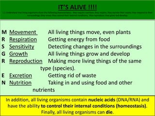
  • 1. IT’S ALIVE !!!! 1.1 Understand that living organisms share the following characteristics: they require nutrition; they respire; they excrete their waste; they respond to their surroundings; they move; they control their internal conditions; they reproduce; they grow and develop. M Movement All living things move, even plants R Respiration Getting energy from food S Sensitivity Detecting changes in the surroundings G Growth All living things grow and develop R Reproduction Making more living things of the same type (species). E Excretion Getting rid of waste N Nutrition Taking in and using food and other nutrients In addition, all living organisms contain nucleic acids (DNA/RNA) and have the ability to control their internal conditions (homeostasis). Finally, all living organisms can die.
  • 2. ALIVE OR NOT ALIVE!! ALIVE OR NOT ALIVE 1.1 Understand that living organisms share the following characteristics: they require nutrition; they respire; they excrete their waste; they respond to their surroundings; they move; they control their internal conditions; they reproduce; they grow and develop.
  • 3. GROUPS OF LIVING ORGANISMS 1.2 describe the common features shared by organisms within the following main groups: plants, animals, fungi, bacteria, protoctists and viruses, and for each group describe examples and their features as follows (details of life cycle and economic importance are not required)
  • 4. PLANTS Plants: These are multicellular organisms; their cells contain chloroplasts and are able to carry out photosynthesis; their cells have cellulose cell walls; they store carbohydrates as starch or sucrose Plants all have the following in common: 1) Multicellular organisms (made of lots of cells) 2) Cells contain chloroplasts and are able to carry out photosynthesis. Photosynthesis takes simple inorganic molecules and turns them into simple sugar (glucose). Glucose can be turned into complex organic molecules such as carbohydrates.
  • 5. PLANTS Plants: These are multicellular organisms; their cells contain chloroplasts and are able to carry out photosynthesis; their cells have cellulose cell walls; they store carbohydrates as starch or sucrose 3) Cells have cellulose cell walls (cellulose is a carbohydrate)
  • 6. PLANTS Plants: These are multicellular organisms; their cells contain chloroplasts and are able to carry out photosynthesis; their cells have cellulose cell walls; they store carbohydrates as starch or sucrose 4) They store carbohydrates as starch. Carbohydrates are polysaccharides (many sugars/glucose linked together) or sucrose. Sucrose is a disaccharide (two sugars linked together).
  • 7. Examples you need to know: 1) flowering plants, such as a cereals A) Maize PLANTS Examples include flowering plants, such as a cereal (for example maize), and a herbaceous legume (for example peas or beans) B) Wheat
  • 8. PLANTS Examples include flowering plants, such as a cereal (for example maize), and a herbaceous legume (for example peas or beans) 2) Herbaceous legume (e.g. peas or beans). Root nodules
  • 9. ANIMALS Animals: These are multicellular organisms; their cells do not contain chloroplasts and are not able to carry out photosynthesis; they have no cell walls; they usually have nervous coordination and are able to move from one place to another; they often store carbohydrate as glycogen 1) Animals include: sponges, molluscs, worms, starfish, insects, crustaceans, fish, amphibians, reptiles, birds, mammals Animals without a backbone (vertebral column) are invertebrates. Animals with a vertebral column are called vertebrates. 2) Animals are Multicellular organisms
  • 10. ANIMALS Animals: These are multicellular organisms; their cells do not contain chloroplasts and are not able to carry out photosynthesis; they have no cell walls; they usually have nervous coordination and are able to move from one place to another; they often store carbohydrate as glycogen 3) They have a nervous system 4) They often store carbohydrate as glycogen in the liver and muscles.
  • 11. ANIMALS Animals: These are multicellular organisms; their cells do not contain chloroplasts and are not able to carry out photosynthesis; they have no cell walls; they usually have nervous coordination and are able to move from one place to another; they often store carbohydrate as glycogen 5) Animal cells do not contain chloroplasts and are not able to carry out photosynthesis 6) Animal cells have no cell walls
  • 12. ANIMALS Animals: These are multicellular organisms; their cells do not contain chloroplasts and are not able to carry out photosynthesis; they have no cell walls; they usually have nervous coordination and are able to move from one place to another; they often store carbohydrate as glycogen
  • 13. ANIMALS Examples include mammals (for example humans) and insects (for example housefly and mosquito) Examples you need to know: 1) Human (mammal)
  • 14. 2) Insects Examples include mammals (for example humans) and insects (for example housefly and mosquito) a) Housefly ANIMALS Why the housefly? - They regurgitate stomach enzymes onto their food - but also because other flies are interesting A) Tsetse fly B) Screw fly C) Drosophila
  • 15. ANIMALS Examples include mammals (for example humans) and insects (for example housefly and mosquito) Lastly….. the use of flies ended the belief in the spontaneous generation of life
  • 16. Examples include mammals (for example humans) and insects (for example housefly and mosquito) b) Mosquito ANIMALS Video Bite So why the Mosquito? (it is really just another fly!) - Complex life cycle - Can be a vector for malaria, yellow fever, abrovirus - Control measures have damaged ecosystems
  • 17. FUNGI Fungi: These are organisms that are not able to carry out photosynthesis; their body is usually organised into a mycelium made from thread-like structures called hyphae, which contain many nuclei; some examples are single-celled; their cells have walls made of chitin; they feed by extracellular secretion of digestive enzymes onto food material and absorption of the organic products; this is known as saprotrophic nutrition; they may store carbohydrate as glycogen Characteristics of Fungi: 1) They feed by saprophytic nutrition (feeds on dead organic material & digestion takes place outside the organism. 2) Fungi feed by excreting extracellular secretions of digestive enzymes onto food and absorbing the digested products. 3) Cells are joined together to form threads, called hyphae. Hyphae contain many nuclei, because they are made from many cells. 4) Hyphae join together to make a network of threads called mycelium
  • 18.
  • 19. FUNGI ARE NOT PLANTS 5) Cells do not contain chloroplasts and are not able to carry out photosynthesis 6) Cell walls are made from a protein called chitin (not cellulose) 7) They store carbohydrates as glycogen (not starch) FUNGI Fungi: These are organisms that are not able to carry out photosynthesis; their body is usually organised into a mycelium made from thread-like structures called hyphae, which contain many nuclei; some examples are single-celled; their cells have walls made of chitin; they feed by extracellular secretion of digestive enzymes onto food material and absorption of the organic products; this is known as saprotrophic nutrition; they may store carbohydrate as glycogen
  • 20. FUNGI Fungi Examples include Mucor , which has the typical fungal hyphal structure, and yeast, which is single-celled Examples you must know: 1) Mucor (bread mold) Why do you need to know about Mucor? - Easy to experiment on in the lab (temperature, moisture) - Mucor must use energy to absorb nutrients in it’s hyphae - Was used to produce penicillin, the first antibiotic.
  • 21. FUNGI Fungi Examples include Mucor , which has the typical fungal hyphal structure, and yeast, which is single-celled 2) Yeast (single celled fungi) Why do you need to know about Yeast? - Easy to experiment on in the lab (temperature & sugar concentration) - Yeast is used in the production of beer & bread
  • 22. BACTERIA Bacteria: These are microscopic single-celled organisms; they have a cell wall, cell membrane, cytoplasm and plasmids; they lack a nucleus but contain a circular chromosome of DNA; some bacteria can carry out photosynthesis but most feed off other living or dead organisms Bacteria Characteristics: 1) Made from single cells 2) Cells do not contain a nucleus. Bacteria cells have a small piece of circular DNA instead of a nucleus (a bacterial chromosome/nucleoid). 2) Bacteria also have DNA in the form of circular plasmids 3) most gain their nutrients by saprophytic nutrition (feed off dead organisms) or by parasitic nutrition (feed off living organisms). Some bacteria can carry out photosynthesis. You need to know their structure: TED
  • 23. BACTERIA Bacteria: These are microscopic single-celled organisms; they have a cell wall, cell membrane, cytoplasm and plasmids; they lack a nucleus but contain a circular chromosome of DNA; some bacteria can carry out photosynthesis but most feed off other living or dead organisms Some pretty pictures of bacteria
  • 24. BACTERIA Examples include Lactobacillus bulgaricus , a rod-shaped bacterium used in the production of yoghurt from milk, and Pneumococcus , a spherical bacterium that acts as the pathogen causing pneumonia Example you need to know: 1) Lactobacillus bulgaricus - Used to make yogurt - Produces lactic acid (excretes) - Rod shaped - Not dangerous to humans Why Lactobacillus bulgaricus? - It is used in how human food production - lowers the pH of milk to inhibit harmful to human bacterial growth
  • 25. Examples include Lactobacillus bulgaricus , a rod-shaped bacterium used in the production of yoghurt from milk, and Pneumococcus , a spherical bacterium 2) Pneumococcus BACTERIA that acts as the pathogen causing pneumonia - pathogen causes pneumonia (inflammation of one or both lungs) - killed with antibiotics - spherical bacteria shape Why Pneumococcus? - reduces surface area of alveoli - use of antibiotics in treatment can lead to resistant strains
  • 26. PROTOCTISTS Protoctists: These are microscopic single-celled organisms. When you don’t fit in scientists drop you into the Protoctists group! Protoctists Characteristics: - most are single celled (exception is seaweeds) - Some have animal-like features - protozoa - Some have plant-like features – algae swim
  • 27. PROTOCTISTS Some, like Amoeba, that live in pond water, have features like an animal cell, while others, like Chlorella, have chloroplasts and are more like plants. A pathogenic example is Plasmodium , responsible for causing malaria Examples you need to know: 1) Amoeba - protozoa - live in pond water - Pathogen amoeba can cause Amoebic dysentery (kills 70,000 people every year) Why Amoeba? - Highlights the need for clean drinking water supplies - Uses amoebiasis to feed (similair to phagocytosis) eating
  • 28. Some, like Amoeba, that live in pond water, have features like an animal cell, while others, like Chlorella, have chloroplasts and are more like plants. A 2) Chlorella PROTOCTISTS pathogenic example is Plasmodium , responsible for causing malaria - ‘more’ like plants than animals - have chloroplasts Why Chlorlla? - both animal and plant characteristics - Possible food source (1940’s solution to world hunger) view
  • 29. 3) Plasmodium - Protozoa - pathogen that causes malaria - uses a mosquito as a vector (transfer) - not killed by antibiotics Why plasmodium? - it is the greatest pathogen killer! TED PROTOCTISTS Some, like Amoeba, that live in pond water, have features like an animal cell, while others, like Chlorella, have chloroplasts and are more like plants. A pathogenic example is Plasmodium , responsible for causing malaria
  • 30. VIRUS Viruses: These are small particles, smaller than bacteria; they are parasitic and can reproduce only inside living cells; they infect every type of living organism. They have a wide variety of shapes and sizes; they have no cellular structure but have a protein coat and contain one type of nucleic acid, either DNA or RNA Viruses Characteristics: 1) Much smaller than bacteria. 1) They are not made from cells (no cellular structures) 3) Totally parasitic and reproduce inside host cells. 3) They infect every type of living cell 3) Have either DNA or RNA as genetic material 3) Have a protein coat
  • 31. VIRUS Viruses: These are small particles, smaller than bacteria; they are parasitic and can reproduce only inside living cells; they infect every type of living organism. They have a wide variety of shapes and sizes; they have no cellular structure but have a protein coat and contain one type of nucleic acid, either DNA or RNA You just have to recognize a virus, but there are several shapes
  • 32. VIRUS Viruses: These are small particles, smaller than bacteria; they are parasitic and can reproduce only inside living cells; they infect every type of living organism. They have a wide variety of shapes and sizes; they have no cellular structure but have a protein coat and contain one type of nucleic acid, either DNA or RNA REVIEW FROM YEAR 8
  • 33. VIRUS Examples include the tobacco mosaic virus that causes discolouring of the leaves of tobacco plants by preventing the formation of chloroplasts, the influenza virus that causes gfluc and the HIV virus that causes AIDS Examples you need to know: 1) Tobacco Mosaic Virus - infects crop plant and prevents formation of chloroplasts - symptoms are discoloration of leaves Why Tobacco Mosaic Virus? - first identification of a virus as a pathogen (1898, Martinus W. Beijerinck) - Genetic engineering
  • 34. VIRUS Examples include the tobacco mosaic virus that causes discolouring of the leaves of tobacco plants by preventing the formation of chloroplasts, the influenza virus that causes gfluc and the HIV virus that causes AIDS 2) Influenza Virus - Viral pathogen causes the flu - Transmitted through air/ contact Why the Flu? - Pandemics (spanish flu) - Mutations leading to new strains - Vaccinations vaccine Flu History
  • 35. Just thought you would like to know.
  • 36. 3) HIV (human immunodeficiency virus) - Pathogen causes AIDS (acquired immunodeficiency syndrome) - Virus attacks white blood cells (immune system) - Host killed by secondary disease - Transmitted by blood to blood contact Why HIV? - Immune system - Mutations VIRUS Examples include the tobacco mosaic virus that causes discolouring of the leaves of tobacco plants by preventing the formation of chloroplasts, the influenza virus that causes gfluc and the HIV virus that causes AIDS
  • 37. PATHOGEN 1.3 recall the term gpathogenc and know that pathogens may be fungi, bacteria, protoctists or viruses. Pathogen: An agent that causes infection or disease, especially a microorganism, such as a bacterium or protozoan, or a virus.
  • 38. LEVELS OF ORGANIZATION 2.1 describe the levels of organisation within organisms: organelles, cells, tissues, organs and systems. Organisms are made from organizations of smaller structures. You need to know the following hierarchy of structures. Organelles - intracellular structures that carry out specific functions within a cell Nucleus Chloroplast Mitochondria Ribosome Vacuole Cells - the basic structural and functional unit from which all biological organisms are made Neurone Skin cell Muscle cell Tissues - a group of specialized cells, which are adapted to carry out a specific function Organs - a collection of two or more tissues, which carries out a specific function or functions Organ Systems - a group of two or more organs Phagocyte Red Blood Cell Muscle Nerves Blood Bone Adipose (Fat) Heart Skin Brain Artery Kidney Pulmonary Cardiac Nervous Endocrine Skeletal
  • 39. LEVELS OF ORGANIZATION 2.1 describe the levels of organisation within organisms: organelles, cells, tissues, organs and systems.
  • 40. CELL STRUCTURE (PLANT & ANIMAL) 2.2 describe cell structures, including the nucleus, cytoplasm, cell membrane, cell wall, chloroplast and vacuole You need to know the differences between plant and animal cells, the functions of the organelles and be able to recognize them in a microscope picture or drawing. Mircro scope
  • 41. CELL STRUCTURE (PLANT & ANIMAL) 2.2 describe cell structures, including the nucleus, cytoplasm, cell membrane, cell wall, chloroplast and vacuole
  • 42. CELL STRUCTURE (PLANT & ANIMAL) 2.3 describe the functions of the nucleus, cytoplasm, cell membrane, cell wall, chloroplast and vacuole Functions of the Organelles (These are the basic definitions you must know) Cytoplasm - site of chemical reactions in the cell Cell Membrane - controls what enters / leaves the cell (selectively permeable) Nucleus - contains nucleic acids, which code for the synthesis of specific proteins. These proteins control all activity in the cell Mitochondrion - site of respiration Chloroplast - site of photosynthesis (contains chlorophyll) Cell Wall - made from cellulose. Strengthens the cell and allows it to be turgid Sap Vacuole - contains the cell sap. Acts as a store of water, or of sugars or, in some cases, of waste products the cell needs to excrete. Helps keep plant cell turgid.
  • 43. PLANTS VS ANIMALS 2.4 compare the structures of plant and animal cells. IF YOU ARE EVER ASKED TO DRAW AND LABLE A CELL IT MUST NOT BE A GENERAL CELL, BUT A SPECIFIC CELL Cell theory
  • 44. CELL STRUCTURE (PLANT & ANIMAL) 2.2 describe cell structures, including the nucleus, cytoplasm, cell membrane, cell wall, chloroplast and vacuole SOME SAMPLE CELL DIAGRAMS: White blood cell SPERM CELL Root hair cell
  • 45. CHEMICAL ELEMENTS OF ORGANIC MOLECULES 2.5 identify the chemical elements present in carbohydrates, proteins and lipids(fats and oils) To be a basic organic molecule you must have: Some have: or even CARBOHYDRATES PROTIENS LIPIDS Carbon Carbon Carbon Hydrogen Hydrogen Hydrogen Oxygen Oxygen Oxygen Nitrogen & Sulphur
  • 46. CHEMICAL ELEMENTS OF ORGANIC MOLECULES 2.5 identify the chemical elements present in carbohydrates, proteins and lipids(fats and oils)
  • 47. Making Complex Organic Structures (molecules) 2.6 describe the structure of carbohydrates, proteins and lipids as large molecules made up from smaller basic units: starch and glycogen from simple sugar; protein from amino acids; lipid from fatty acids and glycerol Components of the main Food Groups: The main food groups are: 1) Carbohydrate 2) Lipids (fats) 3) Proteins DEFINITIONS: Monomer: Single unit Polymer: Two or more monomers chemically combined together All three groups are polymers made from smaller molecules known as monomers. 1) Carbohydrates are large polymer molecules made from one or more monomer sugars. Two carbohydrates you need to know are Starch and Glycogen. Both have glucose as their monomer. 2) Proteins are polymers of Amino Acids (there are 20 amino acids) 3) Lipid polymers are made from one glycerol molecule and three fatty acid molecules joined together. So lipids are made of two different types of monomers.
  • 48. Making Complex Organic Structures (molecules) 2.6 describe the structure of carbohydrates, proteins and lipids as large molecules made up from smaller basic units: starch and glycogen from simple sugar; protein from amino acids; lipid from fatty acids and glycerol CARBOHYDRATE BONDS ARE CALLED: Glycosidic Bonds
  • 49. Making Complex Organic Structures (molecules) 2.6 describe the structure of carbohydrates, proteins and lipids as large molecules made up from smaller basic units: starch and glycogen from simple sugar; protein from amino acids; lipid from fatty acids and glycerol BONDS IN PROTEINS ARE CALLED: Peptide Bonds
  • 50. Making Complex Organic Structures (molecules) 2.6 describe the structure of carbohydrates, proteins and lipids as large molecules made up from smaller basic units: starch and glycogen from simple sugar; protein from amino acids; lipid from fatty acids and glycerol BONDS IN FATTY ACIDS AND GLYCEROL ARE CALLED: Ester Bond
  • 51. REVIEW: Glucose is a Monomer of several Polymers 2.6 describe the structure of carbohydrates, proteins and lipids as largemolecules made up from smaller basic units: starch and glycogen from simple sugar; protein from amino acids; lipid from fatty acids and glycerol Carbohydrate that is the chief form of stored energy in plants Carbohydrate that is the main component of the cell walls of most plants Carbohydrate is stored in the liver and muscles in man and animals
  • 52. Making Cellulose (not required in your syllabus) 2.6 describe the structure of carbohydrates, proteins and lipids as large molecules made up from smaller basic units: starch and glycogen from simple sugar; protein from amino acids; lipid from fatty acids and glycerol
  • 53. Test for Glucose 2.7 describe the tests for glucose and starch NEGATIVE TEST: Blue Solution (No change) POSITIVE TEST: Colour Precipitate (Change) Benedict’s Test: - In test tube with 2 ml of Benedict's reagent. - add 5-6 drops of the test carbohydrate solution and mix well. - Place the test tube in a boiling water bath for 5 minutes. - Observe any change in color or precipitate formation. - Cool the solution. - Observe the colour change from blue to green, yellow, orange or red depending upon the amount of reducing sugar present in the test sample. 0.5% 1% 2%<x
  • 54. Test for Starch 2.7 describe the tests for glucose and starch NEGATIVE TEST: orange/brown Solution (No change) POSITIVE TEST: Black Solution (Change) Iodine Test: - Add 2 drops of iodine solution to about 2 mL of the carbohydrate containing test solution. - A blue-black colour is observed which is indicative of presence of starch.
  • 55. Enzymes AKA Organic Catalysts 2.8 understand the role of enzymes as biological catalysts in metabolic reactions 1) Enzymes are large molecules that speed up the chemical reactions inside cells. 2) Enzymes have a specific job (break/make substances) 3) Enzymes are specific to a particular substrate (protein, carbohydrate, lipid) 4) Enzymes are a type of protein, and like all proteins, they are made from long chains of different amino acids. 5) Enzymes are not used up in the reactions they catalyze (speed up) 6) Enzymes are affected by temperature and pH Enzymes are BIOLOGICAL CATALYSTS
  • 56. So What is an difference between an Inorganic Catalyst and an Enzyme 2.8 understand the role of enzymes as biological catalysts in metabolic reactions Hydrogen peroxide breaks down to water and oxygen hydrogen peroxide water + oxygen manganese oxide 2H2O2 2H2O O2 + The escaping oxygen causes the foaming
  • 57. So What is an difference between an Inorganic Catalyst and an Enzyme 2.8 understand the role of enzymes as biological catalysts in metabolic reactions • They occur inside cells or are secreted by the cells. • Catalase is the enzyme that catalyses the break down of hydrogen peroxide. Catalase
  • 58. Naming Enzymes 2.8 understand the role of enzymes as biological catalysts in metabolic reactions To name an enzyme in most cases just add ‘-ase’ to the ending of the substrate. SUBSTRATE ENZYME Protein Protease Lipid (fats) Lipase Maltose (disaccharide) Maltase Carbohydrate Amylase (it used to be called Other special cases are: Carbohydrase) Specific proteases are Pepsin and Tripsin Catalase increase the rate of H2O2 H20 + O2
  • 59. Enzymes AKA Organic Catalysts 2.8 understand the role of enzymes as biological catalysts in metabolic reactions Enzymes are soluble protein molecules that can speed up chemical reactions in cells. These reactions include : • Respiration • Photosynthesis • Making new proteins For this reason enzymes are called biological catalysts.
  • 60. Enzymes AKA Organic Catalysts 2.8 understand the role of enzymes as biological catalysts in metabolic reactions Each enzyme will only speed up one type of reaction as the shape of the enzyme molecule needs to match the shape of the molecule it reacts with (the substrate molecule). This is called the lock and key model. The part of the enzyme molecule that matches the substrate is called the active site.
  • 61. Rates of enzyme reactions can be measured by recording the time for a substrate to disappear or a product to appear. trypsin Rates of Enzymes 2.8 understand the role of enzymes as biological catalysts in metabolic reactions protein polypeptides white clear Controlled variables: •Volume and concentration of substrate (milk) •Volume and concentration of enzyme (trypsin) •pH (controlled by buffers) •Temperature WHAT KIND OF UNITS WILL RATES OF REACTION HAVE?
  • 62. Temperature’s effect on Enzyme activity 2.9 understand how the functioning of enzymes can be affected by changes in temperature, including changes due to change in active site At low temperatures, enzyme reactions are slow. They speed up as the temperature rises until an optimum temperature is reached. After this point the reaction will slow down and eventually stop. The enzyme activity increases as temperature increases because: 1) More collisions between substrate and enzymes 2) More kinetic energy in each collision between substrate and enzymes 3) More successful collisions because of 1 & 2. The enzyme activity decreasing as temperature increases after a point because: 1) Enzyme’s active site starts to change shape (denature) Enzyme Activity against Temperature Rate Of Reaction Optimum temperature 0 10 20 30 40 50 60 70 Temperature/oC Enzyme is Molecules gain denaturing kinetic energy
  • 63. Temperature’s effect on Enzyme activity 2.9 understand how the functioning of enzymes can be affected by changes in temperature, including changes due to change in active site If the shape of the enzyme changes, its active site may no longer work. We say the enzyme has been denatured. They can be denatured by high temperatures or extremes of pH. Note that it is wrong to say the enzyme has been killed. Although enzymes are made by living things, they are proteins, and not alive. You can investigate the effect of temperature on the enzyme amylase using starch and iodine, putting the mixture in water baths at different temperatures.
  • 64. pH’s effect on Enzyme activity 2.10 understand how the functioning of enzymes can be affected by changes in active site caused by changes in pH (TA) Enzymes and pH Most enzymes work fastest in neutral conditions. Making the solution more acid or alkaline will slow the reaction down. At extremes of pH the reaction will stop altogether. Some enzymes, such as those used in digestion, are adapted to work faster in unusual pH conditions and may have an optimum pH of 2 (very acidic) if they act in the stomach.
  • 65. pH’s effect on Enzyme activity 2.10 understand how the functioning of enzymes can be affected by changes in active site caused by changes in pH (TA) Raising and lowering the pH can: • Make more hydrogen bonds or • Break hydrogen bonds These hydrogen bonds hold the protein’s active site in the correct shape
  • 66. Experiments on Enzymes: TEMPERATURE 2.11 describe experiments to investigate how enzyme activity can be affected by changes in temperature. Amylase Iodine and Starch solution In different temperatures. Measuring: time for iodine test to be negative Yeast and glucose solution vs Temperature. Measuring: CO2 produced (ml) Saliva and Starch solution Vs temperature Measuring: time for iodine test to become negative
  • 67. POSSIBLE CORMMS QUESTIONS TOPICS 2.11 describe experiments to investigate how enzyme activity can be affected by changes in temperature. Enzymes are used in biological washing powders • Proteases break down the coloured, insoluble proteins that cause stains to smaller, colourless soluble polypeptides. • Can wash at lower temperatures Enzymes are used in the food industry • Pectinase break down substances in apple cell walls and enable greater juice extraction. • Lactase breaks down lactose in milk into glucose and galactose. This makes milk drinkable for lactose intolerant people.
  • 68. POSSIBLE CORMMS QUESTIONS TOPICS 2.11 describe experiments to investigate how enzyme activity can be affected by changes in temperature. Enzymes are used in seed germination starch embryo plant amylase secreted maltose
  • 69. Key words catalyst catalyse protein catalase amylase pectinase trypsin pepsin substrate active site product temperature denature enzyme pH optimum lactase protease
  • 70. Movement into and out of a cell DEFINITIONS 2.12 understand definitions of diffusion, osmosis and active transport Diffusion: The net movement of the particles of a gas or a solute from an area of high concentration to an area of low concentration down a concentration gradient. Osmosis: The net movement of water down a concentration gradient from an area of high concentration of water molecules to an area of low concentration of water molecules across a partially permeable membrane. Active transport: The movement of substances against a concentration gradient and/or across a cell membrane, using energy.
  • 71. Movement into and out of a cell Diffusion 2.12 understand definitions of diffusion, osmosis and active transport Diffusion: The net movement of the particles of a gas or a solute from an area of high concentration to an area of low concentration down a concentration gradient. link link
  • 72. Movement into and out of a cell Diffusion 2.12 understand definitions of diffusion, osmosis and active transport In Diffusion experiments you must only change one variable (IV), all other variables must be controlled. Examples are: - Temperature (increases Kinetic energy) - Stirring (increases Kinetic energy) - Surface area of the boundary region - Thickness / distance molecules have to diffuse - The size of the concentration gradient - The surface area to volume ratio
  • 73. Movement into and out of a cell Osmosis 2.12 understand definitions of diffusion, osmosis and active transport Osmosis: The net movement of water down a concentration gradient from an area of high concentration of water molecules to an area of low concentration of water molecules across a partially permeable membrane. Link
  • 74. Movement into and out of a cell Active Transport 2.12 understand definitions of diffusion, osmosis and active transport Active transport: The movement of substances against a concentration gradient and/or across a cell membrane, using energy. This also requires a carrier protein in the cell membrane. link Chemical energy is called ATP.
  • 75. Movement into and out of a cell Review 2.13 understand that movement of substances into and out of cells can be by diffusion, osmosis and active transport In cells molecules can move through the cell membrane by: Diffusion: Small molecules move directly through the cell membrane from high concentration to low concentration. (NO ENERGY REQUIRED) Large molecules move through facilitated diffusion using protein channels from high concentration to low concentration. (NO ENERGY REQUIRED) Osmosis: Water moves from high concentration to low concentration directly through the cell membrane. (NO ENERGY REQUIRED) Active transport: Moves molecules and Ions through the cell membrane from low concentration to high concentration. (ENERGY REQUIRED, CARRIER PROTEIN REQUIRED)
  • 76. TURGID CELLS 2.14 understand the importance in plants of turgid cells as a means of support (TA) EXAMINATION POINTS (step by step) (4 Mark Question) 1) Plant cells are normally turgid (swollen full of water). 2) This is important because it provides strength to plants (rigidity). 3) Plant cells have a cell wall to stop them bursting when turgid. 4) When plant cells start to lose water they become flaccid. 5) Flaccid plants lose their strength and start to wilt. 6) Eventually, flaccid cells become plasmolysed as the cell membrane begins to peel away from the cell wall. 7) This kills the cell.
  • 77. RBC Example (not in syllabus) 2.14 understand the importance in plants of turgid cells as a means of support (TA)
  • 78. Variables affecting movement into and out of cells 2.15 understand the factors that affect the rate of movement of substances into and out of cells, to include the effects of surface area to volume ratio, temperature and concentration gradient VARIABLES THAT AFFECT MOVEMENT RATE OF SUBSTANCES INTO AND OUT OF CELLS: 1) Temperature • As temperature increases movement increases • Eventually increased temperature ruptures the plasma membrane & denatures the enzymes • killing the cell. 2) Concentration Gradient • The higher the concentration gradient of a substance the faster the rate of diffusion • This is only if the substance can cross the plasma membrane (osmosis/water) 3) Surface area/Volume ratio • (Next slide please)
  • 79. Variables affecting movement into and out of cells 2.15 understand the factors that affect the rate of movement of substances into and out of cells, to include the effects of surface area to volume ratio, temperature and concentration gradient If the surface area to volume ratio is too small 1) Living cell can not get nutrients for respiration and growth. 2) Living cells can not remove waste before toxins build up. 3) Cell size is limited by diffusion.
  • 80. Variables affecting movement into and out of cells 2.15 understand the factors that affect the rate of movement of substances into and out of cells, to include the effects of surface area to volume ratio, temperature and concentration gradient WHAT IS THE SURFACE TO VOLUME RATIO’S FOR THESE TWO CELLS?
  • 81. Variables affecting movement into and out of cells 2.15 understand the factors that affect the rate of movement of substances into and out of cells, to include the effects of surface area to volume ratio, temperature and concentration gradient CORMMS QUESTION: Design an experiment that shows how the surface area to volume ratio affects diffusion in agar cubes using a solution of Phenolphthalien (a type of dye). C: O: R: M: M: S:
  • 82. Experiments on Diffusion and Osmosis 2.16 describe experiments to investigate diffusion and osmosis using living and non-living systems. Good examples of diffusion are: - Ink chromatography - The diffusion of KMnO4 crystals (purple) into water - Diffusion of gases in the lung - Diffusion of gases in the leaf - Gas diffusion of Bromine gas Osmosis can be shown by: - Artificially using visking tubing - Potato chips in salt solutions of different concentrations.
  • 84. PLANT NUTRTITION 2.17 describe the process of photosynthesis and understand its importance in the conversion of light energy to chemical energy Nutrition in Plants: Plants are photoautotrophic (i.e. they generate their own “food” using energy from the Sun.) They do this through photosynthesis.
  • 85. Photosynthesis Equation 2.18 write the word equation and the balanced chemical symbol equation for photosynthesis Nutrition in Flowering Plants: The equation for photosynthesis can be written as: -Word equation -Chemical equation In both cases reaction uses a catalyst (chlorophyll)
  • 86. Light ….. Glucose….. ? 2.18 write the word equation and the balanced chemical symbol equation for photosynthesis Through photosynthesis light energy is converted into chemical energy in the bonds in glucose. Plants use glucose for the following; 1) Respiration 2) Stored as Starch 3) Turned into Cellulose (cellulose is a polymer of glucose) 4) Used to make fats and oils
  • 87. Photosynthesis Rate 2.19 understand how varying carbon dioxide concentration, light intensity and temperature affect the rate of photosynthesis At any point the rate of photosynthesis can be increased by adding: 1) More CO2 2) More light WAIT!!!! 3) Heating towards optimum temperature This (photosynthesis is not is catalyzed the by whole enzymes). story
  • 88. Limiting Factors 2.19 understand how varying carbon dioxide concentration, light intensity and temperature affect the rate of photosynthesis a)At a certain point the addition of MORE (light & CO2) will not increase the rate of photosynthesis any further. b)This is because a second factor is limiting the rate of photosynthesis. c)Adding more of the rate-limiting factor will increase the rate further until another factor becomes limiting.
  • 89. Drawing the Graph 2.19 understand how varying carbon dioxide concentration, light intensity and temperature affect the rate of photosynthesis The addition of MORE (light & CO2) will not increase the rate of photosynthesis after reaching a rate limiting factor. What about Temperature?
  • 90. ?Temperature? 2.19 understand how varying carbon dioxide concentration, light intensity and temperature affect the rate of photosynthesis
  • 91. ?Temperature? 2.19 understand how varying carbon dioxide concentration, light intensity and temperature affect the rate of photosynthesis Without enough light, a plant cannot photosynthesize very quickly, even if there is plenty of water and carbon dioxide. 1) Increasing the temperature will boost the speed (rate) of photosynthesis. 2) Increasing the intensity will boost the speed (rate) of photosynthesis.
  • 92. Changing the Limiting Factor 2.19 understand how varying carbon dioxide concentration, light intensity and temperature affect the rate of photosynthesis Adding more of the rate-limiting factor increases the rate further…….............until another factor becomes limiting.
  • 93. What about Water? 2.19 understand how varying carbon dioxide concentration, light intensity and temperature affect the rate of photosynthesis Water is not seen as a limiting factor. Plants have enough water in their tissues for photosynthesis. If they do not have enough water the plant will wilt and die anyway. Very sad, but very true.
  • 94. Leaf Structure 2.20 describe the structure of the leaf and explain how it is adapted for photosynthesis You need to know the parts of the leaf and their adaptations. DO NOT DRAW THIS DIAGRAM
  • 95. SIMPLE CROSS SECTIONAL LEAF DIAGRAM 2.20 describe the structure of the leaf and explain how it is adapted for photosynthesis
  • 96. More Complicated Cross Section 2.20 describe the structure of the leaf and explain how it is adapted for photosynthesis
  • 97. In Real Life 2.20 describe the structure of the leaf and explain how it is adapted for photosynthesis
  • 98. LABEL 2.20 describe the structure of the leaf and explain how it is adapted for photosynthesis
  • 99. Adaptation 2.20 describe the structure of the leaf and explain how it is adapted for photosynthesis
  • 100. Which Tissues Are Missing? 2.20 describe the structure of the leaf and explain how it is adapted for photosynthesis Please add into your notes any tissue missing and write in their functions: 1) Xylem 2) Phloem 3) Vascular Bundle 4) Spongy Mesophyll
  • 101. Minerals for Nutrition 2.21 understand that plants require mineral ions for growth and that magnesium ions are needed for chlorophyll and nitrate ions are needed for amino acids In addition to water and CO2 plants also need specific minerals; • Nitrate – used to make amino acids for use in plant proteins Magnesium – forms part of the chlorophyll molecule • Potassium - essential for cell membranes • Phosphate - essential part of DNA and cell membranes
  • 102. Experiment we CAN NOT do!
  • 103. EXPERIMENTS WE CAN DO 2.22 describe experiments to investigate photosynthesis, showing the evolution of oxygen from a water plant, the production of starch and the requirements of light, carbon dioxide and chlorophyll
  • 104. Using Pond Weed 2.22 describe experiments to investigate photosynthesis, showing the evolution of oxygen from a water plant, the production of starch and the requirements of light, carbon dioxide and chlorophyll You must know an experiment that shows how the rate of photosynthesis is affected by rate-limiting factors. Example: Use pond weed (Elodea) which produces bubbles of O2 as it photosynthesizes. 1) The rate of bubble production is proportional to the rate of photosynthesis. 2) When you add light or give it more CO2, the rate of bubble production increases. Watch out:  Cut Elodea underwater or air bubbles will form in xylem  Make sure the O2 is a result of light and not temperature  The examiner may ask for a better way to measure O2 production
  • 105. Set up for Photosynthesis Rate Vs Light intensity 2.22 describe experiments to investigate photosynthesis, showing the evolution of oxygen from a water plant, the production of starch and the requirements of light, carbon dioxide and chlorophyll Change: Light intensity (distance of lamp from Elodea) Measure: Number of bubbles per minute
  • 106. Setup for Photosynthsis Rate Vs CO2 Concentration 2.22 describe experiments to investigate photosynthesis, showing the evolution of oxygen from a water plant, the production of starch and the requirements of light, carbon dioxide and chlorophyll Change: Concentration of Sodium Hydrogen Carbonate Solution (CO2) Measure: Number of bubbles per minute
  • 107. Testing Photosynthesis by Starch 2.22 describe experiments to investigate photosynthesis, showing the evolution of oxygen from a water plant, the production of starch and the requirements of light, carbon dioxide and chlorophyll You need to know an experiment that proves that light and CO2 are essential for the production of starch. A good example is the Geranium plant. It’s leaves normally turn blue-black in the presence of iodine solution showing starch is present (you have to boil it in ethanol first to remove the chlorophyll to show the colour).
  • 108. Testing Photosynthesis by Starch 2.22 describe experiments to investigate photosynthesis, showing the evolution of oxygen from a water plant, the production of starch and the requirements of light, carbon dioxide and chlorophyll Negative Test: Reddish / Brown Positive Test: Blue / Black Safety: Why is it dangerous to boil ethanol directly with a Bunsen Burner instead of using a water bath?
  • 109. Destarching 2.22 describe experiments to investigate photosynthesis, showing the evolution of oxygen from a water plant, the production of starch and the requirements of light, carbon dioxide and chlorophyll You will want to destarch a leaf for this experiment. To remove the starch (destarch) 1) put the poor plant in a dark room for 24 hours. 2) No light means no photosynthesis, no photosynthesis means no glucose produced, no glucose produced means no starch stored in the leaf. Sadly the leaf still needs to respire so it will break all the previously stored starch back into glucose to use in respiration. No more starch, poor leaf…
  • 110. Destarching 2.22 describe experiments to investigate photosynthesis, showing the evolution of oxygen from a water plant, the production of starch and the requirements of light, carbon dioxide and chlorophyll However, if one leaf is put in aluminium foil and another is kept with lime water both do not turn blue-black. Both CO2 and light are essential for starch production and, therefore, essential for photosynthesis.
  • 111. Nutrition in Humans Syllabus points 2.23 – 2.32
  • 112. Balanced Diet 2.23 understand that a balanced diet should include appropriate proportions of carbohydrate, protein, lipid, vitamins, minerals, water and dietary fibre(TA) A diet that contains adequate amounts of all the necessary nutrients required for healthy growth and activity. A balanced diet is one that contains all the ingredients needed for our body to healthily continue its day to day functions in the most efficient way.
  • 113. Balanced Diet 2.23 understand that a balanced diet should include appropriate proportions of carbohydrate, protein, lipid, vitamins, minerals, water and dietary fibre(TA)
  • 114. Balanced Diet 2.23 understand that a balanced diet should include appropriate proportions of carbohydrate, protein, lipid, vitamins, minerals, water and dietary fibre(TA) 72% of our body is WATER. We contain so much water because water: -Distributes essential nutrients to cells, such as minerals, vitamins and glucose as part of the plasma in our blood -Is an integral part of urine and faeces, which removes waste from our body -Is needed for sweat (sweat is essential in controlling our internal body temperature)
  • 115. What do you have to eat 2.24 identify sources and describe functions of carbohydrate, protein, lipid (fats and oils), vitamins A, C and D, and the mineral ions calcium and iron, water and dietary fibre as components of the diet Component Function Example of sources Carbohydrate Short-term chemical energy Bread, potatoes Lipids (fats and oils) Long-term chemical energy Bacon, beef Protein Growth & Repair Fish, egg Vitamin A Eyesight Carrots, fish liver oil Vitamin C Healthy skin + gums Oranges Vitamin D Absorb Ca (calcium) Sunlight Mineral ions – Fe (iron) Making haemoglobin in RBC Spinach, animal liver Mineral ions – Ca (calcium) Strong bones and teeth milk Dietary fiber Peristalsis Vegetables, cereal Water Transport system To sweat All chemical reactions occur in solution inside cells Fruits like watermelon
  • 116. Not all bodies are Energy (J) Equal 2.25 understand that energy requirements vary with activity levels, age and pregnancy (TA) Person Energy needed per day (kJ) Newborn baby 2000 Age 2 5000 Age 6 7500 Gril age 12-14 9000 Boy age 12-14 11000 Girl age 15-17 9000 Boy age 15-17 12000 Female office worker 9500 Male office worker 10500 Heavy manual worker 15000 Pregnant woman 10000 Breast-feeding woman 11300
  • 117. Not all bodies are Energy (J) Equal 2.25 understand that energy requirements vary with activity levels, age and pregnancy (TA) The two groups that provide energy (through respiration) are lipids and carbohydrates. Per mass lipids have about 10x more energy in them than carbohydrates. The energy in food is measured in Calories (equivalent to 4.2 kJ). If Males need to consume 2500 Calories a day and Females need to consume 2000 Calories a day how many kJ do they need to consume in a day? If: Fat: 1 gram = 9 calories Carbohydrates: 1 gram = 4 calories How many grams of each do you need to supply your energy for the day?
  • 118. Not all bodies are Energy (J) Equal 2.25 understand that energy requirements vary with activity levels, age and pregnancy (TA) Energy requirements vary according to several factors: • Age: growing people require more energy than others. • Gender: on average, males require more energy than females. • Pregnancy: pregnant women require more energy to nourish themselves and the baby. • Activity levels: more active people require more energy as they use up more energy throughout the day.
  • 119. Name that structure 2.26 describe the structures of the human alimentary canal and describe the functions of the mouth, oesophagus, stomach, small intestine, large intestine and pancreas game
  • 120. Describe the function 2.26 describe the structures of the human alimentary canal and describe the functions of the mouth, oesophagus, stomach, small intestine, large intestine and pancreas • Functions Mouth • Physical digestion by teeth • Salivary glands produce saliva  moistens food  making it easier to be swallowed • Chemical digestion by amylase breaks down starch into maltose Oesophagus • Food is moved by peristalsis Stomach • Produces HCl & protease (pepsin) enzymes Small intestine • Produces carbohydrase (maltase), protease (trypsin) & lipase enzymes • Absorbs digested food Large intestine • Absorbs water Pancreas • Produces carbohydrase (maltase), protease (trypsin) & lipase enzymes
  • 121. Flow Chart the Process 2.27 understand the processes of ingestion, digestion, absorption, assimilation and egestion Ingestion • Taking food into the body Digestion • The breakdown of large insoluble molecules into small soluble molecules so they can be absorbed into the blood Absorption • The process of absorbing nutrients into the body after digestion Assimilation • Using food molecules to build new molecules Egestion • Getting rid of undigested/unwan ted food
  • 122. Flow Chart the Process 2.27 understand the processes of ingestion, digestion, absorption, assimilation and egestion Digestion can be mechanical or chemical Mechanical Digestion: digestion by physically breaking food into smaller pieces (i.e. not using enzymes). Carried out by; • mouth and teeth chewing food • stomach churning food Chemical Digestion: digestion using enzymes
  • 123. Peristalsis 2.28 explain how and why food is moved through the gut by peristalsis Food is moved the digestive system by a process known as peristalsis. This is the contractions of two sets of muscles in the walls of the gut. 1) One set runs along the gut 2) The other set circles it. Their wave-like contractions create a squeezing action, moving down the gut. ani
  • 124. Digestive Enzymes 2.29 understand the role of digestive enzymes, to include the digestion of starch to glucose by amylase and maltase, the digestion of proteins to amino acids by proteases and the digestion of lipids to fatty acids and glycerol by lipases Enzymes and digestion The enzymes involved in respiration, photosynthesis and protein synthesis work inside cells. Other enzymes are produced by specialised cells and released from them these are digestive enzymes. They pass out into the gut, where they catalyse the breakdown of food molecules. Different enzymes (Different enzymes catalyse different digestion reactions) Amylase Starch → sugars Amylase catalyses the breakdown of starch into sugars in the mouth and small intestine Protease Proteins → amino acids Proteases catalyse the breakdown of proteins into amino acids in the stomach and small intestine Lipase Lipids → fatty acids + glycerol Lipases catalyse the breakdown of fats and oils into fatty acids and glycerol in the small intestine
  • 125. Bile is not so Vile 2.30 understand that bile is produced by the liver and stored in the gall bladder, and understand the role of bile in neutralising stomach acid and emulsifying lipids After the stomach, food travels to the small intestine. The enzymes in the small intestine work best in alkaline conditions, but the food is acidic after being in the stomach. • Bile is alkaline substance • Bile is produced by the liver • Bile is stored in the gall bladder. • Bile is secreted into the small intestine, where it emulsifies fats This is important, because it provides a larger surface area in which the lipases can work.
  • 126. Silli Villi 2.31 describe the structure of a villus and explain how this helps absorption of the products of digestion in the small intestine The Villus is the location of Absorption of small soluble nutrients into to blood.
  • 127.
  • 128. How much energy is in that crisp? 2.32 describe an experiment to investigate the energy content in a food sample.(TA) You need to know an experiment that can show how much energy there is in food. Burn a sample of food and use it to heat a fixed volume of water. Record the change in temperature of the water and use the equation below to find out the energy the food gave to the water; Energy = change in temp. x volume of water x 4.2J/g/°C Problem is that not all the food will burn. To control this, you measure the start and end mass of the food and calculate the mass that actually burned. To standardize this, you can divide your calculated energy value by the change in mass to give you the change in mass per gram of food (which will allow you to compare values fairly between different food samples)
  • 129. How much energy is in that peanut? 2.32 describe an experiment to investigate the energy content in a food sample.(TA) There are problems with using this system: Heat from food item does not heat water Not all the food burns Water looses heat to environment So what is the solution?
  • 130. Respiration releases ENERGY 2.33 understand that the process of respiration releases energy in living organisms
  • 131. Aerobic and Anerobic 2.34 describe the differences between aerobic and anaerobic respiration
  • 132. Word and Chemical Equation 2.35 write the word equation and the balanced chemical symbol equation for aerobic respiration in living organisms
  • 133. Anaerobic Repiration Word Equation 2.36 write the word equation for anaerobic respiration in plants and in animals
  • 134. Experiment for Respiration 2.37 describe experiments to investigate the evolution of carbon dioxide and heat from respiring seeds or other suitable living organisms.
  • 135. Gas Exchange 2.38 understand the role of diffusion in gas exchange
  • 136. GAS EXCHANGE IN PLANTS 2.39 understand gas exchange (of carbon dioxide and oxygen) in relation to respiration and photosynthesis CO2 and O2 diffuse in and out of leaves through stomata. CO2 is used in photosynthesis and produced by respiration, whereas O2 is used in respiration and produced in photosynthesis!
  • 137. Photosynthesis & Respiration 2.39 understand gas exchange (of carbon dioxide and oxygen) in relation to respiration and photosynthesis Both processes run all the time. So the net amount of glucose the plant produces (i.e. the amount it gets to use for growth etc) is governed by the formula; Net Glucose = Total production – Amount used in respiration The Compensation Point is defined as; 0 = Total production – Amount used in respiration Or Photosynthesis = Respiration
  • 138. Light Intensity & Gas Exchange 2.40 understand that respiration continues during the day and night, but that the net exchange of carbon dioxide and oxygen depends on the intensity of light (TA) The amount of glucose the plant uses in respiration in nearly constant. However, glucose production by photosynthesis is not. It is dependent on the rate-limiting factors (i.e. light intensity, CO2 level, water availability, temperature etc). At night photosynthesis is virtually zero. (Net Carbon Dioxide production) In the day the photosynthesis is large. (Net Oxygen production)
  • 139. Light Intensity & Gas Exchange 2.40 understand that respiration continues during the day and night, but that the net exchange of carbon dioxide and oxygen depends on the intensity of light (TA)
  • 140. Leaf Structure and Photosynthesis 2.41 explain how the structure of the leaf is adapted for gas exchange
  • 141. Stomata and Gas Exchange 2.42 describe the role of stomata in gas exchange In sunlight the guard cell becomes turgid Turgid guard cells open the stoma Increases gas exchange Low light causes guard cells to become flaccid Flaccid guard cells close the stoma Decreases water loss
  • 142. Stomata and Gas Exchange 2.42 describe the role of stomata in gas exchange Exp 1 Exp 2
  • 143. Stomata and Gas Exchange 2.42 describe the role of stomata in gas exchange Potometer 1) You must cut the shoots under water and you must assemble the potometer under water. If air gets into the xylem vessels of the plant, it can form air locks which will prevent the plant taking up water and so prevent steady transpiration. 1) Check all seals are airtight – coat seals with Vaseline jelly 3) The potometers should be left for the leaves to dry. Alternatively dry the leaves gently with a paper towel. The potometer will not work properly until any excess water on the leaves has evaporated or been removed. 2) Adding food colouring to the water makes it easier to see the air bubble in the capillary tube.
  • 144. Stomata and Gas Exchange 2.42 describe the role of stomata in gas exchange
  • 145. Experiment to Know 2.43 describe experiments to investigate the effect of light on net gas exchange from a leaf, using hydrogen-carbonate indicator (TA) An experiment which will show the effect of light intensity on the rate of gas exchange. - Seal two leaves (still attached to the plant) in separate plastic bags with some bicarbonate indicator solution. - One of the bags is black and the other is translucent. - The leaf in the black bag produces CO2 via respiration and the colour of the bicarbonate indicator changes quickly to yellow. - The leaf in the translucent bag produces O2 via photosynthesis and the bicarbonate indicator solution changes to red slowly. Bicarbonate Indicator colours: Red in the absence of CO2 Yellow in the presence of CO2
  • 146. Gas Exchange in Humans Syllabus points 2.44 – 2.48
  • 147. Gas Exchange in Humans Syllabus points 2.44 – 2.48
  • 148. Gas Exchange in Humans Syllabus points 2.44 – 2.48
  • 149. Gas Exchange in Humans Syllabus points 2.44 – 2.48
  • 150. Transport syllabus points 2.49 – 2.50
  • 151. Transport syllabus points 2.49 – 2.50
  • 152. Plant Transport (2.51 – 2.56) 2.51 describe the role of phloem in transporting sucrose and amino acids between the leaves and other parts of the plant
  • 153. Plant Transport (2.51 – 2.56) 2.51 describe the role of phloem in transporting sucrose and amino acids between the leaves and other parts of the plant
  • 154. Plant Transport (2.51 – 2.56) 2.51 describe the role of phloem in transporting sucrose and amino acids between the leaves and other parts of the plant
  • 155. Plant Transport (2.51 – 2.56) 2.51 describe the role of phloem in transporting sucrose and amino acids between the leaves and other parts of the plant
  • 156. Plant Transport (2.51 – 2.56) 2.51 describe the role of phloem in transporting sucrose and amino acids between the leaves and other parts of the plant
  • 157. Transport in Humans (2.57 – 2.66)
  • 158. Transport in Humans (2.57 – 2.66)
  • 159. Transport in Humans (2.57 – 2.66)
  • 160. Transport in Humans (2.57 – 2.66)
  • 161. Transport in Humans (2.57 – 2.66)
  • 162. Transport in Humans (2.57 – 2.66)
  • 163. Transport in Humans (2.57 – 2.66)
  • 164. Transport in Humans (2.57 – 2.66)
  • 165. Transport in Humans (2.57 – 2.66)
  • 166. Excretion in Plants 2.67 understand the origin of carbon dioxide and oxygen as waste products of metabolism and their loss from the stomata of a leaf
  • 167. Excretion in Humans (2.68 – 2.76) 2.68 recall that the lungs, kidneys and skin are organs of excretion Humans have 3 main excretory organs 1) Lungs – excrete CO2 and H2O 2) Skin – excretes H2O 3) Kidneys – excrete H2O, urea, excess minerals and other wastes. Parts game
  • 168. UREA 2.68 recall that the lungs, kidneys and skin are organs of excretion What’s urea? (not technically on syllabus) • All organisms produce ammonia as they metabolize nutrients (protein digestion/amino acids) • Ammonia is a nitrogenous waste that is toxic and must be removed from the body Solution: the liver turns the ammonia into urea, which is harmless. Therefore urea is a product of the metabolism of amino acids.
  • 169. UREA 2.68 recall that the lungs, kidneys and skin are organs of excretion Many land animals and some bony fish (amphibians, mammals) dilute the toxic ammonia with water. This substance is called Urea & is filtered out by the kidneys. - The problem is they do lose water in the process - Requires Energy
  • 170. Excretion & Osmoregulation 2.69 understand how the kidney carries out its roles of excretion and osmoregulation The Kidney: The functional unit of the kidney is the nephron. There are millions of nephrons in a single kidney. Nephrons have 2 jobs; Excretion - filtering the blood and reclaiming the “good bits” & removing waste Osmoregulation - balancing the water level of the body (water homeostasis)
  • 171. Excretion System 2.70 describe the structure of the urinary system, including the kidneys, ureters, bladder and urethra
  • 172. Excretion System 2.70 describe the structure of the urinary system, including the kidneys, ureters, bladder and urethra
  • 173. Kidney Nephron and the Kidney 2.71 describe the structure of a nephron, to include Bowmancs capsule and glomerulus, convoluted tubules, loop of Henlé and collecting duct
  • 174. Nephron and capilliaries 2.71 describe the structure of a nephron, to include Bowmancs capsule and glomerulus, convoluted tubules, loop of Henlé and collecting duct
  • 175. How the Nephron works 2.71 describe the structure of a nephron, to include Bowmancs capsule and glomerulus, convoluted tubules, loop of Henlé and collecting duct 2.74 understand that selective reabsorption of glucose occurs at the proximal convoluted tubule How the nephron works: 1) Dirty blood enters the kidney via the afferent artery 2) The artery splits up into a ball of capillaries, called the glomerulus 3) The blood is under high pressure, so all small substances are forced out of the holes in the capillary walls. Only large proteins and cells stay behind. 4) The small substances (glucose, minerals, urea, water etc) move into the bowman’s capsule, which wraps around the glomerulus 5) The capsule leads into the PCT, which re-absorbs all glucose via active transport (i.e. it selectively removes the glucose from the nephron and returns it to the blood)
  • 176. How the Nephron works 2.71 describe the structure of a nephron, to include Bowmancs capsule and glomerulus, convoluted tubules, loop of Henlé and collecting duct 6) The PCT leads to the Loop of Henlé, which re-absorbs the water by osmosis 7) The Loop leads to the DCT, which re-absorbs all minerals, amino acids and other “useful” substances by active transport 8) The remaining fluid (containing excess water, excess minerals and urea) passes into the collecting duct 9) The collecting ducts from other nephrons join and form the ureter, which leads to the bladder 10) The fluid is now called urine and is stored in the bladder for excretion 11) The bladder takes the urine to the outside world via the urethra This is the first role of the nephron (it’s role in excretion). Remember, the nephron has a second role in osmoregulation.
  • 177. Excretion in Humans (2.68 – 2.76) 2.71 describe the structure of a nephron, to include Bowmancs capsule and glomerulus, convoluted tubules, loop of Henlé and collecting duct Kidney
  • 178. Excretion in Humans (2.68 – 2.76) 2.72 describe ultrafiltration in the Bowmancs capsule and the composition of the glomerular filtrate The glomerulus filters blood and produces glomerular filtrate. This filtrate contains: water, glucose, salts and urea (amino acids). (Large molecules such as protein are too large to fit through the blood capillary walls.)
  • 179. Excretion in Humans (2.68 – 2.76) 2.73 understand that water is reabsorbed into the blood from the collecting duct Teach Blood water levels are sensed by the hypothalamus in the brain. When water levels are too low, the hypothalamus tells the pituitary gland (also in the brain) to release the hormone Anti-Diuretic Hormone (ADH)
  • 180. Excretion in Humans (2.68 – 2.76) 2.73 understand that water is reabsorbed into the blood from the collecting duct 2.75 describe the role of ADH in regulating the water content of the blood When blood water levels are too low; 1) Hypothalamus detects 2) Pituitary gland releases ADH into bloodstream 3) ADH travels all over the body 4) Only the cells in the collecting duct of the nephrons of the kidney have receptors for ADH, so only they respond to the hormone 5) The collecting duct becomes more permeable 6) Water is draw out of the collecting duct back into the blood 7) Water levels return to normal BBC
  • 181. Review of Urine 2.76 understand that urine contains water, urea and salts. The waste, consisting of: • excess water • excess salts • urea is urine. This process can be summarized in three important steps: Revision 1) Ultra-Filtration - where lots of water, ions, urea and sugar are squeezed from the blood into the tubules. 2) Selective reabsorption – the useful substances (ions and sugars) are reabsorbed back into the blood from the tubules. The amount of water in the blood is regulated here to maintain it at a constant rate. This is known as ‘osmoregulation’. 3) Excretion of waste - urea and excess water and ions travel to the bladder as urine, to be released from the body.
  • 182. Coordination and Response Homeostasis: the maintenance of a constant internal environment
  • 183. The S in MRS GREN 2.77 understand that organisms are able to respond to changes in their environment M Movement All living things move, even plants R Respiration Getting energy from food S Sensitivity Detecting changes in the surroundings G Growth All living things grow R Reproduction Making more living things of the same type E Excretion Getting rid of waste N Nutrition Taking in and using food SENSITIVITY A stimulus is a change in the environment of an organism. Animals respond to a stimulus in order to keep themselves in favourable conditions. Examples of this include: • moving to somewhere warmer if they are too cold • moving towards food if they are hungry • moving away from danger to protect themselves Animals that do not respond to a stimulus do not survive for long.
  • 184. Homeostasis 2.78 understand that homeostasis is the maintenance of a constant internal environment and that body water content and body temperature are both examples of homeostasis All organisms try and maintain a constant internal environment. This is called homeostasis. Examples of homeostasis include: 1) The regulation of water levels. 2) The regulation of body temperature.
  • 185. Stimulus > Receptor> Coordination > Effector > Response 2.79 understand that a coordinated response requires a stimulus, a receptor and an effector • Both systems respond to stimuli (i.e. events that change the internal environment). • Both systems have a: 1) Receptor, which detects the stimulus. 2) Effector, which carries out a response to correct the effect of the stimulus. The message from detector to effector is carried either via an electrical nerve impulse or as a hormone, depending which homeostatic system is being used.
  • 186. STIMULI 2.79 understand that a coordinated response requires a stimulus, a receptor and an effector
  • 187. Examples of Receptors and Effectors 2.79 understand that a coordinated response requires a stimulus, a receptor and an effector Receptor ORGAN RECEPTOR skin Temperature Receptor skin Pressure / Pain Receptor Brain (hypothalamus) Water Concentration Receptor Eye (retina) Light Receptors (Rods & Cones) Effectors ORGAN EFFECTOR Heart Muscle cell Skin Sweat gland Kidney Collecting duct walls
  • 188. STIMULI 2.79 understand that a coordinated response requires a stimulus, a receptor and an effector Receptors detect Stimuli. Some examples are: a) Light Eye (retina) b) Sound Ear (hearing) c) Movement (K.E) Ear (balance) / Skin d) Chemical Nose / Tongue e) Heat Skin
  • 189. Response in Plants 2.80 understand that plants respond to stimuli Plants also respond to stimuli. As plants don’t have nerves their responses are limited to hormones only. Plants respond to the following stimuli: • Gravity: Roots grow towards gravitational pull and stems grow away. This is Geotropism. • Water: Roots grow towards water. This is Hydrotropism. • Light: Shoots grow towards light. This is Phototropism.
  • 190. GEOTROPISM 2.81 describe the geotropic responses of roots and stems Shoot tips and Root tips respond to GRAVITY. • Shoot tips grow away from gravity (Negative Geotropism) • Root tips grow in the direction of gravity (Positive Geotropism)
  • 191. WHY You only need to know that plants respond to a chemical hormone called AUXIN BUT if you want more……… 2.81 describe the geotropic responses of roots and stems
  • 192. 2.81 describe the geotropic responses of roots and stems
  • 193. 2.81 describe the geotropic responses of roots and stems You do need to know some fun experiments with plants
  • 194. 2.81 describe the geotropic responses of roots and stems What happens when you grow a plant in space?
  • 195. Positive Phototropism 2.82 describe positive phototropism of stems Positive Phototropism is controlled by hormones released by the growing tip of the shoot. (Only the tip makes the hormone) If you remove the tip, the shoot stops growing. The hormone made by the tip is called Auxin (which part of the plant would be controlled by Negative Phototropism?)
  • 196. 2.82 describe positive phototropism of stems
  • 197. MORE EXPERIMENTS 2.82 describe positive phototropism of stems
  • 198. 2.82 describe positive phototropism of stems What if you grow a plant in the dark?
  • 199. Response Systems 2.83 describe how responses can be controlled by nervous or by hormonal communication and understand the differences between the two systems Humans have two systems which carry out detection and response: • Nervous System – Chemical electrical system using Neurons Example: Iris dilation, movement, ventilation • Endocrine System –Chemical system using proteins called hormones in the blood. Example: Osmoregulation, Fight or Flight, Menstruation.
  • 200. Differences 2.83 describe how responses can be controlled by nervous or by hormonal communication and understand the differences between the two systems Nervous System Endocrine System Works by nerve impulses (has chemicals in synapses though) Works by hormones transmitted in blood stream Travel fast and usually have ‘instant’ effect Travel slowly and may take longer to ask Response is short lived Response is usually longer lasting Impulse act on individual cells (localised effect) Widespread effects on different organs (still only work on cells/organs with correct receptors)
  • 201. Nervous System 2.84 understand that the central nervous system consists of the brain and spinal cord and is linked to sense organs by nerves The Central Nervous System (CNS) consists of 1) the brain 2) the spinal cord There are also Peripheral Nerves System (PNS) 1) Sensory Nerves/organs (e.g. pain receptors in skin, or photoreceptors in the eye) 2) nerves that link brain and sense organs 3) Motor Nerves
  • 202. 2.85 understand that stimulation of receptors in the sense organs sends electrical impulses along nerves into and out of the central nervous Stimulation of the sense organs results in an electrical signal (a nerve impulse) being sent along the nerve to the brain. Nerve impulses are very quick (~120m/s), allowing rapid responses to the stimulus. nerve impulse Nerve Impulse synaptic cleft receptor system, resulting in rapid responses
  • 203. DIFFUSION AGAIN?? 2.85 understand that stimulation of receptors in the sense organs sends electrical impulses along nerves into and out of the central nervous system, resulting in rapid responses (You do not have to know the terms just the ideas) 1) An electrical impulse travels along a nerve ending. 2) This triggers the nerve-ending of a neuron to release chemical messengers called neurotransmitters. 3) These chemicals diffuse across the synapse (the gap) and bind with receptor molecules on the membrane of the next neuron. 4) The receptor molecules on the second neuron bind only to the specific chemicals released from the first neuron. This stimulates the second neuron to transmit the electrical impulse.
  • 204. REFLEXS 2.86 describe the structure and functioning of a simple reflex arc illustrated by the withdrawal of a finger from a hot object Some sense organs are not connected directly to the brain. This is a defence mechanism allowing almost instant responses to threatening or dangerous stimuli (e.g. pain/hot object). These instant responses are controlled by nerves in the spine, rather than the brain and are called reflexes
  • 205. REFLEX ARC 2.86 describe the structure and functioning of a simple reflex arc illustrated by the withdrawal of a finger from a hot object Relay Neuron
  • 206. STEP BY STEP REFLEX ARC 2.86 describe the structure and functioning of a simple reflex arc illustrated by the withdrawal of a finger from a hot object 1) A stimulus is detected by a receptor 2) The receptor initiates a nerve impulse in the sensory nerve 3) The sensory nerve (which runs from the receptor to the spine) passes the message/impulse onto an interneuron/Relay Neuron in the spine 4) The interneuron/relay neuron passes the message on the a motor nerve 5) The motor nerve (which runs from the spine to a muscle in the same limb as the receptor) passes the message onto the effector muscle 6) The effector muscle carries out the response (muscle contraction).
  • 207. Reflex Arc Timing 2.86 describe the structure and functioning of a simple reflex arc illustrated by the withdrawal of a finger from a hot object The entire process (stimulus to response) happens in less than a second and does not involve the brain. The purpose of the interneuron is also to inform the brain of what has happened. Reflex Summary 1) Immediate response to stimulus 2) Automatic reactions 3) Limits damage to organism
  • 209. EyE WebSites 2.87 describe the structure and function of the eye as a receptor a) Label the eye http://www.kscience.co.uk/animations/eye_drag.htm b) Function of the eye http://www.kscience.co.uk/animations/eye_function_drag.htm c) Label the eye http://www.freezeray.com/flashFiles/eye.htm d) Light and the eye http://www.kscience.co.uk/animations/eye.htm e) Fun with your blind spot http://www.med.yale.edu/neurobio/mccormick/fill_in_seminar/figure1.htm
  • 210. EYE DIAGRAM 2.87 describe the structure and function of the eye as a receptor Conjunctiva
  • 211. Define these Terms 2.87 describe the structure and function of the eye as a receptor • Ciliary Body (muscle) • Iris • Pupil • Cornea • Lens • Suspensory Ligament • Sclera • Choroid • Retina • Fovea • Optic Disk (blind spot) • Optic Nerve • Conjunctiva Contracts to Relaxes to change shape of lens Coloured muscular part of eye that controls pupils size Opening in the iris that lets light through to the retina Transparent front part of the eye that refracts light Transparent part of the eye that bends light (refracts) Ligament that controls the shape of the lens Dense white covering of the eye Vascular membrane of the eyeball between the sclera and the retina & stops light reflecting around in the eye Light sensitive membrane that converts light to electrical signals to the brain Part of the retina with a high density of cones for very sharp vision Area of retina that is insensitive to light Where electrical impulses from retina travel to brain helps lubricate the eye by producing mucus and tears
  • 212. RODS & CONES 2.87 describe the structure and function of the eye as a receptor Light is detected by photoreceptors in the eye. These receptors form the retina (the inner lining of the eye). There are two types of photoreceptor Rods, which see only in & & Cones, which see in either red, blue or green (3 types of cones) Hint: Cone and Colour both start with ‘C’
  • 213. Automatic Reflexs 2.88 understand the function of the eye in focusing near and distant objects, and in responding to changes in light intensity (TA) There are two types of reflex you need to know about in the eye 1) Responding to different light levels 2) Focusing the eye
  • 214. Responding to Light Levels (Antagonistic Muscle Pairs at Work) 2.88 understand the function of the eye in focusing near and distant objects, and in responding to changes in light intensity (TA) Condition (Stimulus) Bright light Dim light Receptors More photoreceptors stimulated Less photoreceptors stimulated Impulses More impulses sent to the brain via optic nerve Fewer impulses sent to the brain via optic nerve Effectors Radial muscles of the iris relax Radial muscles of the iris contract Circular muscles of the iris contract Circular muscles of the iris relax Gross Effect Pupil constricts (becomes smaller) Pupil dilates (becomes larger) Less light enters the eye More light enters the eye
  • 215. FOCUSING THE EYE 2.88 understand the function of the eye in focusing near and distant objects, and in responding to changes in light intensity (TA) Cilary Muscle & Suspensory Ligaments Ciliary fibers are also know as Suspensory ligaments Cilary Muscle Contracted & Suspensory Ligament Relaxed Cilary Muscle Relaxed & Suspensory Ligament Contracted
  • 216. 2.88 understand the function of the eye in focusing near and distant objects, and in responding to changes in light intensity (TA) Distant Focus Cilary Muscle relax ACCOMODATION (Focusing) Suspensory Ligaments contract Lens is pulled Lens is thin Light rays bent slightly Light rays focus on retina Close Focus Cilary Muscle contract Suspensory Ligaments relax Lens is not pulled Lens is fat Light rays are bent strongly Light rays focus on retina
  • 217. Controlling Skin Temperature 2.89 describe the role of the skin in temperature regulation, with reference to sweating, vasoconstriction and vasodilation (TA) Too HOT Too COLD When you are HOT the following happens (controlled by reflexes) When you are cold the following happens (controlled by reflexes) 1) Hairs on skin lie flat (less insulating air trapped) 1) Hairs on skin stand up (more insulating air trapped) 2) Sweating starts 2) Sweating stops 3) Blood is diverted close to the surface of the skin (more heat radiation) 3) Shivering starts, so muscles respire more, producing more heat. 4) Blood is diverted away from the surface of the skin (less heat radiation)
  • 218. 2.89 describe the role of the skin in temperature regulation, with reference to sweating, vasoconstriction and vasodilation (TA)
  • 219. Blood is diverted 2.89 describe the role of the skin in temperature regulation, with reference to sweating, vasoconstriction and vasodilation (TA) Arterioles in the skin can open and close in response to nerve messages. Vasoconstriction – arteriole closes Vasodilation – arteriole opens
  • 220. Hormones involved in Coordination 2.90 understand the sources, roles and effects of the following hormones: ADH, adrenaline, insulin, testosterone, progesterone and oestrogen. Hormone Source Effect ADH Pituitary Regulated blood osmoregulation Adrenaline Adrenal glands Increases heart rate and breathing rate during exercise (more O2 for respiration) Insulin Pancreas Decreases blood glucose level after a meal. Glucose converted to Glycogen and stored in liver. Testosterone Testes Triggers puberty in boys (secondary sexual characteristics) Progesterone Ovaries Maintains uterus lining and (indirectly) causes menstruation Oestrogen Ovaries Triggers puberty in girls. Stimulates growth of uterus lining each month and (indirectly) causes ovulation
  • 221. REPRODUCTION 3.1 understand the differences between sexual and asexual reproduction There are two types of reproduction; • Sexual: reproduction in which two gametes (sex cells) fuse to create a new offspring that is genetically different to the parents. Two parents are involved. • Asexual: reproduction without fusion of gametes. It involves one parent only and produces offspring that are genetically identical to the parent (clones).
  • 222. Fertilization 3.2 understand that fertilisation involves the fusion of a male and female gamete to produce a zygote that undergoes cell division and develops into an embryo Definitions • Fertilization: • Zygote: • Embryo: A male and a female gamete fuse to form a zygote a cell that is the result of fertilization. It will divide by mitosis to form an embryo An organism in its early stages of development, especially before it has reached a distinctively recognizable form.
  • 223. Reproduction in Plants (3.3 - 3.7)
  • 224. Reproduction in Plants (3.3 - 3.7)
  • 225. Reproduction in Plants (3.3 - 3.7)
  • 226. Reproduction in Plants (3.3 - 3.7)
  • 227. Structure and Function of Reproductive systems 3.8 describe the structure and explain the function of the male and female reproductive systems
  • 228. Structure and Function of Reproductive systems 3.8 describe the structure and explain the function of the male and female reproductive systems
  • 229. Structure and Function of Reproductive systems 3.8 describe the structure and explain the function of the male and female reproductive systems DRAW IN YOUR OWN DIAGRAM LABLE
  • 230. MENSTRUAL CYCLE 3.9 understand the roles of oestrogen and progesterone in the menstrual cycle The menstrual cycle in women is a recurring process, taking around 28 days. During the process, the lining of the uterus - womb - is prepared for pregnancy. If pregnancy does not happen, the lining is then shed at menstruation. Several hormones control this cycle. Oestrogen The hormone oestrogen is secreted by the ovaries. Oestrogen makes two things happen: it stops more ovum being matured it causes the thinkening of the uterus lining Progesterone Progesterone is a hormone secreted by ovaries. It maintains the lining of the uterus during the middle part of the menstrual cycle and during pregnancy.
  • 231. MENSTRUAL CYCLE 3.9 understand the roles of oestrogen and progesterone in the menstrual cycle
  • 232. MENSTRUAL CYCLE 3.9 understand the roles of oestrogen and progesterone in the menstrual cycle
  • 233. PLACENTA 3.10 describe the role of the placenta in the nutrition of the developing embryo (TA) Diffuse from foetus to mother: 1) CO2 2) water, 3) Urea Diffuse from mother to foetus: 1) O2 2) glucose, 3) amino acids, 4) minerals
  • 234. PLACENTA 3.10 describe the role of the placenta in the nutrition of the developing embryo (TA) The placenta is adapted for diffusion in much the same way as other exchange organs: 1. Huge surface area (it has lots of villi-like projections) 2. Only a few cells thick 3. Blood supplies keep the concentration gradients high 4. Counter-current system
  • 235. AMNIOTIC FLUID 3.11 understand how the developing embryo is protected by amniotic fluid (TA)
  • 236. SECONDARY SEXUAL CHARACTERISTICS 3.12 understand the roles of oestrogen and testosterone in the development of secondary sexual characteristics.
  • 237. INHERITANCE (Syllabus points 3.13 – 3.33)
  • 238. REPRODUCTION (review) 3.1 understand the differences between sexual and asexual reproduction There are two types of reproduction; • Sexual: reproduction in which two gametes (sex cells) fuse to create a new offspring that is genetically different to the parents. Two parents are involved. • Asexual: reproduction without fusion of gametes. It involves one parent only and produces offspring that are genetically identical to the parent (clones).
  • 239. Fertilization (review) 3.2 understand that fertilisation involves the fusion of a male and female gamete to produce a zygote that undergoes cell division and develops into an embryo Definitions • Fertilization: • Zygote: • Embryo: A male and a female gamete fuse to form a zygote a cell that is the result of fertilization. It will divide by mitosis to form an embryo An organism in its early stages of development, especially before it has reached a distinctively recognizable form.
  • 240. Fertilization, Zygote, Embryo(Review) 3.2 understand that fertilisation involves the fusion of a male and female gamete to produce a zygote that undergoes cell division and develops into an embryo
  • 241. Genes are on Chromosomes 3.13 understand that the nucleus of a cell contains chromosomes on which genes are located The nucleus of every cell contains DNA. The DNA is organized in genes and the genes are located on Chromosomes. The best way to think about it is like a library…. video press
  • 242. 3.13 understand that the nucleus of a cell contains chromosomes on which genes are located LIBRARY Books Chapters Words Letters Nucleus Chromosome (23 pairs) Gene (makes one protein) Group of 3 letters DNA letters (A, C, T, G)
  • 243. 3.13 understand that the nucleus of a cell contains chromosomes on which genes are located
  • 244. Genes Make a SPECIFIC Protein 3.14 understand that a gene is a section of a molecule of DNA and that a gene codes for a specific protein 1) Genes are written in DNA code. 2) The code can be translated into amino acids. 3) Amino Acids are linked together to make proteins. ONE Gene codes for ONE specific protein
  • 245. Genes Make a SPECIFIC Protein 3.14 understand that a gene is a section of a molecule of DNA and that a gene codes for a specific protein A three-base sequence codes for each amino acid. base sequence amino acid
  • 246. Genes Make a SPECIFIC Protein 3.14 understand that a gene is a section of a molecule of DNA and that a gene codes for a specific protein Genes don’t actually make proteins – they just contain the instructions on how to make them. DNA stays in the nucleus but proteins are built in the cell’s cytoplasm.
  • 247. 3.14 understand that a gene is a section of a molecule of DNA and that a gene codes for a specific protein So Your Genes code for Your Proteins
  • 248. WHAT IS DNA? 3.15 describe a DNA molecule as two strands coiled to form a double helix, the strands being linked by a series of paired bases: adenine (A) with thymine (T), and cytosine (C) with guanine (G) DNA is a very long molecule. It is shaped like a twisted ladder. Two long strands make the backbones and are connected by rungs or links.
  • 249. BASE PAIRS 3.15 describe a DNA molecule as two strands coiled to form a double helix, the strands being linked by a series of paired bases: adenine (A) with thymine (T), and cytosine (C) with guanine (G) The Strands are connected by BASE PAIRS • Adenine (A) • Thymine (T) • Cytosine (C) • Guanine (G) The bases only match: A-T C-G
  • 250. Genes come in Variations 3.16 understand that genes exist in alternative forms called alleles which give rise to differences in inherited characteristics Sometimes more than one version of a gene occurs. The different versions are called alleles (i.e. we all have the gene for iris pigment (protein), but there are different colours of iris pigment, same gene but different alleles)
  • 251. Alleles give rise to Variation 3.16 understand that genes exist in alternative forms called alleles which give rise to differences in inherited characteristics over view
  • 252. Alleles give rise to Variation (2) 3.16 understand that genes exist in alternative forms called alleles which give rise to differences in inherited characteristics Alleles give rise to a range of different inherited characteristics in a population. These can include in humans: Eye Colour Skin Colour Hitch Hikers Thumb Rolling of the tongue Earlobe shape Blood Type Many many others………..
  • 253. DEFINITIONS OF INHERITANCE TERMS 3.17 understand the meaning of the terms: dominant, recessive, homozygous, heterozygous, phenotype, genotype and co-dominance Dominant: A gene allele that ‘expresses’ over another allele in homozygous and heterozyogus pairs. Shown in phenotype. b B Recessive: A gene allele that only ‘expresses’ when it is matched with another recessive allele and never when matched with a dominant allele. Homozygous Recessive. Shown in phenotype b b
  • 254. DEFINITIONS OF INHERITANCE TERMS 3.17 understand the meaning of the terms: dominant, recessive, homozygous, heterozygous, phenotype, genotype and co-dominance Homozygous: having identical alleles at corresponding chromosome Loci (Gene Location). Heterozygous: having dissimilar alleles at corresponding chromosomal Loci. b B B B b b
  • 255. DEFINITIONS OF INHERITANCE TERMS 3.17 understand the meaning of the terms: dominant, recessive, homozygous, heterozygous, phenotype, genotype and co-dominance
  • 256. DEFINITIONS OF INHERITANCE TERMS 3.17 understand the meaning of the terms: dominant, recessive, homozygous, heterozygous, phenotype, genotype and co-dominance
  • 257. DEFINITIONS OF INHERITANCE TERMS 3.17 understand the meaning of the terms: dominant, recessive, homozygous, heterozygous, phenotype, genotype and co-dominance Phenotype: the set of observable characteristics Geneotype of an individual resulting from the interaction of its genotype with the environment Genotype: The genetic makeup of a cell, an organism, or an individual with reference to a specific characteristic. Environemnt Phenotype
  • 258. DEFINITIONS OF INHERITANCE TERMS 3.17 understand the meaning of the terms: dominant, recessive, homozygous, heterozygous, phenotype, genotype and co-dominance
  • 259. 3.17 understand the meaning of the terms: dominant, recessive, homozygous, heterozygous, phenotype, genotype and co-dominance
  • 260. DEFINITIONS OF INHERITANCE TERMS 3.17 understand the meaning of the terms: dominant, recessive, homozygous, heterozygous, phenotype, genotype and co-dominance Codominance: A single gene has more than one dominant allele and both genes are expressed. The meaning of the prefix "co-" is "together". Cooperate = work together. Coexist = exist together. Cohabitat = habitat together. When writing alleles remember: All alleles are CAPITAL letters *I remember codominance in the form of an example like so: red x ---> r d & h t s o t d
  • 262. Genetic Diagrams - Generations 3.18 describe patterns of monohybrid inheritance using a genetic diagram Generations There are the parents, then their offspring, and their offspring, etc. etc. Each generation has a name. The first plants or animals bred together are called the Parental generation, or P1 generation. Their offspring are called the First Filial generation, or F1 generation. Their offspring are called the Second Filial generation, or F2 generation. And so on. And so on.
  • 263. Genetic Diagrams – Punnett Squares 3.18 describe patterns of monohybrid inheritance using a genetic diagram SOME SIMPLE EXAMPLES OF WHAT YOU CAN USE A PUNNETT SQUARE FOR SEED COLOUR FLOWER COLOUR GENDER press
  • 264. Genetic Diagrams – Punnett Squares 3.18 describe patterns of monohybrid inheritance using a genetic diagram P1 P1 P1 Genotype of F2 press
  • 265. Genetic Diagrams – Punnett Squares 3.18 describe patterns of monohybrid inheritance using a genetic diagram How to diagram patterns in monohybrid inheritance: 1) Phenotype of Parents P1 2) Genotype of Parents 3) Gametes Produced 4) Genotype of F1 (you may need a Punnett square) 5) Phenotype of F1 6) Gametes from F1 produced 7) Genotype of F2 (you may need a Punnett square) 8) Phenotype of F2 9) What are the ratios of F2 Phenotype and Genotypes
  • 266. Genetic Diagrams – Crossing 3.18 describe patterns of monohybrid inheritance using a genetic diagram To cross two tall plants 1. The allele for tallness is H and is dominant to that for smallness, h. 2. If the two plants are heterozygous, they will have a genotype, which contains the alleles Hh. 3. Gametes of individuals contain half of the chromosomes. So only one of the alleles will be present in each gamete cell. So there will be 3 tall plants for every 1 small plant. Or to put it another way, there is a 75% chance that each F1 (offspring) plant will be tall. press
  • 267. TESTCROSS 3.18 describe patterns of monohybrid inheritance using a genetic diagram Geneticists use the testcross to determine unknown Genotypes A testcross can reveal an unknown genotype 1. Mate an individual of unknown genotype and a homozygous-recessive individual 2. In a test cross you breed an organism showing the dominant features with one showing the recessive feature 3. Each of the two possible genotypes (homozygous or heterozygous) gives a different phenotypic ratio in the F1 generation
  • 268. TESTCROSS 3.18 describe patterns of monohybrid inheritance using a genetic diagram
  • 269. Pedigree Charts 3.19 understand how to interpret family pedigrees A pedigree is a chart of the genetic history of family over several generations. Constructing a Pedigree • Female • Male Connecting Pedigree Symbols • Married Couple • Siblings • Fraternal twins • Identical twins • Not Affected • Affected • Deceased
  • 270. Example (Dominant or Recessive) 3.19 understand how to interpret family pedigrees Is the Affected allele Dominant or Recessive? Affected Unaffected aa AA RECESSIVE Aa Aa Aa aa aa aa Aa Aa aa aa aa
  • 271. Example (Dominant or Recessive) 3.19 understand how to interpret family pedigrees Is the Affected allele Dominant or Recessive? Affected Unaffected Aa aa Aa Aa DOMINANT aa aa Aa Aa Aa AA AA aa Aa
  • 272. Interpreting a Pedigree Chart (hard) 3.19 understand how to interpret family pedigrees Determine if the pedigree chart shows: • An autosomal disease -The disease Allele is not on Sex Chromosome (X Y) -The disease Allele can be dominant or recessive • X-linked disease -The disease Allele is found on X Sex Chromosome -( X = Normal Allele, Xr = Disease Recessive Allele) press If it is a 50/50 ratio between men and women. The disorder is autosomal. Most of the males in the pedigree are affected. The disorder is X-linked press
  • 273. Interpreting a Pedigree Chart (additional) 3.19 understand how to interpret family pedigrees Sex Linked diseases can include: • Hemophilia (Xr) - Recessive • Colour blindness (Xr) - Recessive The phenotype of a Carrier is “NOT DISEASED”