Perrault & Hahn 2014 Addressing the Instability of DNA Nanostrctures in Tissu...
Kinesin Mutation Alters Pectin Seed Coat Morphology June 2015
1. Kinesin Mutation Alters Pectin Seed Coat Morphology
Introduction:
Pectin is an important component of cell walls that help provide
cell wall flexibility. Microtubules have been shown to mark the
secretion of the pectin seed coat, however the complete
mechanism of pectin secretion has yet to be completely
elucidated. Kinesins are the molecular motors that transport
cargoes along microtubule tracks to necessary locations, and as
such are good candidates for pectin secretion. We analyzed a
kinesin knock out mutant in terms of its pectin mucilage secretion.
Methods
Seed Prep: Arabadopsis thaliana seeds were soaked in EDTA for two hours
prior to staining. They were then stained in Ruthenium Red (RR) in water for
one hour. Seeds were destained over three water washes.
Imaging: Seeds were mounted onto a glass microscope slide and soaked in
water sufficient to immerse all seeds. No coverslip was used as the size of the
seeds would cause bubbles that would lead to distorted imaging. Seeds were
photographed using an iPhone 5 camera with no zoom at the 10x objective,
using a 10x eyepiece.
Data analysis: Quantitative data was obtained via Image J analysis of our
images. We used a micrometer to determine the number of pixels per 100 µm.
This enabled us to measure the number of pixels in Image J, and convert that
value to µm in Microsoft Excel. Seed length was measured as the micropylar
axis. Seed width was measured by bisecting the micropylar axis. Pectin depth
was determined by measuring the depth of the pectin projections at the
midpoint of the seed. Comparisons between seed type A2 and B2 were done
via SPSS statistical analyzer. A T-Test was done to establish statistical
significance of the results. A P-value of <0.05 was considered statistically
significant.
Eric Lauterbach, Kyle Shanafelt, Jessica Lucas
Results:
Figure 4: Seed dimensions of Arabadopsis thaliana stained with Ruthenium
Red. Seed length (t=2.3 p=.029) and seed width (t=6.49 p<.0001) were both
greater in the B2 seeds than the A2 seeds. Pectin depth was greater in the A2
seeds than the B2 seeds (t=29.7 p=.0001)
Discussion:
Our data indicate that the B2 seeds are still functional in bringing some pectin to
the cell wall to form the projections that are visible with the Ruthenium Red
staining. However, the pectin fails to form the “halo” structure of the wild-type
seed. Since microtubules have been implicated in pectin secretion, they could be
a cause of the abnormal phenotype seen in this Ruthenium Red staining.
Aside from error imparted by using different cameras, lighting, and measuring
techniques, it is possible that the mutant seeds were measured as larger
because of the presence of the pectin coat in the wild-type. While measuring the
wild-type seeds, it is possible that the true boundaries of the seed were obscured
by the seed coat, making the seeds appear smaller.
Future Directions:
We will immunostain for microtubules in the mutant seeds to determine if the
abnormal pectin secretion is a result of disorganized microtubule action.
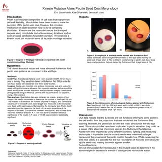
Arabidopsis
seeds
Ruthenium
Red stain
A B
References:
Mcfarlane, Heather E., Robin E. Young, Geoffrey O. Wasteneys, and A. Lacey Samuels. "Cortical
Microtubules Mark the Mucilage Secretion Domain of the Plasma Membrane in Arabidopsis Seed
Coat Cells." Planta 227.6 (2008): 1363-375. Web.
Hypothesis:
The kinesin knockout mutant will have abnormal Ruthenium Red
pectin stain patterns as compared to the wild type.
Figure 3. Examples of A. thaliana seeds stained with Ruthenium Red.
Seeds stained for pectin using Ruthenium Red. a) Wild-type seed showing full
seed-coat. Image taken at 10x. b) Mutant seed showing no pectin coat. Seed does
have small projections that are stained by Ruthenium Red. Image taken at 10x.
Figure 1: Diagram of Wild type hydrated seed (center) with pectin
containing mucilage (Outside)
Figure 2: Diagram of staining method
Pectin Stained
Seeds
Visualize on
Scope