Mammalian cell culture growth response to animal serum
SWE poster
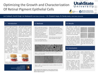
1. Optimizing the Growth and Characterization
Of Retinal Pigment Epithelial Cells
Lori Caldwell, Harshit Singh, Ian Wadsworth Utah State University | Dr. Elizabeth Vargis, Dr. Randy Lewis, Utah State University
Study conducted with funding from a Career Starter Grant from
Knights Templar Eye Foundation and an Undergraduate Creative
Research Opportunity grant.
Lori Caldwell
Utah State University
Department of Biological Engineering
12lcaldwell@gmail.com
I. Introduction
ARPE-19 cells were cultured in
standard T25 cell culture flasks
until a concentration of 4.5 x
10^5 cells/ mL was reached
(Figure 3). Cells were
maintained in this phase of
research using DMEM-F12
nutrient medium with 10% FBS
and 10,000 units/ mL penicillin.
9.5 cm2 six well plates were
coated with spider silk protein
(Figure 2); cells were then
seeded onto the six well plates.
The cells were maintained using
the same media for the first
two days, then changed to a 5%
FBS solution in order to prevent
cell overgrowth.
Cell confluency was measured
daily using light microscopy.
II. Methods
Cells grown on spider silk
showed similar characteristics
to those grown on standard
tissue culture plates. Cells did
not pigment in the time period
measured, but had similar
confluency and morphology.
The use of spider silk protein
makes it difficult to distinguish
cells, but cells were measured
to have 75% confluency at day
10, which was 4 days slower
than cells grown in standard
culture plates (Figure 4).
III. Results
The Spider silk protein shows
promise as a surface to grow
RPE cells on. Further research
may show advantages in cell
characterization using spider
silk compared to culture plates.
For future research, run trials
using a variety of spider silk
proteins, possibly with other
protein additives to mimic
Bruch’s Membrane more
accurately. Furthermore, use
nano-scale imaging techniques
such as SEM to study the
surface characteristics of the
Bruch’s membrane substitute
• Collagen I-V, Fibronectin,
Vascular Endothelial
Growth Factor (VEG-F),
and RGD (Arginine –
Glycine – Asparagine).
• Layer proteins according
to physiology of Bruch’s
Membrane
Lastly, future research may also
involve growing cells on micro-
patterns of 10-100 nm sizes to
promote characterization.
IV. Conclusions
Figure 4 – Time lapse of RPE cell
growth on spider silk protein matrix
over 10 days. Top left – day 2, top
right – day 4, bottom left – day 6,
bottom right – day 10. Scale = 50
μm.
Figure 1 – Schematic of cell layers comparing normal function (left)
to a patient affected by AMD (right).
The Retinal Pigment Epithelium
(RPE) is a single layer of cells
that supports photoreceptors
(rods and cones) by providing
nutrients and filtering waste
products. RPE cells grow on the
acellular Bruch’s Membrane,
which sits directly superior to
the Choroid layer, and have a
characteristic dark pigment
and grow in tight polygonal cell
junctions. These tight junctions
provide a blood vitreous
barrier that prevents large
molecules from entering the
eye while the pigment works to
absorb excess light.
Bruch’s Membrane fails to
perform its designed function
in age-related macular
degeneration (Figure 1). In this
disease, Bruch’s membrane
retains excess amounts of the
lipid drusen which is thought
to lead to reduced RPE cell
function by limiting the
nutrients available to them
from nearby blood vessels and
causing an excess waste
buildup leading to premature
photoreceptor cell death.
The Center for Disease Control
reports 1.8 million Americans
over the age of 40 affected by
AMD with 7.3 million at risk for
developing the disease. This
makes it the leading cause of
permanent vision loss in all
developed nations, and also
accounts for nearly 10% of
vision loss across the entire
world.
Figure 2 - The above images show spider silk proteins M4M5, M4, and
FLYS3 proteins respectively.
Figure 3 – ARPE – 19 cell growth after 12 weeks on M4M5, M4, and FLYS3
proteins left to right.