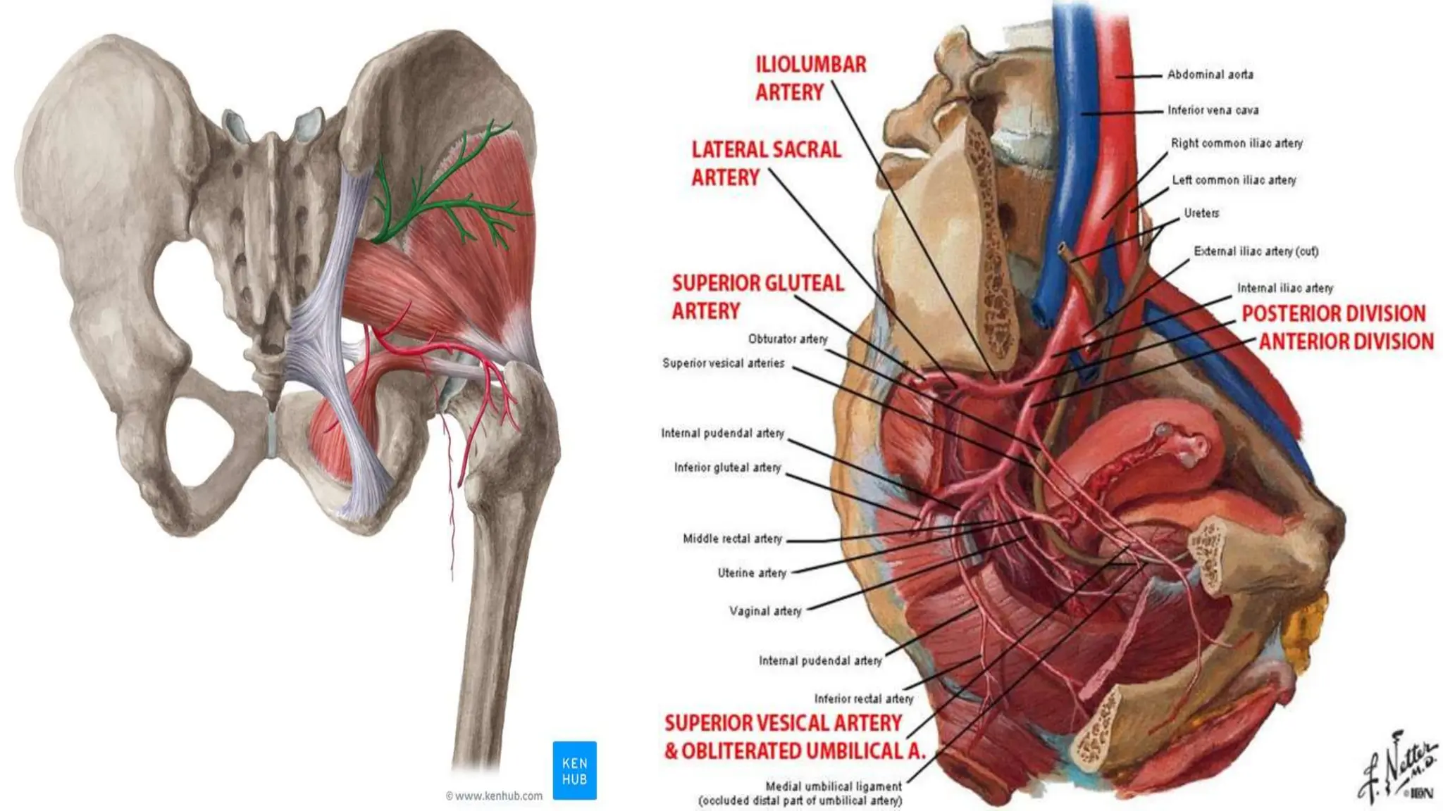
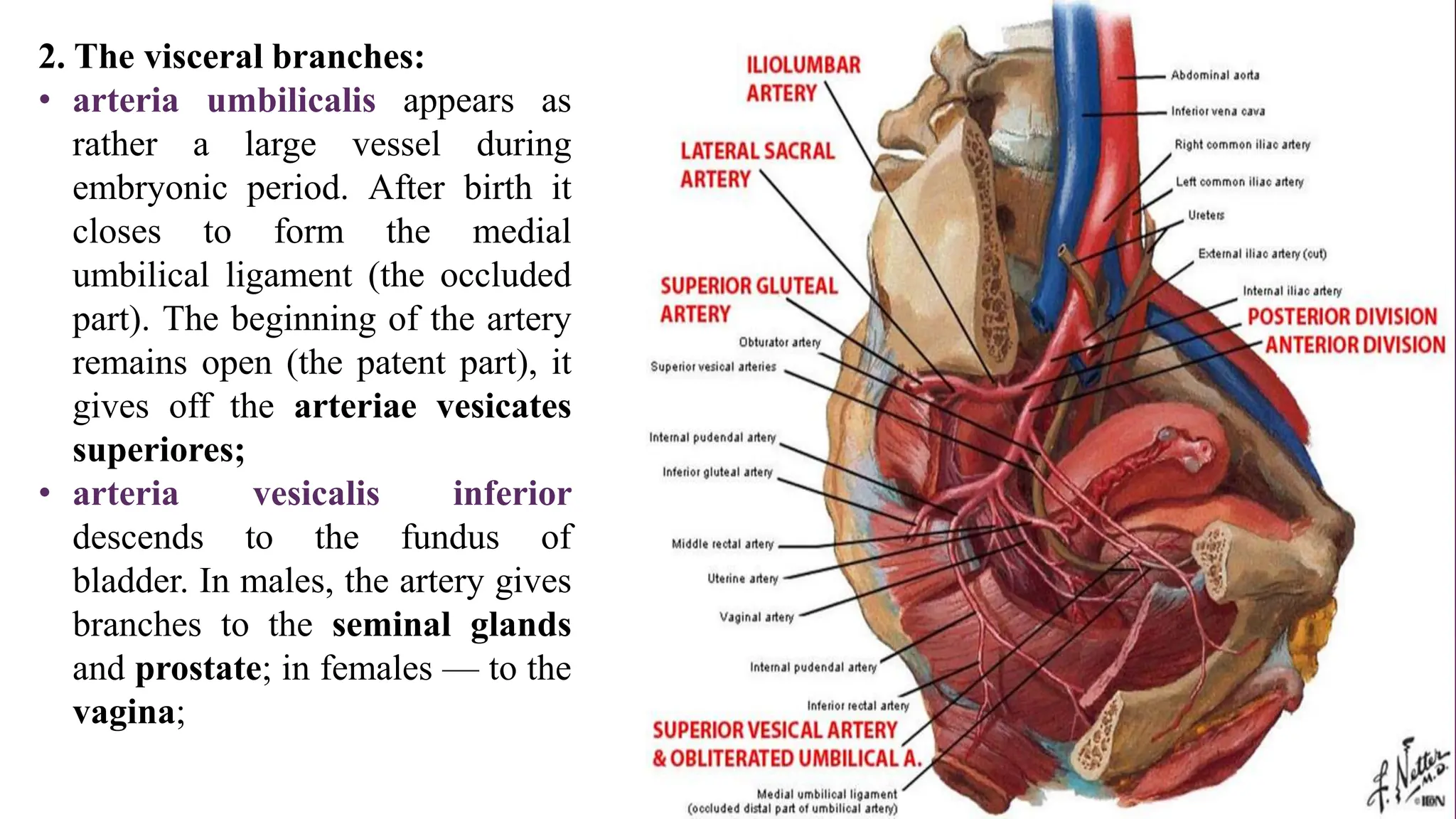
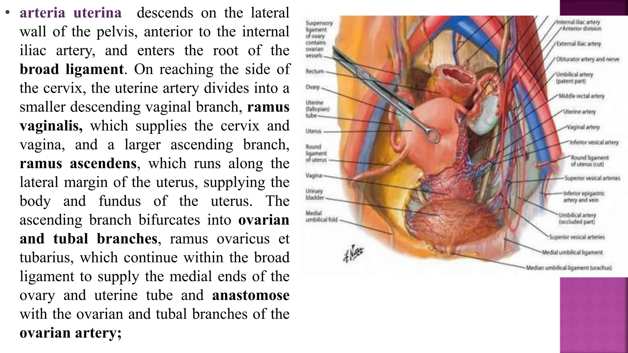
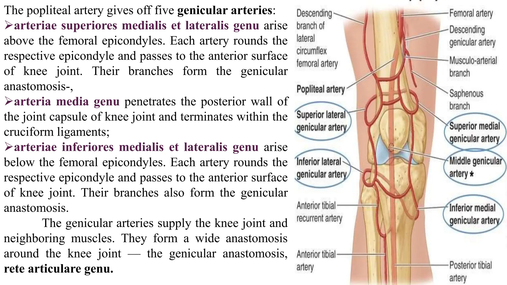
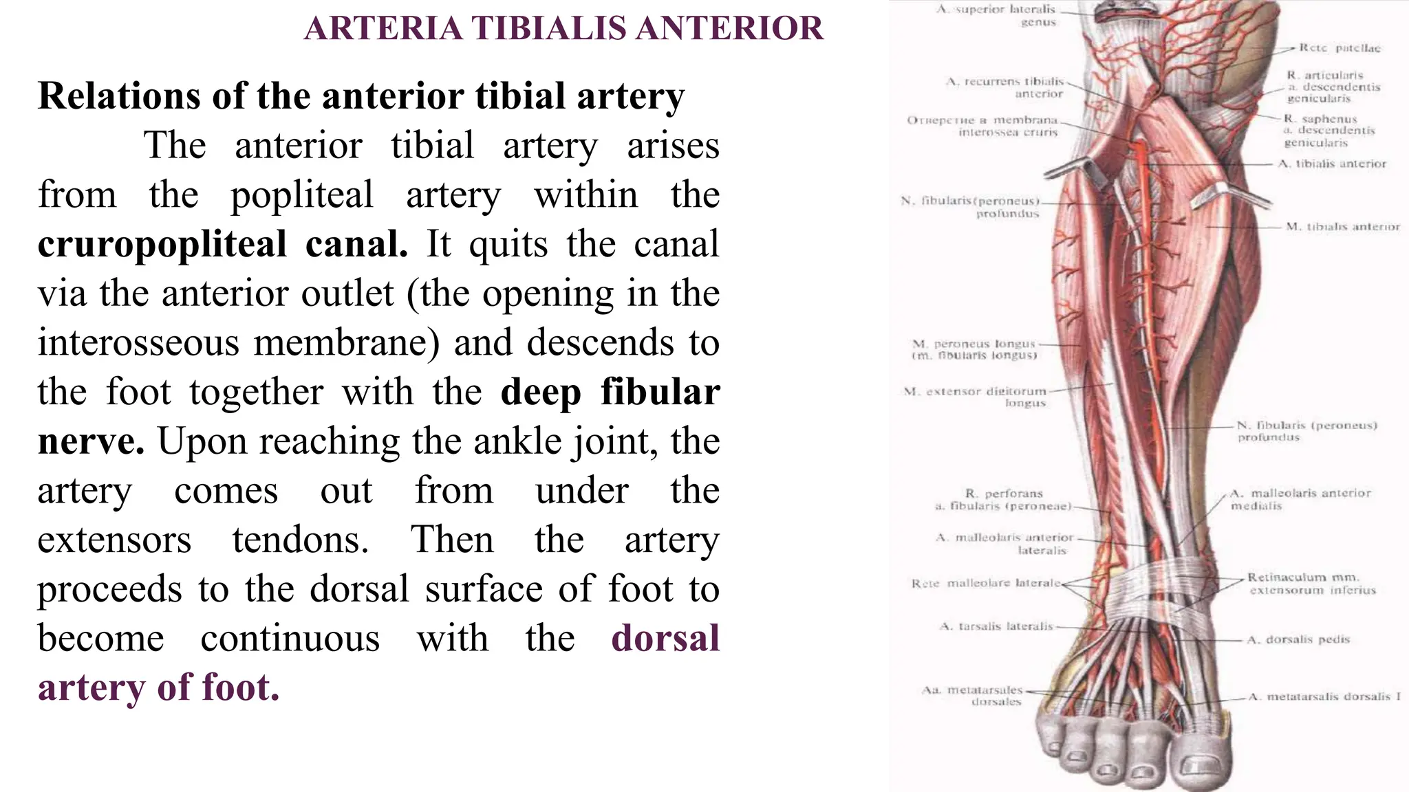
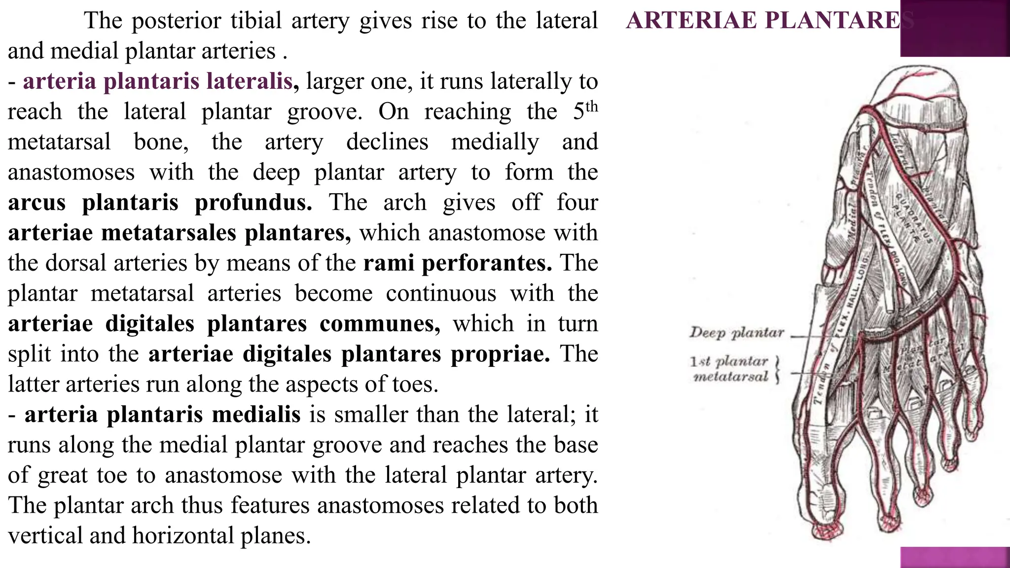
SlideShare a Scribd company logo
1 of 26
THE COMMON ILIAC ARTERY, ARTERIA ILIACA
COMMUNIS
The abdominal aorta diverges at sharp angle
(60-70°) to give rise to the common iliac arteries.
Each artery descends laterally to reach the respective
sacroiliac joint. There, the arteries give rise to the
• external and
• internal iliac arteries.
ARTERIA ILIACA INTERNA
Relations of the internal iliac artery
The internal iliac artery arises from the common iliac
artery and descends to the lesser pelvis. The artery is
the principal nourishing vessel for this region. The
artery gives off numerous branches, both
Parietal
visceral.
1. The parietal branches:
• arteria obturatoria reaches the thigh region
via the obturator canal. The artery supplies
the adductors of thigh and the hip joint. The
hip joint receives blood from the acetabular
branch, ramus acetabularis, which passes
within the ligament of head of femur. The
obturator artery anastomoses with the
inferior epigastric artery via the pubic
branch, ramus pubicus. In 25% of individ-
uals, this anastomosis is quite well
developed; in fact, the obturator artery in this
case arises from the inferior hypogastric
artery. This variant is of surgical concern be-
cause the artery passes next to the internal
femoral ring and thus can be damaged during
surgery (“corona mortis”);
• arteria iliolumbalis ascends
posteriorly to the related
muscles, the ilium and the
spinal cord (the spinal branches,
rami spinales);
• arteriae sacrales laterales run
to the sacrum and the spinal
cord (the spinal branches);
• arteria glutea superior leaves the
lesser pelvis cavity via the
suprapiriform foramen. Within the
gluteal region the artery gives off the
branches to supply the gluteus medius
and minimus;
• arteria glutea inferior leaves the
lesser pelvis cavity via the
infrapiriform foramen. Within the
gluteal region, the artery supplies the
gluteus maximus and other related
muscles.
2. The visceral branches:
• arteria umbilicalis appears as
rather a large vessel during
embryonic period. After birth it
closes to form the medial
umbilical ligament (the occluded
part). The beginning of the artery
remains open (the patent part), it
gives off the arteriae vesicates
superiores;
• arteria vesicalis inferior
descends to the fundus of
bladder. In males, the artery gives
branches to the seminal glands
and prostate; in females — to the
vagina;
• arteria uterina descends on the lateral
wall of the pelvis, anterior to the internal
iliac artery, and enters the root of the
broad ligament. On reaching the side of
the cervix, the uterine artery divides into a
smaller descending vaginal branch, ramus
vaginalis, which supplies the cervix and
vagina, and a larger ascending branch,
ramus ascendens, which runs along the
lateral margin of the uterus, supplying the
body and fundus of the uterus. The
ascending branch bifurcates into ovarian
and tubal branches, ramus ovaricus et
tubarius, which continue within the broad
ligament to supply the medial ends of the
ovary and uterine tube and anastomose
with the ovarian and tubal branches of the
ovarian artery;
• arteria rectalis media may arise independently
from the internal iliac artery, or it may arise in
common with the inferior vesical artery or the
internal pudendal artery. The middle rectal artery
supplies the inferior part of the rectum, anas-
tomosing with the superior and inferior rectal
arteries, supplying the seminal glands and
prostate (or the vagina);
• Arteria pudenda interna passes infero-
laterally, anterior to the piriformis muscle and
sacral plexus. It leaves the pelvis between the
piriformis and the coccygeus muscles by passing
through the inferior part of the greater sciatic
foramen. The internal pudendal artery then
passes around the posterior aspect of the ischial
spine or the sacrospinous ligament and enters the
ischio-anal fossa through the lesser sciatic fo-
ramen.
• Within the ischio-anal fossa, the artery gives
the branches as follows:
a) arteria rectalis inferior passes to the anus;
b) arteria bulbi penis supplies the respective
bulb of penis;
c) arteria dorsalis penis (clitoridis) passes
below skin (to the penis or clitoris respectively);
d) arteria profunda penis (clitoridis) take the
same route as the latter arteries yet deeper;
e) arteria perinealis supplies the perineal
muscles and skin;
f) rami labiales posteriores and the rami
scrotales posteriores supply the respective
external genitals.
ARTERIA ILIACA EXTERNA
Relations of the external iliac
artery
The external iliac artery descends
on the medial aspect of the psoas
major and quits the lesser pelvis
via the vascular space. Within the
femoral triangle, the artery
becomes
continuous with the femoral
artery.
The external iliac artery gives the branches as
follows:
• arteria epigastrica inferior arises from the
main trunk above the inguinal ligament and
then ascends medially along the internal
surface of the anterior abdominal wall
occupying the lateral umbilical ligament.
Then the artery enters the rectus sheath and
ascends along its posterior surface to reach
the umbilical ring. Here it anastomoses with
the superior hypogastric artery. In the
beginning, the artery gives off anastomotic
branch (the pubic branch, ramus pubicus) to
the obturator artery;
• arteria circumflexa ilium profunda runs
laterally along the inguinal ligament and the
iliac crest. It supplies the iliacus and the
muscles of abdominal wall.
Relations of the femoral artery
The femoral artery arises directly from the
external iliac artery. The arteries are delimited
by the inferior border of the inguinal
ligament. On leaving the vascular space, the
artery appears within the femoral triangle
together with the femoral nerve (found laterally)
and the femoral vein (found medially).
Pulsation of the artery is palpable below the
inguinal ligament in the area related to the
vascular space. Within the femoral triangle, the
artery runs along the iliopectineal groove and
then along the femoral groove. From the femo-
ral groove, the artery proceeds to the adductor
canal, which leads it to the popliteal fossa.
Here it becomes continuous with the popliteal
artery.
THE FEMORALARTERY
The greatest branch of the femoral artery is the deep artery of thigh.
arteria profunda femoris is nearly as wide as the femoral artery
(5-7 mm). It arises 4-5 cm below the inguinal ligament and
descends posteriorly running laterally from the femoral artery. It is
the main artery, which supplies the thigh. Its shorter trunk splits
into the following branches:
•arteria circumflexa femoris medialis passes in between the il-
iopsoas and the pectineus muscles to enter deep into the adductors
of thigh. The artery gives off the deep, the ascending and the trans
verse branches, ramus profundus, ascendens et transversus. The
branches supply the muscles of thigh and the hip joint and
anastomose with the obturator and inferior gluteal arteries;
•arteria cicumflexa femoris lateralis gives off the ascending and
descending branches. The ascending branch anastomoses with the
gluteal arteries;
•arteriae perforantes rather wide, they penetrate the tendons of
the adductors and proceed to the posterior femoral area. There they
supply the related muscles and the femur. The arteries reach the
knee joint and anastomose with the branches of the popliteal artery.
The perforating arteries are important for collateral circulation.
Other branches
Apart from the deep artery of thigh, the femoral artery
gives some smaller arteries:
•arteria epigastrica superficialis arises below the
inguinal ligament and ascends to the umbilical region.
There it anastomoses with the superior epigastric
artery.
•arteria circumflexa ilium superficialis also arises
below the inguinal ligament and passes laterally to
reach the anterior superior iliac spine. The artery
supplies the neighboring soft tissues;
•arteriae pudendae externae (superficiales et
profundae) are small branches that supply the scrotum
(or the labia majores);
•arteria descendens genu arises within the adductor
canal. Together with the saphenous nerve, the artery
leaves the canal via the anterior opening and terminates
at the knee joint (it forms the genicular anastomosis).
THE FEMORALARTERY
THE POPLITEALARTERY, ARTERIA
POPLITEA
Relations of the popliteal artery
The popliteal artery is a direct
continuation of the femoral artery. It
occupies the popliteal fossa together with
the vein of the same name (it runs
laterally and posteriorly). Upon reaching
the leg region, the artery enters the
cruropopliteal canal and gives off its
two terminal branches — the
anterior tibial
posterior tibial arteries.
The branches of the popliteal artery
The popliteal artery gives off five genicular arteries:
arteriae superiores medialis et lateralis genu arise
above the femoral epicondyles. Each artery rounds the
respective epicondyle and passes to the anterior surface
of knee joint. Their branches form the genicular
anastomosis-,
arteria media genu penetrates the posterior wall of
the joint capsule of knee joint and terminates within the
cruciform ligaments;
arteriae inferiores medialis et lateralis genu arise
below the femoral epicondyles. Each artery rounds the
respective epicondyle and passes to the anterior surface
of knee joint. Their branches also form the genicular
anastomosis.
The genicular arteries supply the knee joint and
neighboring muscles. They form a wide anastomosis
around the knee joint — the genicular anastomosis,
rete articulare genu.
ARTERIA TIBIALIS ANTERIOR
Relations of the anterior tibial artery
The anterior tibial artery arises
from the popliteal artery within the
cruropopliteal canal. It quits the canal
via the anterior outlet (the opening in the
interosseous membrane) and descends to
the foot together with the deep fibular
nerve. Upon reaching the ankle joint, the
artery comes out from under the
extensors tendons. Then the artery
proceeds to the dorsal surface of foot to
become continuous with the dorsal
artery of foot.
The branches of the anterior tibial artery
Apart from numerous muscular branches, the
artery gives off other branches as follows:
•arteria recurrens tibialis posterior
becomes evident on the posterior surface of
leg; it ascends to join the genicular
anastomosis;
•arteria recurrens tibialis anterior arises
opposite to the latter artery. It also joins the
genicular anastomosis;
•arteriae malleolares anteriores (medialis
et lateralis) descend to the respective ankles.
They participate in formation of the medial
and lateral malleolar networks.
Relations of the dorsal artery of
foot
The dorsal artery of foot is a
direct continuation of the anterior
tibial artery. It runs between the
tendons of the extensor hallucis
longus and the extensor digitorum
longus. At the first intermetatarsal
space, the artery gives off its
terminal branches — the
deep plantar artery and the
first metatarsal artery.
ARTERIA DORSALIS
PEDIS
The branches of the dorsal artery of foot
The dorsal artery of foot also gives the following branches:
-arteriae tarsales laterales et mediates run to the respective
aspects of foot;
-arteria arcuata arises at the bases of metatarsals and runs
laterally to anastomose with the lateral tarsal artery. The arch
formed gives off the, arteriae metatarsals dorsales (2
through 5), which split into the arteriae digitales dorsales;
the first dorsal metatarsal artery arises directly from the
dorsal artery of foot. It gives off three dorsal digital arteries
to both aspects of the great toe and to the medial aspect of
the second toe;
-arteria plantaris profunda is the second terminal branch of
the dorsal artery of foot. It penetrates the muscles of the first
intermetatarsal space and proceeds to the plantar surface of
foot; there it anastomoses with the lateral plantar artery to
form the arcus plantaris profundus.
Relations of the posterior tibial
artery
The larger posterior tibial artery arises
immediately from the popliteal artery. The
artery occupies the cruropopliteal canal
together with the tibial nerve. Within the
canal, the artery runs along the deep muscles
of leg anterior to the soleus. The artery quits
the canal and passes medially from the
calcaneal tendon immediately below the skin
and fascia. Here, one can palpate pulsation of
the artery. The artery then rounds the medial
malleolus (in canalis maleolaris), passes under
the flexor retinaculum and eventually appears
on the plantar surface of foot. There it gives
the lateral and medial plantar arteries.
ARTERIA TIBIALIS POSTERIOR
The branches of posterior tibial artery
The artery gives numerous muscular branches all the way
down to the foot.
- arteria circumflexa fibulae becomes evident
on the posterior surface of leg; it ascends to join the
genicular anastomosis;
- At the ankle joint, the artery gives the rami
malleolaris mediates and rami calcanei.
- The greatest branch given is the fibular artery.
The fibular artery, arteria peronea is quite a large branch
that becomes evident in the upper portion of leg. The
artery runs laterally, along the fibula and below the flexor
hallucis longus. Then it descends to the calcaneus and
terminates at the calcaneal anastomosis, rete calcaneum.
At the lateral malleolus, the artery gives the ramus
malleolaris lateralis that joins the lateral malleolar
network. At the ankle joint, the artery gives off the per-
forating branch, ramus perforans that traverses the
interosseous membrane to anastomose with the branches
of the anterior tibial artery.
ARTERIAE PLANTARES
The posterior tibial artery gives rise to the lateral
and medial plantar arteries .
- arteria plantaris lateralis, larger one, it runs laterally to
reach the lateral plantar groove. On reaching the 5th
metatarsal bone, the artery declines medially and
anastomoses with the deep plantar artery to form the
arcus plantaris profundus. The arch gives off four
arteriae metatarsales plantares, which anastomose with
the dorsal arteries by means of the rami perforantes. The
plantar metatarsal arteries become continuous with the
arteriae digitales plantares communes, which in turn
split into the arteriae digitales plantares propriae. The
latter arteries run along the aspects of toes.
- arteria plantaris medialis is smaller than the lateral; it
runs along the medial plantar groove and reaches the base
of great toe to anastomose with the lateral plantar artery.
The plantar arch thus features anastomoses related to both
vertical and horizontal planes.

More Related Content

What's hot

Review of pelvic_anatomy_by- dr. armaan singh
Review of pelvic_anatomy_by- dr. armaan singhReview of pelvic_anatomy_by- dr. armaan singh
Review of pelvic_anatomy_by- dr. armaan singhDr. Armaan Singh
 
Development of Midgut (Special Embryology)
Development of Midgut (Special Embryology)Development of Midgut (Special Embryology)
Development of Midgut (Special Embryology)Dr. Sherif Fahmy
 
Anatomy of inguinal canal - Dr nesar Ahmad
Anatomy of inguinal canal -  Dr nesar AhmadAnatomy of inguinal canal -  Dr nesar Ahmad
Anatomy of inguinal canal - Dr nesar AhmadStudent
 
210 lower limb rs updated
210 lower limb rs updated210 lower limb rs updated
210 lower limb rs updatedAHS_anatomy2
 
Gluteal region complete
Gluteal region completeGluteal region complete
Gluteal region completeLucidante1
 
Development of Liver and Gall bladder
Development of Liver and Gall bladderDevelopment of Liver and Gall bladder
Development of Liver and Gall bladdertypetnt
 
Pelvic vessels,nerves and lymph nodes.
Pelvic vessels,nerves and lymph nodes.Pelvic vessels,nerves and lymph nodes.
Pelvic vessels,nerves and lymph nodes.Abdirahman Śïçïïd
 
CLINICAL ANATOMY OF PELVIS
CLINICAL ANATOMY OF PELVISCLINICAL ANATOMY OF PELVIS
CLINICAL ANATOMY OF PELVISNitesh Sharma
 
Vessels of the pelvis
Vessels of the pelvisVessels of the pelvis
Vessels of the pelvisLow Yousi
 
The iliac artery
The iliac arteryThe iliac artery
The iliac arterykumarshaman
 
The Pelvis - Bones and Joints
The Pelvis - Bones and JointsThe Pelvis - Bones and Joints
The Pelvis - Bones and Jointsmeducationdotnet
 
Development of stomach
Development of stomachDevelopment of stomach
Development of stomachdrasarma1947
 

What's hot (20)

Review of pelvic_anatomy_by- dr. armaan singh
Review of pelvic_anatomy_by- dr. armaan singhReview of pelvic_anatomy_by- dr. armaan singh
Review of pelvic_anatomy_by- dr. armaan singh
 
Development of Midgut (Special Embryology)
Development of Midgut (Special Embryology)Development of Midgut (Special Embryology)
Development of Midgut (Special Embryology)
 
Anatomy of inguinal canal - Dr nesar Ahmad
Anatomy of inguinal canal -  Dr nesar AhmadAnatomy of inguinal canal -  Dr nesar Ahmad
Anatomy of inguinal canal - Dr nesar Ahmad
 
Arm & Cubital Fossa.pptx
Arm & Cubital Fossa.pptxArm & Cubital Fossa.pptx
Arm & Cubital Fossa.pptx
 
210 lower limb rs updated
210 lower limb rs updated210 lower limb rs updated
210 lower limb rs updated
 
Gluteal region complete
Gluteal region completeGluteal region complete
Gluteal region complete
 
Development of Liver and Gall bladder
Development of Liver and Gall bladderDevelopment of Liver and Gall bladder
Development of Liver and Gall bladder
 
Anatomy of duodenum and pancreas
Anatomy of duodenum and pancreas Anatomy of duodenum and pancreas
Anatomy of duodenum and pancreas
 
Dr.gamal presentation
Dr.gamal presentationDr.gamal presentation
Dr.gamal presentation
 
Pelvic vessels,nerves and lymph nodes.
Pelvic vessels,nerves and lymph nodes.Pelvic vessels,nerves and lymph nodes.
Pelvic vessels,nerves and lymph nodes.
 
Front of forearm
Front of forearmFront of forearm
Front of forearm
 
Veins of the neck
Veins of the neckVeins of the neck
Veins of the neck
 
4.gluteal region
4.gluteal region4.gluteal region
4.gluteal region
 
CLINICAL ANATOMY OF PELVIS
CLINICAL ANATOMY OF PELVISCLINICAL ANATOMY OF PELVIS
CLINICAL ANATOMY OF PELVIS
 
Vessels of the pelvis
Vessels of the pelvisVessels of the pelvis
Vessels of the pelvis
 
Popliteal fossa
Popliteal fossaPopliteal fossa
Popliteal fossa
 
The iliac artery
The iliac arteryThe iliac artery
The iliac artery
 
The Pelvis - Bones and Joints
The Pelvis - Bones and JointsThe Pelvis - Bones and Joints
The Pelvis - Bones and Joints
 
Anatomy of pharynx
Anatomy of pharynxAnatomy of pharynx
Anatomy of pharynx
 
Development of stomach
Development of stomachDevelopment of stomach
Development of stomach
 

Similar to Common iliac artery

Lower limb 7_101921.pptx
Lower limb 7_101921.pptxLower limb 7_101921.pptx
Lower limb 7_101921.pptxZakaryaKhan12
 
Vessels of the lower limb.ppt
Vessels of the lower limb.pptVessels of the lower limb.ppt
Vessels of the lower limb.pptTofikMohammed3
 
Common Iliac Artery Anatomy.pptx
Common Iliac Artery Anatomy.pptxCommon Iliac Artery Anatomy.pptx
Common Iliac Artery Anatomy.pptxglendalourdes03
 
Arterial Supply and Venous Drainage of Pelvis.pptx
Arterial Supply and Venous Drainage of Pelvis.pptxArterial Supply and Venous Drainage of Pelvis.pptx
Arterial Supply and Venous Drainage of Pelvis.pptxMathew Joseph
 
Blood Supply, Venous Drainage and Lymphatic Drainage of Lower Limb
Blood Supply, Venous Drainage and Lymphatic Drainage of Lower LimbBlood Supply, Venous Drainage and Lymphatic Drainage of Lower Limb
Blood Supply, Venous Drainage and Lymphatic Drainage of Lower LimbSado Anatomist
 
bloodvesselsoflowerlimb-230302034633-5d194285.pdf
bloodvesselsoflowerlimb-230302034633-5d194285.pdfbloodvesselsoflowerlimb-230302034633-5d194285.pdf
bloodvesselsoflowerlimb-230302034633-5d194285.pdfdoctor / pediatrician
 
Blood Vessels of Lower Limb.pptx
Blood Vessels of Lower Limb.pptxBlood Vessels of Lower Limb.pptx
Blood Vessels of Lower Limb.pptxMathew Joseph
 
Review of Gait, Locomotion & Lower Limbs
Review of Gait, Locomotion & Lower LimbsReview of Gait, Locomotion & Lower Limbs
Review of Gait, Locomotion & Lower LimbsDrSaeed Shafi
 
ARTERIAL SUPPLY OF UPPER LIMBThe arterial supply to the .docx
ARTERIAL SUPPLY OF UPPER LIMBThe arterial supply to the .docxARTERIAL SUPPLY OF UPPER LIMBThe arterial supply to the .docx
ARTERIAL SUPPLY OF UPPER LIMBThe arterial supply to the .docxrossskuddershamus
 
Blood vessel, Innervation and lymph system of lower limb
Blood vessel, Innervation and lymph system of lower limbBlood vessel, Innervation and lymph system of lower limb
Blood vessel, Innervation and lymph system of lower limbEneutron
 
lower limb blood and nerve supply..pptx
lower limb blood and nerve supply..pptxlower limb blood and nerve supply..pptx
lower limb blood and nerve supply..pptxMeetVaghasiya20
 
Anterior cmpartment of thigh
Anterior  cmpartment of thighAnterior  cmpartment of thigh
Anterior cmpartment of thighAtifRaza11
 
Subclavian artery and it's branches
Subclavian artery and it's branchesSubclavian artery and it's branches
Subclavian artery and it's branchesHarshal Shinde
 
Vessels of upper limb..pptx
Vessels of upper limb..pptxVessels of upper limb..pptx
Vessels of upper limb..pptxvrajpatel154117
 
Blood supply of upper limb
Blood supply of upper limbBlood supply of upper limb
Blood supply of upper limbIdris Siddiqui
 
Blood vessels of upper limb bsc nursing
Blood vessels of upper limb bsc nursingBlood vessels of upper limb bsc nursing
Blood vessels of upper limb bsc nursingKAVIN6369950450
 

Similar to Common iliac artery (20)

Lower limb 7_101921.pptx
Lower limb 7_101921.pptxLower limb 7_101921.pptx
Lower limb 7_101921.pptx
 
Vessels of the lower limb.ppt
Vessels of the lower limb.pptVessels of the lower limb.ppt
Vessels of the lower limb.ppt
 
Common Iliac Artery Anatomy.pptx
Common Iliac Artery Anatomy.pptxCommon Iliac Artery Anatomy.pptx
Common Iliac Artery Anatomy.pptx
 
Arterial Supply and Venous Drainage of Pelvis.pptx
Arterial Supply and Venous Drainage of Pelvis.pptxArterial Supply and Venous Drainage of Pelvis.pptx
Arterial Supply and Venous Drainage of Pelvis.pptx
 
Blood Supply, Venous Drainage and Lymphatic Drainage of Lower Limb
Blood Supply, Venous Drainage and Lymphatic Drainage of Lower LimbBlood Supply, Venous Drainage and Lymphatic Drainage of Lower Limb
Blood Supply, Venous Drainage and Lymphatic Drainage of Lower Limb
 
bloodvesselsoflowerlimb-230302034633-5d194285.pdf
bloodvesselsoflowerlimb-230302034633-5d194285.pdfbloodvesselsoflowerlimb-230302034633-5d194285.pdf
bloodvesselsoflowerlimb-230302034633-5d194285.pdf
 
Blood Vessels of Lower Limb.pptx
Blood Vessels of Lower Limb.pptxBlood Vessels of Lower Limb.pptx
Blood Vessels of Lower Limb.pptx
 
Review of Gait, Locomotion & Lower Limbs
Review of Gait, Locomotion & Lower LimbsReview of Gait, Locomotion & Lower Limbs
Review of Gait, Locomotion & Lower Limbs
 
ARTERIAL SUPPLY OF UPPER LIMBThe arterial supply to the .docx
ARTERIAL SUPPLY OF UPPER LIMBThe arterial supply to the .docxARTERIAL SUPPLY OF UPPER LIMBThe arterial supply to the .docx
ARTERIAL SUPPLY OF UPPER LIMBThe arterial supply to the .docx
 
Blood vessel, Innervation and lymph system of lower limb
Blood vessel, Innervation and lymph system of lower limbBlood vessel, Innervation and lymph system of lower limb
Blood vessel, Innervation and lymph system of lower limb
 
anatomy of thigh
anatomy of thighanatomy of thigh
anatomy of thigh
 
lower limb blood and nerve supply..pptx
lower limb blood and nerve supply..pptxlower limb blood and nerve supply..pptx
lower limb blood and nerve supply..pptx
 
Anterior cmpartment of thigh
Anterior  cmpartment of thighAnterior  cmpartment of thigh
Anterior cmpartment of thigh
 
Blood supply of female pelvis.
Blood supply of female pelvis.Blood supply of female pelvis.
Blood supply of female pelvis.
 
2. front of the thigh ii
2. front of the thigh ii2. front of the thigh ii
2. front of the thigh ii
 
Subclavian artery and it's branches
Subclavian artery and it's branchesSubclavian artery and it's branches
Subclavian artery and it's branches
 
Arteries of upper limb
Arteries of upper limbArteries of upper limb
Arteries of upper limb
 
Vessels of upper limb..pptx
Vessels of upper limb..pptxVessels of upper limb..pptx
Vessels of upper limb..pptx
 
Blood supply of upper limb
Blood supply of upper limbBlood supply of upper limb
Blood supply of upper limb
 
Blood vessels of upper limb bsc nursing
Blood vessels of upper limb bsc nursingBlood vessels of upper limb bsc nursing
Blood vessels of upper limb bsc nursing
 

Recently uploaded

Class 11th Physics NEET formula sheet pdf
Class 11th Physics NEET formula sheet pdfClass 11th Physics NEET formula sheet pdf
Class 11th Physics NEET formula sheet pdfAyushMahapatra5
 
microwave assisted reaction. General introduction
microwave assisted reaction. General introductionmicrowave assisted reaction. General introduction
microwave assisted reaction. General introductionMaksud Ahmed
 
APM Welcome, APM North West Network Conference, Synergies Across Sectors
APM Welcome, APM North West Network Conference, Synergies Across SectorsAPM Welcome, APM North West Network Conference, Synergies Across Sectors
APM Welcome, APM North West Network Conference, Synergies Across SectorsAssociation for Project Management
 
Measures of Dispersion and Variability: Range, QD, AD and SD
Measures of Dispersion and Variability: Range, QD, AD and SDMeasures of Dispersion and Variability: Range, QD, AD and SD
Measures of Dispersion and Variability: Range, QD, AD and SDThiyagu K
 
1029 - Danh muc Sach Giao Khoa 10 . pdf
1029 -  Danh muc Sach Giao Khoa 10 . pdf1029 -  Danh muc Sach Giao Khoa 10 . pdf
1029 - Danh muc Sach Giao Khoa 10 . pdfQucHHunhnh
 
Measures of Central Tendency: Mean, Median and Mode
Measures of Central Tendency: Mean, Median and ModeMeasures of Central Tendency: Mean, Median and Mode
Measures of Central Tendency: Mean, Median and ModeThiyagu K
 
9548086042 for call girls in Indira Nagar with room service
9548086042  for call girls in Indira Nagar  with room service9548086042  for call girls in Indira Nagar  with room service
9548086042 for call girls in Indira Nagar with room servicediscovermytutordmt
 
Sports & Fitness Value Added Course FY..
Sports & Fitness Value Added Course FY..Sports & Fitness Value Added Course FY..
Sports & Fitness Value Added Course FY..Disha Kariya
 
Russian Escort Service in Delhi 11k Hotel Foreigner Russian Call Girls in Delhi
Russian Escort Service in Delhi 11k Hotel Foreigner Russian Call Girls in DelhiRussian Escort Service in Delhi 11k Hotel Foreigner Russian Call Girls in Delhi
Russian Escort Service in Delhi 11k Hotel Foreigner Russian Call Girls in Delhikauryashika82
 
Software Engineering Methodologies (overview)
Software Engineering Methodologies (overview)Software Engineering Methodologies (overview)
Software Engineering Methodologies (overview)eniolaolutunde
 
Call Girls in Dwarka Mor Delhi Contact Us 9654467111
Call Girls in Dwarka Mor Delhi Contact Us 9654467111Call Girls in Dwarka Mor Delhi Contact Us 9654467111
Call Girls in Dwarka Mor Delhi Contact Us 9654467111Sapana Sha
 
IGNOU MSCCFT and PGDCFT Exam Question Pattern: MCFT003 Counselling and Family...
IGNOU MSCCFT and PGDCFT Exam Question Pattern: MCFT003 Counselling and Family...IGNOU MSCCFT and PGDCFT Exam Question Pattern: MCFT003 Counselling and Family...
IGNOU MSCCFT and PGDCFT Exam Question Pattern: MCFT003 Counselling and Family...PsychoTech Services
 
Paris 2024 Olympic Geographies - an activity
Paris 2024 Olympic Geographies - an activityParis 2024 Olympic Geographies - an activity
Paris 2024 Olympic Geographies - an activityGeoBlogs
 
SOCIAL AND HISTORICAL CONTEXT - LFTVD.pptx
SOCIAL AND HISTORICAL CONTEXT - LFTVD.pptxSOCIAL AND HISTORICAL CONTEXT - LFTVD.pptx
SOCIAL AND HISTORICAL CONTEXT - LFTVD.pptxiammrhaywood
 
Accessible design: Minimum effort, maximum impact
Accessible design: Minimum effort, maximum impactAccessible design: Minimum effort, maximum impact
Accessible design: Minimum effort, maximum impactdawncurless
 
fourth grading exam for kindergarten in writing
fourth grading exam for kindergarten in writingfourth grading exam for kindergarten in writing
fourth grading exam for kindergarten in writingTeacherCyreneCayanan
 
Q4-W6-Restating Informational Text Grade 3
Q4-W6-Restating Informational Text Grade 3Q4-W6-Restating Informational Text Grade 3
Q4-W6-Restating Informational Text Grade 3JemimahLaneBuaron
 
BASLIQ CURRENT LOOKBOOK LOOKBOOK(1) (1).pdf
BASLIQ CURRENT LOOKBOOK  LOOKBOOK(1) (1).pdfBASLIQ CURRENT LOOKBOOK  LOOKBOOK(1) (1).pdf
BASLIQ CURRENT LOOKBOOK LOOKBOOK(1) (1).pdfSoniaTolstoy
 

Recently uploaded (20)

Class 11th Physics NEET formula sheet pdf
Class 11th Physics NEET formula sheet pdfClass 11th Physics NEET formula sheet pdf
Class 11th Physics NEET formula sheet pdf
 
microwave assisted reaction. General introduction
microwave assisted reaction. General introductionmicrowave assisted reaction. General introduction
microwave assisted reaction. General introduction
 
APM Welcome, APM North West Network Conference, Synergies Across Sectors
APM Welcome, APM North West Network Conference, Synergies Across SectorsAPM Welcome, APM North West Network Conference, Synergies Across Sectors
APM Welcome, APM North West Network Conference, Synergies Across Sectors
 
Measures of Dispersion and Variability: Range, QD, AD and SD
Measures of Dispersion and Variability: Range, QD, AD and SDMeasures of Dispersion and Variability: Range, QD, AD and SD
Measures of Dispersion and Variability: Range, QD, AD and SD
 
1029 - Danh muc Sach Giao Khoa 10 . pdf
1029 -  Danh muc Sach Giao Khoa 10 . pdf1029 -  Danh muc Sach Giao Khoa 10 . pdf
1029 - Danh muc Sach Giao Khoa 10 . pdf
 
Measures of Central Tendency: Mean, Median and Mode
Measures of Central Tendency: Mean, Median and ModeMeasures of Central Tendency: Mean, Median and Mode
Measures of Central Tendency: Mean, Median and Mode
 
9548086042 for call girls in Indira Nagar with room service
9548086042  for call girls in Indira Nagar  with room service9548086042  for call girls in Indira Nagar  with room service
9548086042 for call girls in Indira Nagar with room service
 
Sports & Fitness Value Added Course FY..
Sports & Fitness Value Added Course FY..Sports & Fitness Value Added Course FY..
Sports & Fitness Value Added Course FY..
 
Russian Escort Service in Delhi 11k Hotel Foreigner Russian Call Girls in Delhi
Russian Escort Service in Delhi 11k Hotel Foreigner Russian Call Girls in DelhiRussian Escort Service in Delhi 11k Hotel Foreigner Russian Call Girls in Delhi
Russian Escort Service in Delhi 11k Hotel Foreigner Russian Call Girls in Delhi
 
Software Engineering Methodologies (overview)
Software Engineering Methodologies (overview)Software Engineering Methodologies (overview)
Software Engineering Methodologies (overview)
 
Código Creativo y Arte de Software | Unidad 1
Código Creativo y Arte de Software | Unidad 1Código Creativo y Arte de Software | Unidad 1
Código Creativo y Arte de Software | Unidad 1
 
Call Girls in Dwarka Mor Delhi Contact Us 9654467111
Call Girls in Dwarka Mor Delhi Contact Us 9654467111Call Girls in Dwarka Mor Delhi Contact Us 9654467111
Call Girls in Dwarka Mor Delhi Contact Us 9654467111
 
IGNOU MSCCFT and PGDCFT Exam Question Pattern: MCFT003 Counselling and Family...
IGNOU MSCCFT and PGDCFT Exam Question Pattern: MCFT003 Counselling and Family...IGNOU MSCCFT and PGDCFT Exam Question Pattern: MCFT003 Counselling and Family...
IGNOU MSCCFT and PGDCFT Exam Question Pattern: MCFT003 Counselling and Family...
 
Paris 2024 Olympic Geographies - an activity
Paris 2024 Olympic Geographies - an activityParis 2024 Olympic Geographies - an activity
Paris 2024 Olympic Geographies - an activity
 
SOCIAL AND HISTORICAL CONTEXT - LFTVD.pptx
SOCIAL AND HISTORICAL CONTEXT - LFTVD.pptxSOCIAL AND HISTORICAL CONTEXT - LFTVD.pptx
SOCIAL AND HISTORICAL CONTEXT - LFTVD.pptx
 
Accessible design: Minimum effort, maximum impact
Accessible design: Minimum effort, maximum impactAccessible design: Minimum effort, maximum impact
Accessible design: Minimum effort, maximum impact
 
fourth grading exam for kindergarten in writing
fourth grading exam for kindergarten in writingfourth grading exam for kindergarten in writing
fourth grading exam for kindergarten in writing
 
INDIA QUIZ 2024 RLAC DELHI UNIVERSITY.pptx
INDIA QUIZ 2024 RLAC DELHI UNIVERSITY.pptxINDIA QUIZ 2024 RLAC DELHI UNIVERSITY.pptx
INDIA QUIZ 2024 RLAC DELHI UNIVERSITY.pptx
 
Q4-W6-Restating Informational Text Grade 3
Q4-W6-Restating Informational Text Grade 3Q4-W6-Restating Informational Text Grade 3
Q4-W6-Restating Informational Text Grade 3
 
BASLIQ CURRENT LOOKBOOK LOOKBOOK(1) (1).pdf
BASLIQ CURRENT LOOKBOOK  LOOKBOOK(1) (1).pdfBASLIQ CURRENT LOOKBOOK  LOOKBOOK(1) (1).pdf
BASLIQ CURRENT LOOKBOOK LOOKBOOK(1) (1).pdf
 

Common iliac artery

  • 1. THE COMMON ILIAC ARTERY, ARTERIA ILIACA COMMUNIS The abdominal aorta diverges at sharp angle (60-70°) to give rise to the common iliac arteries. Each artery descends laterally to reach the respective sacroiliac joint. There, the arteries give rise to the • external and • internal iliac arteries. ARTERIA ILIACA INTERNA Relations of the internal iliac artery The internal iliac artery arises from the common iliac artery and descends to the lesser pelvis. The artery is the principal nourishing vessel for this region. The artery gives off numerous branches, both Parietal visceral.
  • 2. 1. The parietal branches: • arteria obturatoria reaches the thigh region via the obturator canal. The artery supplies the adductors of thigh and the hip joint. The hip joint receives blood from the acetabular branch, ramus acetabularis, which passes within the ligament of head of femur. The obturator artery anastomoses with the inferior epigastric artery via the pubic branch, ramus pubicus. In 25% of individ- uals, this anastomosis is quite well developed; in fact, the obturator artery in this case arises from the inferior hypogastric artery. This variant is of surgical concern be- cause the artery passes next to the internal femoral ring and thus can be damaged during surgery (“corona mortis”);
  • 3.
  • 4. • arteria iliolumbalis ascends posteriorly to the related muscles, the ilium and the spinal cord (the spinal branches, rami spinales); • arteriae sacrales laterales run to the sacrum and the spinal cord (the spinal branches);
  • 5. • arteria glutea superior leaves the lesser pelvis cavity via the suprapiriform foramen. Within the gluteal region the artery gives off the branches to supply the gluteus medius and minimus; • arteria glutea inferior leaves the lesser pelvis cavity via the infrapiriform foramen. Within the gluteal region, the artery supplies the gluteus maximus and other related muscles.
  • 6.
  • 7. 2. The visceral branches: • arteria umbilicalis appears as rather a large vessel during embryonic period. After birth it closes to form the medial umbilical ligament (the occluded part). The beginning of the artery remains open (the patent part), it gives off the arteriae vesicates superiores; • arteria vesicalis inferior descends to the fundus of bladder. In males, the artery gives branches to the seminal glands and prostate; in females — to the vagina;
  • 8. • arteria uterina descends on the lateral wall of the pelvis, anterior to the internal iliac artery, and enters the root of the broad ligament. On reaching the side of the cervix, the uterine artery divides into a smaller descending vaginal branch, ramus vaginalis, which supplies the cervix and vagina, and a larger ascending branch, ramus ascendens, which runs along the lateral margin of the uterus, supplying the body and fundus of the uterus. The ascending branch bifurcates into ovarian and tubal branches, ramus ovaricus et tubarius, which continue within the broad ligament to supply the medial ends of the ovary and uterine tube and anastomose with the ovarian and tubal branches of the ovarian artery;
  • 9. • arteria rectalis media may arise independently from the internal iliac artery, or it may arise in common with the inferior vesical artery or the internal pudendal artery. The middle rectal artery supplies the inferior part of the rectum, anas- tomosing with the superior and inferior rectal arteries, supplying the seminal glands and prostate (or the vagina); • Arteria pudenda interna passes infero- laterally, anterior to the piriformis muscle and sacral plexus. It leaves the pelvis between the piriformis and the coccygeus muscles by passing through the inferior part of the greater sciatic foramen. The internal pudendal artery then passes around the posterior aspect of the ischial spine or the sacrospinous ligament and enters the ischio-anal fossa through the lesser sciatic fo- ramen.
  • 10. • Within the ischio-anal fossa, the artery gives the branches as follows: a) arteria rectalis inferior passes to the anus; b) arteria bulbi penis supplies the respective bulb of penis; c) arteria dorsalis penis (clitoridis) passes below skin (to the penis or clitoris respectively); d) arteria profunda penis (clitoridis) take the same route as the latter arteries yet deeper; e) arteria perinealis supplies the perineal muscles and skin; f) rami labiales posteriores and the rami scrotales posteriores supply the respective external genitals.
  • 11.
  • 12. ARTERIA ILIACA EXTERNA Relations of the external iliac artery The external iliac artery descends on the medial aspect of the psoas major and quits the lesser pelvis via the vascular space. Within the femoral triangle, the artery becomes continuous with the femoral artery.
  • 13. The external iliac artery gives the branches as follows: • arteria epigastrica inferior arises from the main trunk above the inguinal ligament and then ascends medially along the internal surface of the anterior abdominal wall occupying the lateral umbilical ligament. Then the artery enters the rectus sheath and ascends along its posterior surface to reach the umbilical ring. Here it anastomoses with the superior hypogastric artery. In the beginning, the artery gives off anastomotic branch (the pubic branch, ramus pubicus) to the obturator artery; • arteria circumflexa ilium profunda runs laterally along the inguinal ligament and the iliac crest. It supplies the iliacus and the muscles of abdominal wall.
  • 14.
  • 15. Relations of the femoral artery The femoral artery arises directly from the external iliac artery. The arteries are delimited by the inferior border of the inguinal ligament. On leaving the vascular space, the artery appears within the femoral triangle together with the femoral nerve (found laterally) and the femoral vein (found medially). Pulsation of the artery is palpable below the inguinal ligament in the area related to the vascular space. Within the femoral triangle, the artery runs along the iliopectineal groove and then along the femoral groove. From the femo- ral groove, the artery proceeds to the adductor canal, which leads it to the popliteal fossa. Here it becomes continuous with the popliteal artery. THE FEMORALARTERY
  • 16. The greatest branch of the femoral artery is the deep artery of thigh. arteria profunda femoris is nearly as wide as the femoral artery (5-7 mm). It arises 4-5 cm below the inguinal ligament and descends posteriorly running laterally from the femoral artery. It is the main artery, which supplies the thigh. Its shorter trunk splits into the following branches: •arteria circumflexa femoris medialis passes in between the il- iopsoas and the pectineus muscles to enter deep into the adductors of thigh. The artery gives off the deep, the ascending and the trans verse branches, ramus profundus, ascendens et transversus. The branches supply the muscles of thigh and the hip joint and anastomose with the obturator and inferior gluteal arteries; •arteria cicumflexa femoris lateralis gives off the ascending and descending branches. The ascending branch anastomoses with the gluteal arteries; •arteriae perforantes rather wide, they penetrate the tendons of the adductors and proceed to the posterior femoral area. There they supply the related muscles and the femur. The arteries reach the knee joint and anastomose with the branches of the popliteal artery. The perforating arteries are important for collateral circulation.
  • 17. Other branches Apart from the deep artery of thigh, the femoral artery gives some smaller arteries: •arteria epigastrica superficialis arises below the inguinal ligament and ascends to the umbilical region. There it anastomoses with the superior epigastric artery. •arteria circumflexa ilium superficialis also arises below the inguinal ligament and passes laterally to reach the anterior superior iliac spine. The artery supplies the neighboring soft tissues; •arteriae pudendae externae (superficiales et profundae) are small branches that supply the scrotum (or the labia majores); •arteria descendens genu arises within the adductor canal. Together with the saphenous nerve, the artery leaves the canal via the anterior opening and terminates at the knee joint (it forms the genicular anastomosis). THE FEMORALARTERY
  • 18. THE POPLITEALARTERY, ARTERIA POPLITEA Relations of the popliteal artery The popliteal artery is a direct continuation of the femoral artery. It occupies the popliteal fossa together with the vein of the same name (it runs laterally and posteriorly). Upon reaching the leg region, the artery enters the cruropopliteal canal and gives off its two terminal branches — the anterior tibial posterior tibial arteries. The branches of the popliteal artery
  • 19. The popliteal artery gives off five genicular arteries: arteriae superiores medialis et lateralis genu arise above the femoral epicondyles. Each artery rounds the respective epicondyle and passes to the anterior surface of knee joint. Their branches form the genicular anastomosis-, arteria media genu penetrates the posterior wall of the joint capsule of knee joint and terminates within the cruciform ligaments; arteriae inferiores medialis et lateralis genu arise below the femoral epicondyles. Each artery rounds the respective epicondyle and passes to the anterior surface of knee joint. Their branches also form the genicular anastomosis. The genicular arteries supply the knee joint and neighboring muscles. They form a wide anastomosis around the knee joint — the genicular anastomosis, rete articulare genu.
  • 20. ARTERIA TIBIALIS ANTERIOR Relations of the anterior tibial artery The anterior tibial artery arises from the popliteal artery within the cruropopliteal canal. It quits the canal via the anterior outlet (the opening in the interosseous membrane) and descends to the foot together with the deep fibular nerve. Upon reaching the ankle joint, the artery comes out from under the extensors tendons. Then the artery proceeds to the dorsal surface of foot to become continuous with the dorsal artery of foot.
  • 21. The branches of the anterior tibial artery Apart from numerous muscular branches, the artery gives off other branches as follows: •arteria recurrens tibialis posterior becomes evident on the posterior surface of leg; it ascends to join the genicular anastomosis; •arteria recurrens tibialis anterior arises opposite to the latter artery. It also joins the genicular anastomosis; •arteriae malleolares anteriores (medialis et lateralis) descend to the respective ankles. They participate in formation of the medial and lateral malleolar networks.
  • 22. Relations of the dorsal artery of foot The dorsal artery of foot is a direct continuation of the anterior tibial artery. It runs between the tendons of the extensor hallucis longus and the extensor digitorum longus. At the first intermetatarsal space, the artery gives off its terminal branches — the deep plantar artery and the first metatarsal artery. ARTERIA DORSALIS PEDIS
  • 23. The branches of the dorsal artery of foot The dorsal artery of foot also gives the following branches: -arteriae tarsales laterales et mediates run to the respective aspects of foot; -arteria arcuata arises at the bases of metatarsals and runs laterally to anastomose with the lateral tarsal artery. The arch formed gives off the, arteriae metatarsals dorsales (2 through 5), which split into the arteriae digitales dorsales; the first dorsal metatarsal artery arises directly from the dorsal artery of foot. It gives off three dorsal digital arteries to both aspects of the great toe and to the medial aspect of the second toe; -arteria plantaris profunda is the second terminal branch of the dorsal artery of foot. It penetrates the muscles of the first intermetatarsal space and proceeds to the plantar surface of foot; there it anastomoses with the lateral plantar artery to form the arcus plantaris profundus.
  • 24. Relations of the posterior tibial artery The larger posterior tibial artery arises immediately from the popliteal artery. The artery occupies the cruropopliteal canal together with the tibial nerve. Within the canal, the artery runs along the deep muscles of leg anterior to the soleus. The artery quits the canal and passes medially from the calcaneal tendon immediately below the skin and fascia. Here, one can palpate pulsation of the artery. The artery then rounds the medial malleolus (in canalis maleolaris), passes under the flexor retinaculum and eventually appears on the plantar surface of foot. There it gives the lateral and medial plantar arteries. ARTERIA TIBIALIS POSTERIOR
  • 25. The branches of posterior tibial artery The artery gives numerous muscular branches all the way down to the foot. - arteria circumflexa fibulae becomes evident on the posterior surface of leg; it ascends to join the genicular anastomosis; - At the ankle joint, the artery gives the rami malleolaris mediates and rami calcanei. - The greatest branch given is the fibular artery. The fibular artery, arteria peronea is quite a large branch that becomes evident in the upper portion of leg. The artery runs laterally, along the fibula and below the flexor hallucis longus. Then it descends to the calcaneus and terminates at the calcaneal anastomosis, rete calcaneum. At the lateral malleolus, the artery gives the ramus malleolaris lateralis that joins the lateral malleolar network. At the ankle joint, the artery gives off the per- forating branch, ramus perforans that traverses the interosseous membrane to anastomose with the branches of the anterior tibial artery.
  • 26. ARTERIAE PLANTARES The posterior tibial artery gives rise to the lateral and medial plantar arteries . - arteria plantaris lateralis, larger one, it runs laterally to reach the lateral plantar groove. On reaching the 5th metatarsal bone, the artery declines medially and anastomoses with the deep plantar artery to form the arcus plantaris profundus. The arch gives off four arteriae metatarsales plantares, which anastomose with the dorsal arteries by means of the rami perforantes. The plantar metatarsal arteries become continuous with the arteriae digitales plantares communes, which in turn split into the arteriae digitales plantares propriae. The latter arteries run along the aspects of toes. - arteria plantaris medialis is smaller than the lateral; it runs along the medial plantar groove and reaches the base of great toe to anastomose with the lateral plantar artery. The plantar arch thus features anastomoses related to both vertical and horizontal planes.