
Recommended
More Related Content
Similar to The Anatomy of the Human Heart
Similar to The Anatomy of the Human Heart (20)
Recently uploaded
Recently uploaded (20)
The Anatomy of the Human Heart
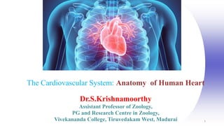
- 1. 1 The Cardiovascular System: Anatomy of Human Heart Dr.S.Krishnamoorthy Assistant Professor of Zoology, PG and Research Centre in Zoology, Vivekananda College, Tiruvedakam West, Madurai
- 2. Introduction Human heart is basically a biological pump that circulates blood in the human body. The heart is the size of a human fist and is located between the lungs in the thoracic cavity, slightly towards the left of the sternum (breastbone). It forms the cardiovascular system together with blood vessels, veins and capillaries. The blood is carried through arteries, arterioles and capillaries to the body whereas, blood is returned to the heart through venules and veins. The human heart is made of four chambers i.e., two upper left and right atria and two lower left and right ventricle.
- 4. Chambers of Heart To avoid the mix of blood rich in oxygen and carbon dioxide. Human heart has four chambers. There is a partition between the two chambers which does not allow the mix of blood. Right Atrium: These are called as blood receiving chambers. It receives blood from veins and pump it to right ventricle. Right Ventricle: It receives blood from the right atrium and pump it to the lungs. Left Atrium: It receives oxygenated blood from the lungs and pump it to the left ventricle. Left Ventricle: It pumps blood to the rest of the body.
- 5. 5 Coverings of the Heart: Anatomy Pericardium – a double-walled sac around the heart composed of: 1. A superficial fibrous pericardium 2. A deep two-layer serous pericardium a. The parietal layer lines the internal surface of the fibrous pericardium b. The visceral layer or epicardium lines the surface of the heart They are separated by the fluid-filled pericardial cavity
- 6. 6 Pericardial Layers of the Heart
- 8. Nodal tissues in heart There is a thin wall called interatrial septum that separates the right and left atria. Also, there is a thick wall called as inter ventricular septum that separates the left and right ventricles. The same side of the atria and ventricle are separated by fibrous tissue known as atrio-ventricular septum. There is an opening between the right atrium and right ventricle guarded by valves known as flaps or cusps. The tissue present in the upper right atrium is called as sinoatrial node( SAN). Whereas, the other tissue is seen in the left corner of the right atrium known as atrio-ventricular node (AVN).
- 9. Nodal tissues of heart
- 10. Blood Circulation in Human Heart
- 13. Facts about Human Heart Some of the facts about the human heart are as follows: The size of the human heart is similar to size of closed fist. The weight of the heart is around 280-340 grams in men and 230-280 gram in female. The heart beat in human is about 1,00,000 times per day. For adult, heart beat is 60-80 times per minute. The newborns heart beats are much faster then adults which is around 70-190 per minute.
Editor's Notes
- 1