SlideShare a Scribd company logo
1 of 33
1578185 - McGraw-Hill Professional ©
CHAPTER 236
Pleural Diseases
Carlos E. Kummerfeldt, MD
Nicholas J. Pastis, MD
John T. Huggins, MD
Key Clinical Questions
When does a pleural effusion require drainage?
Why make the distinction between transudates and exudates?
How does the pleural fluid analysis assist in guiding your
differential diagnosis?
When is a chest tube placement indicated?
When should a pulmonary consultation be obtained?
EPIDEMIOLOGY
The seven leading causes of pleural effusions in the United
States, in descending order
include: (1) congestive heart failure; (2) bacterial pneumonia;
(3) malignancy; (4)
pulmonary embolism; (5) viral disease; (6) postcoronary artery
bypass surgery; and (7)
1578185 - McGraw-Hill Professional ©
cirrhosis with ascites. Pneumothorax in the hospitalized patient
is most commonly found
in (1) blunt trauma (35%); (2) transthoracic needle aspiration
biopsies (25%); (3) pleural
biopsies (8%); (4) transbronchial lung biopsies (6%); (5)
mechanically ventilated patients
(4%); (6) thoracentesis (2%); and (7) central line insertions
(1%-2%). Spontaneous
pneumothorax occurs in about 15,000 cases per year in the
United States: primary
spontaneous pneumothorax occurs in adults with no underlying
lung disease, whereas
secondary spontaneous pneumothorax occurs in older adults
with underlying lung
disease, most commonly with chronic obstructive pulmonary
disease.
PLEURAL EFFUSIONS
A thoracentesis should be performed in most patients with a
pleural effusion (Table 236-
1). Thoracentesis should be performed in patients with likely
heart failure if the pleural
effusion is unilateral, if one side is greater than the other, or if
there is suspicion for a dual
diagnosis. The major risks and complications of thoracentesis
include the following: (1)
pneumothorax; (2) bleeding; (3) infection; and (4) procedural
related pain. There are no
evidence based guidelines in patients with coagulopathies, and
there are reports of
thoracentesis being performed in patients with an elevated
international normalized ratio,
uremia, thrombocytopenia or on oral antiplatelet or
anticoagulation therapies. The benefits
of correcting coagulopathy with transfusions or by withholding
antiplatelet or
anticoagulation medications should be weighed against the risks
in the individual patient
and should always be discussed with the patient. Ultrasound
should be performed in all
patients undergoing thoracentesis to less the risk of
complications associated with a blind
tap. Ultrasound can identify the pleural fluid and other
underlying anatomical structures,
estimate the size of the effusion, and determine the presence of
underlying septations or
complexity that may indicate the presence of loculations (Figure
236-1) (see Chapter 124
[Thoracentesis]).
TABLE 236-1 Indications for Thoracentesis
1. Pleural effusion size ≥1 cm on chest radiography, ultrasound
or computed tomography
(CT)
2. Fever
3. Pleuritic chest pain
4. Dyspnea
5. Suspected hospital acquired infection
6. Evidence of loculation, complexity, or septations on imaging
7. Evidence of mediastinal shift, complete hemithorax
opacification or large effusion on
imaging
file://view/books/9780071843140/epub/EPUB/xhtml/156_Chapt
er124.html
1578185 - McGraw-Hill Professional ©
Figure 236-1 Ultrasound images show a large simple pleural
effusion and a complex
pleural effusion with septations. (A) Large simple anechoic
pleural effusion causing
atelectasis of the right lower lobe (arrow). The bright white line
on the right (arrowheads)
represents the diaphragm; the atelectatic lung is on the left and
lower parts of the image.
(B) Complex pleural effusion with septations demonstrating
loculations and complexity
(arrow). The small lines between the diaphragm and
consolidated lung represent
septations. (Images courtesy of John T Huggins, MD.)
PLEURAL FLUID ANALYSIS
Pleural fluid analysis is essential to determine the cause of the
effusion. The hospitalist
should familiarize themselves with the pleural fluid tests
routinely ordered and the
diagnostic clues they provide. The pleural fluid appearance,
color, and even its smell may
provide clues as to the diagnosis. Table 236-2 shows the
differential diagnosis based on
the appearance of the fluid. The following tests should always
be obtained: protein,
lactate dehydrogenase (LDH), pH, glucose, white cell count and
differential, cytology and
cultures. Other fluid tests that may assist in confirming a
suspected diagnosis in selected
patients include fluid amylase, triglyceride, cholesterol,
adenosine deaminase (ADA),
rheumatoid factor, and antinuclear antibody. Tables 236-3
through 236-5 summarize the
use of these tests to narrow the differential diagnosis.
TABLE 236-2 Differential Diagnosis of Pleural Fluid Based on
Appearance
Fluid Appearance Differential Diagnosis
Light yellow Transudate
Urinothorax (smells like urine)
Exudate
Dark yellow or serous Exudate
Turbid Parapneumonic effusion, chylothorax, cholesterol
effusion
Purulent Empyema (putrid smell)
Milky Chylothorax
Bloody Parapneumonic, malignancy, hemothorax
Clear or watery Cerebrospinal fluid leak, peritoneal dialysis,
extravascular
1578185 - McGraw-Hill Professional ©
migration of central venous catheter
Satin sheen Cholesterol effusion
TABLE 236-3 Routine Pleural Fluid Tests Used for Analyzing
Pleural Fluid
Test Comment
Protein Elevated in exudates; >5.0 g/dL associated with
tuberculosis;
very low <0.5 g/dL seen in urinothorax, CSF leak, or
extravascular migration of central venous catheter
Lactate dehydrogenase Elevated in exudates; if increasing with
serial thoracentesis,
indicates worsening degree of pleural space inflammation
pH* Low pH >7.2-7.3 associated with: (1) complicated
parapneumonic effusion; (2) esophageal rupture; (3)
rheumatoid and lupus pleuritis; (4) tuberculosis; (5)
malignancy; (6) hemothorax; (7) urinothorax
Glucose Low glucose typically <60 mg/dL associated with: (1)
parapneumonic effusion; (2) malignancy; (3) tuberculosis; (4)
rheumatoid pleuritis (lupus has normal glucose); (5)
hemothorax. When <40 mg/dL and presence of infection,
chest tube insertion is indicated
Cytology Positive for malignancy in up to 60%; yield increases
with
repeat thoracentesis
Culture Yield increases when using blood culture bottles
(aerobic and
anaerobic); mycobacterial and fungal cultures useful when
undiagnosed exudate present
*The pH should ideally be measured in a heparinized syringe,
placed on ice if not immediately
processed and analyzed in a blood gas machine.
LIGHT’S CRITERIA
Distinction between whether the effusion is an exudate or
transudate these two categories
may assist in determining the etiology of the effusion (Figure
236-2). Light’s criteria is a
set of three characteristics that compare the following:
1578185 - McGraw-Hill Professional ©
Figure 236-2 Approach to the hospitalized patient with a
pleural effusion. CABG, coronary
artery bypass graft; CHF, congestive heart failure; CSF,
cerebrospinal fluid; SF-A, serum to
pleural fluid albumin gradient; SF-P, serum to pleural fluid
protein gradient; smx, syndrome;
TB, tuberculosis.
1. pleural fluid to serum protein ratio >0.5;
2. pleural fluid to serum lactate dehydrogenase >0.6; or
3. pleural fluid LDH > two-thirds of the upper normal limit for
serum using an “or” rule.
The pleural fluid is classified as an exudate if one of the three
criteria is met. The
pleural fluid is a transudate if none of the three criteria are met.
Light’s criteria may misclassify some transudates as exudates.
This commonly occurs
in patients with pleural effusions due to congestive heart failure
that have received diuretic
therapy. In this setting, correct classification may be possible
by applying the serum to
pleural fluid protein and albumin gradients. If the difference
between the serum to pleural
fluid protein is >3.1 g/dL or the serum to pleural fluid albumin
is >1.2 g/dL, then the
effusion is reclassified as a transudate (see Figure 236-2).
Transudates occur as a consequence of changes in the
hydrostatic or oncotic forces
within the pleural space. The resulting pleural fluid is low in
protein and LDH content. The
two most commonly encountered transudates in the hospital are
congestive heart failure
and liver cirrhosis with ascites resulting in hepatic hydrothorax.
HEART FAILURE
Systolic and diastolic heart failure represents the most common
cause of pleural effusion
encountered in the hospital. Fluid accumulates in the pleural
space by moving from the
lung interstitium across leaky mesothelial cells. The triad of
clinical signs and symptoms
(dyspnea, orthopnea, lower-extremity edema), bilateral pleural
effusions, and
1578185 - McGraw-Hill Professional ©
cardiomegaly on chest radiography establish the diagnosis. The
majority of these
effusions resolve with diuretic therapy and do not require
thoracentesis for diagnosis (see
Chapter 129 [Heart Failure]).
A thoracentesis is indicated if no cardiomegaly is appreciated
on chest radiography, if
the effusion is unilateral, or if the patient meets one of the
criteria listed in Table 236-1.
Thoracentesis may be indicated if dyspnea does not resolve with
diuretic therapy after a
few days or the effusion is large or does not appear to resolve.
Thoracentesis should be
performed if another concomitant cause for an effusion (dual
diagnosis) is suspected or if
one of the effusions is significantly greater than the other. The
hospitalist should keep in
mind that about 80% of patients have bilateral effusions, 15% to
20% a unilateral right
side effusion, and only 5% to 10% have a unilateral left side
effusion. Pulmonary
consultation should be considered in the above settings or if a
patient presents with
refractory symptomatic pleural effusions despite optimal
diuretic therapy. In such extreme
cases, an indwelling pleural catheter or talc pleurodesis may be
considered for palliative
measures.
HEPATIC HYDROTHORAX
Hepatic hydrothorax is the second most commonly encountered
cause of a transudate in
the hospitalized patient. It is estimated that about 6% of
patients with cirrhosis develop
this complication. About 80% of the effusions develop on the
right side, 17% on the left,
and 3% occur bilaterally. Ascitic fluid moves via diaphragmatic
pores and defects into the
pleural space resulting in fluid accumulation. In addition, the
negative pressure gradient
between the pleural and peritoneal cavities favors movement of
fluid into the pleural
space. Hepatic hydrothorax may occur in patients without
ascites if all the ascites has
moved into the pleural cavity.
Thoracentesis should always be performed to exclude
spontaneous bacterial pleuritis,
defined as the presence of a positive bacterial culture, a pleural
fluid neutrophil count >250
cells/μL, and absence of empyema or pneumonia with
parapneumonic effusion. Culture
negative spontaneous bacterial pleuritis occurs if pleural fluid
cultures do not grow any
microorganisms and the fluid neutrophil count is >500 cells/μL.
A diagnostic
thoracentesis should be performed in all cases of ascites and
hepatic hydrothorax in
patients presenting with fever, even when spontaneous bacterial
peritonitis is excluded,
due to the presence of hematogenous spread. Antibiotic therapy
is the treatment of
choice. Chest tube insertion or indwelling pleural catheters
should be avoided as they
result in persistent fluid drainage and protein loss that l eads to
malnourishment and
higher rates of infection.
Definitive treatment of hepatic hydrothorax should target
control of the ascites in
consultation with both pulmonary and liver specialists (see
Chapter 160 [Cirrhosis and Its
Complications]).
OTHER TRANSUDATES
Less common transudates include nephrotic syndrome,
urinothorax, peritoneal dialysis,
trapped lung, myxedema, pericarditis, and cerebrospinal fluid
leak (Table 236-6).
Pulmonary consultation should be sought whenever the cause of
a transudate remains
unclear.
EXUDATES
file://view/books/9780071843140/epub/EPUB/xhtml/163_Chapt
er129.html
file://view/books/9780071843140/epub/EPUB/xhtml/198_Chapt
er160.html
1578185 - McGraw-Hill Professional ©
Exudates occur as a consequence of pleural membrane
inflammation and disruption. The
resulting pleural fluid is high in protein and LDH content.
Exudates result from disruption
of the pleural membranes due to inflammation (parapneumonic
effusions), direct injury, or
invasion as with malignancy (Table 236-7). Initial diagnosis
will not establish a diagnosis
in about 20% of exudates. When the exudate does not resolve
spontaneously or if
malignancy is being considered, pulmonary consultation should
be obtained to assist with
appropriate workup that may include pleural biopsy by either
medical or surgical
thoracoscopy.
PARAPNEUMONIC EFFUSIONS
About 40% of bacterial pneumonias are complicated by the
development of a
parapneumonic effusion. Three stages develop: (1) exudative;
(2) fibrinopurulent; and (3)
organized. During the first exudative stage, increased
permeability in the visceral pleura
results in pleural fluid formation characterized by high protein
content but normal glucose,
pH, and LDH. Bacterial invasion during the second
fibrinopurulent stage results in
leukocyte, bacteria and cell debris accumulation. Pleural fluid
continues to accumulate,
and fibrin deposits in the visceral and parietal pleura with
resulting loculations. Anaerobic
utilization of glucose results in a lower glucose and pH levels,
and cell lysis results in
increased LDH levels. During the third and final organized
stage, pus formation occurs
from cellular debris resulting in empyema formation and pleural
thickening.
Unless the effusion is small in size (<1 cm when measured from
the inner border of the
chest wall), the majority of parapneumonic or suspected
parapneumonic effusions require
thoracentesis. Pleural fluid analysis will determine if chest tube
drainage is required (see
Figure 236-3). Complicated parapneumonic effusions and all
empyemas require chest
tube drainage. Pulmonary consultation is recommended when
chest tube drainage is
indicated for evaluation of intrapleural tissue plasminogen
activator (t-PA) combined with
DNase administration. Intrapleural t-PA with DNase has been
shown to reduce hospital
stay as well as surgical referrals. Surgical drainage via video-
assisted thoracoscopic
surgery (VATS) or open thoracotomy should be considered
when there is ongoing sepsis,
fever and infection despite appropriate antibiotics or chest tube
drainage.
1578185 - McGraw-Hill Professional ©
Figure 236-3 Management of parapneumonic effusions.
MALIGNANT EFFUSIONS
Malignant effusions represent the second most common cause of
exudates after
parapneumonic effusions, affecting about 200,000 persons per
year in the United States.
Most patients present with dyspnea, cough and less often chest
pain. In order of
frequency, the most common causes of tumors leading to
development of malignant
pleural effusion include: (1) lung, most commonly
adenocarcinoma (38%); (2) breast
(17%); (3) lymphoma (12%); (4) genitourinary (9%); (5)
gastrointestinal (7%); other (7%)
and unknown cause (10%).
Malignant effusions develop as a consequence of both an
increased amount of fluid
entry and a decreased amount of fluid exit from the pleural
cavity. Factors that lead to an
increased amount of fluid entry include: (1) direct pleural and
pulmonary vessel invasion
with increased permeability; (2) increased hydrostatic pressures
due to venous
obstruction; (3) increased vascular endothelial growth factor
(VEGF) formation by some
tumors; and (4) in some occasions, disruption of lymphatic
vessels leading to chyle
accumulation. Factors that lead to a decreased amount of fluid
exit include: (1) lymphatic
obstruction in the parietal pleura or mediastinal lymph nodes;
(2) decreased intrapleural
pressure from atelectasis formation; and (3) increased central
venous pressure if
underlying thrombosis is present.
Pleural fluid may demonstrate a serous appearing or bloody
effusion. Fluid analysis
varies but typically shows an elevated LDH due to a high cell
turnover with lysis, a
differential showing lymphocyte predominance, and glucose and
pH may be low. Fluid
cytology may be positive in up to 60% of cases. If measured,
amylase may be elevated in
about 10% of the cases. A chylothorax may be present. If
pleural fluid cytology is negative
1578185 - McGraw-Hill Professional ©
or indeterminate on initial thoracentesis and a malignant
diagnosis is highly suspected, a
repeat thoracentesis with cytology is recommended to increase
the diagnostic yield.
Pulmonary consultation should be sought at this time in order to
assist with diagnosis.
Malignant effusions are the most common cause of near
complete hemithorax
opacification on chest imaging (see Figure 236-4). Contralateral
shift of the mediastinum
usually indicates a large effusion rather than a large mass. If a
large effusion does not
result in contralateral mediastinal shift, then the lung may be
unable to expand.
Unexpandable lung or the inability of the lung to fully expand
to the chest wall results
from the following: (1) trapped lung; (2) visceral pleural
inflammation or invasion causing
lung entrapment; (3) endobronchial obstruction; and/or (4)
chronic atelectasis. Tumor
causing endobronchial obstruction and atelectasis may require
bronchoscopy; visceral
pleural thickening from direct tumor invasion can be better
visualized via a contrast chest
CT (see Figure 236-5).
Figure 236-4 Postero-anterior chest imaging shows a large left-
sided pleural effusion with
mediastinal shift to the contralateral side. Note the right
tracheal deviation, near-complete
left hemithorax opacification. The patient had a malignant
bloody effusion due to lung
adenocarcinoma. (Image courtesy of Sharon Jessie, Radiology
Department, TJ Samson
Community Hospital.)
1578185 - McGraw-Hill Professional ©
Figure 236-5 Chest ultrasound shows a pleural opacity
consistent with tumor invasion
(arrow). (Image courtesy of Sharon Jessie, Radiology
Department, TJ Samson Community
Hospital.)
The goal of therapy of malignant pleural effusions is directed
toward palliation of
symptoms. Due to rapid reaccumulation and symptom
recurrence, repeated thoracentesis
is not recommended in the majority of cases. A chest physi cian
should be consulted to
recommend the most appropriate treatment, based on the
individual circumstances:
1) Breast, small cell lung cancer and lymphoma are
chemosensitive and respond well
to chemotherapy.
2) Talc pleurodesis via chest tube or thoracoscopy may be
considered if no evidence of
unexpandable lung.
3) Indwelling pleural catheter insertion (such as PleurX®
catheters) if unexpandable
lung present; about 50% to 60% of effusions resolve after
indwelling catheter has
been inserted, with subsequent catheter removal and no
evidence of recurrence.
4) Thoracic duct ligation or a pleuroperitoneal shunt with pump
system is
recommended in the presence of chylothorax; indwelling pleural
catheters may
result in protein and lymphocyte depletion with subsequent
malnourishment and
infections.
PULMONARY EMBOLISM
It is estimated that about 30% of patients with pulmonary
embolism have an associated
pleural effusion. The effusion may be unilateral or bilateral.
Computed tomography
imaging with contrast can identify segmental or subsegmental
filling defects consistent
with embolic disease. Treatment of the pulmonary embolism
results in resolution of the
associated effusion (see Chapter 115 [Advanced Cardiothoracic
Imaging]).
POSTCORONARY ARTERY BYPASS SURGERY
About 10% of patients who undergo CABG develop a large
pleural effusion within 1 month
after the surgery most commonly in the left hemithorax,
although it may be bilateral with
file://view/books/9780071843140/epub/EPUB/xhtml/146_Chapt
er115.html
1578185 - McGraw-Hill Professional ©
the left effusion usually larger than the right. The effusion is
typically bloody as the result
of bleeding from the internal mammary harvest site. The cell
count has a lymphocyte
predominance. One or two therapeutic thoracentesis are required
as treatment. Persistence
of pleural effusion for greater than 6 months post-CABG is
usually due to the presence of
a trapped lung. Most often the effusions are transudative and are
not associated with
respiratory symptoms. However, surgical decortication should
be considered if the trapped
lung causes a large effusion.
POSTCARDIAC INJURY SYNDROME
Postcardiac injury syndrome (previously known as Dressler
syndrome) occurs after
myocardial infarction, cardiac surgery, pacemaker implantation
or blunt chest trauma. It is
characterized by the presence of fever, chest pain, a new
pericardial friction rub and
effusion, and in about 70% of cases small bilateral pleural
effusions. Postcardiac injury
syndrome may develop between 3 weeks and up to a year after
cardiac injury. Postcardiac
injury syndrome is usually treated with aspirin, colchicine, or
indomethacin and in severe
cases corticosteroids.
TUBERCULOSIS
Although uncommon in the United States, tuberculous pleuritis
may result in serious
health consequences both to the patient and from a public health
perspective if not
recognized. There is a 50% probability of developing active
tuberculosis within 5 years if
the patient does not receive antituberculous therapy.
Tuberculous pleuritis may be a
consequence of primary infection that occurred 3 to 6 months
prior, or due to reactivation.
Pleural fluid cultures are negative nearly 80% of the time, and a
tuberculin skin test may be
negative in up to one-third of patients.
About 67% of patients present with an acute clinical
presentation that includes cough,
dyspnea and chest pain; these symptoms may be confused with
pneumonia and a
parapneumonic effusion. Less commonly, patients may present
with a chronic illness and
a unilateral effusion. Pleural fluid analysis shows lymphocyte
predominance. If
thoracentesis is done in early stages, pleural fluid may show
neutrophil predominance. A
very high protein level of greater than 5.0 g/dL is highly
suggestive of the diagnosis (Table
236-4). Pulmonary consultation may assist in recommending
specific diagnostic pleural
tests such as adenosine deaminase (see Table 236-6),
polymerase chain reaction for
mycobacterial DNA and pleural fluid interferon-γ. Induced
sputum smear and culture will
be positive in half of the patients. Pleural biopsy should be
obtained if suspicion is high
and exudative effusion has not resolved. Tuberculous pleuritis
typically resolves in several
months regardless if tuberculosis treatment is given; however, if
tuberculosis treatment is
not provided, these patients have a high risk for relapse (see
Chapter 200 [Tuberculosis]).
TABLE 236-4 Differential Diagnosis Based on the Pleural
Fluid Cell Count Differential
Neutrophil
Predominance
Lymphocyte
Predominance Eosinophil Predominance
Parapneumonic Tuberculosis Pulmonary embolism
Empyema Malignancy Asbestos exposure
Pulmonary embolism Lymphoma Hemothorax
file://view/books/9780071843140/epub/EPUB/xhtml/242_Chapt
er200.html
1578185 - McGraw-Hill Professional ©
Acute pancreatitis Sarcoidosis Drug induced
Intra-abdominal abscess Rheumatoid pleuritis Fungal infections
Bilio-pleural fistula Postcoronary artery
bypass surgery
Eosinophilic granulomatosis with
polyangiitis (formerly Churg-
Strauss)
Uremia Parasite infections
Chylothorax
TABLE 236-5 Special Pleural Fluid Tests Used for Analyzing
Pleural Fluid
Test Comment
Albumin Useful when suspected transudate is misclassified as
exudate
(see Figure 236-2); if SF-A gradient >1.2 g/dL, re-classify
effusion as transudate
Amylase Elevated in: (1) esophageal perforation; (2)
pancreatitis (3)
pancreatico-pleural fistulas; (4) malignancy
Triglyceride Elevated to >110 mg/dL in chylothorax
Cholesterol Elevated to >250 mg/dL in cholesterol effusion due
to
tuberculosis, rheumatoid pleuritis, trauma, or parasitic
infection
Hematocrit Hemothorax if fluid to peripheral blood hematocrit
ratio >50%
Adenosine deaminase Elevated in patients with tuberculosis;
tuberculosis excluded if
<40 U/L
Rheumatoid factor May be elevated to ≥1:320 in rheumatoid
pleuritis
Antinuclear antibody Elevated to >1:40 in lupus pleuritis
Creatinine Elevated to higher level than serum in urinothorax
SF-A, serum to fluid albumin gradient.
TABLE 236-6 Other Less Common Causes of Transudates
Cause Characteristics Imaging
Nephrotic syndrome Due to decreased oncotic pressure from
urine protein loss and increased
intravascular hydrostatic pressure from salt
retention
Usually bilateral
effusions
Urinothorax Due to renal obstruction resulting in
retroperitoneal urine collection and drainage
across pressure gradient into pleural cavity;
creatinine level in pleural fluid higher than
serum
Effusion on same
side as obstruction
1578185 - McGraw-Hill Professional ©
Peritoneal dialysis Leakage of dialysate rich in glucose from
peritoneal cavity through diaphragmatic
defects into pleural space; high glucose and
low protein fluid
Usually right sided;
may be bilateral
Trapped lung Old inflammation resulting in fibrous
membrane with visceral pleural thickening
that causes inability of lung to fully re-
expand, increasing negative pressure within
pleural space; pleural manometry
recommended to establish diagnosis
Unilateral effusion;
chest CT with air
contrast shows
visceral pleural
thickening
Myxedema Forms from decreased lymphatic drainage Bilateral;
concomitant
pericardial effusion
many times
Constrictive
pericarditis
Increased pulmonary and systemic capillary
pressures result in fluid formation
Bilateral; may be
unilateral
Cerebrospinal fluid
leak
Fistula formation between CSF and pleural
cavity from surgery, trauma, or shunts; low
protein and LDH in fluid; measurement of
β2-transferrin virtually diagnostic
Unilateral
CT, computed tomography; LDH, lactate dehydrogenase.
TABLE 236-7 Causes of Pleural Effusions
Exudates Transudates
Common
Parapneumonic
Malignancy
Common
Congestive heart failure
Liver cirrhosis
Less common
Tuberculosis
Pulmonary embolism
Postcoronary artery bypass surgery
Chylothorax
Pseudo-chylothorax
Hemothorax
Uremia
Rheumatoid pleuritis
Lupus (drug induced or systemic)
Less common
Nephrotic syndrome
Urinothorax
Peritoneal dialysis
Trapped lung
Atelectasis
Uncommon
Asbestos exposure
Drug induced
Uncommon
Cerebrospinal fluid leak
Constrictive pericarditis
1578185 - McGraw-Hill Professional ©
Yellow-nail syndrome
Esophageal perforation
Pancreatitis
Postabdominal surgery
Bilio-pleural fistula
Sarcoidosis
Myxedema
Pulmonary veno-occlusive disease
Central venous occlusion
Extravascular migration of central venous
catheter
Glycinothorax
HEMOTHORAX
Hemothorax or the presence of blood in the pleural cavity is
defined as a pleural fluid
hematocrit that is at least 50% that of blood. Table 236-8
summarizes the causes of
hemothorax encountered in the hospital. Bleeding may be
significant and lead to
hemodynamic compromise and cardiovascular collapse if not
recognized quickly.
Clinicians should always consider hemothorax in the situations
listed in Table 236-8.
Management requires chest tube insertion in all cases in order
to quantify the rate of
bleeding and prevent any of the following complications: (1)
retention of clot; (2) infection;
and (3) fibrothorax. Thoracic surgical consultation is
recommended if chest tube output is
greater than 200 mL/h and there are no signs of slowing.
Persistence of blood in the
pleural space increases the risk for fibrothorax or trapped lung.
TABLE 236-8 Causes of Hemothorax
Traumatic Iatrogenic Nontraumatic
Penetrating injury Thoracic surgery (heart or
lung)
Malignant effusion
Nonpenetrating injury Central vein perforation
after central line insertion
Anticoagulation therapy
Thoracentesis Ruptured aortic aneurysm
Chest tube insertion Arterio-venous malformation
Lung biopsy Hematological disorder (ie,
hemophilia, thrombocytopenia)
Transbronchial biopsy Intrapleural fibrinolytics
Catamenial
CHYLOTHORAX AND CHOLESTEROL EFFUSIONS
Chylothorax is the accumulation of lipid from chyle in the
pleural space due to disruption
or obstruction of the thoracic duct. Chylothoraces may be
unilateral or bilateral, depending
on the level at which the thoracic duct disruption occurs: right
sided if the disruption
occurs below the fourth to sixth thoracic vertebrae, left sided or
bilateral if the disruption
occurs at this level or above. The pleural fluid has a
characteristic milky appearance but
may mimic that of empyema. Centrifugation of the fluid will
result in layering and
deposition of cellular debris at the bottom in empyema, whereas
in chylothorax the
1578185 - McGraw-Hill Professional ©
appearance will remain the same. Pleural fluid analysis shows
lymphocyte predominance,
a relatively low LDH and high protein (protein discordance)
level. Triglyceride levels are
greater than 110 mg/dL (see Table 236-6). Chylomicrons should
be measured if the
triglyceride level falls between 50 and 110 mg/dL. Table 236-9
lists the causes of
chylothorax encountered in the hospital.
TABLE 236-9 Causes of Chylothorax
Traumatic Nontraumatic
Iatrogenic
Surgery
Radiotherapy
Endoscopy
Tumors
Lymphoma
Metastatic pleural tumors
Noniatrogenic
Chest wall trauma
Childbirth
Lymphatic involvement
Lymphangioleiomyomatosis
Tuberous sclerosis
Amyloidosis
Yellow-nail syndrome
Sarcoidosis
Filariasis
Dasatinib and tyrosine kinase inhibitors
Gorham syndrome
Venous pressure
Mediastinal fibrosis
Superior vena cava thrombosis
Chylous ascites
A pulmonary consultation is recommended in all cases of
chylothoraces to tailor the
most appropriate therapy according to the etiology. A diet rich
in medium-chain
triglycerides may reduce the flow of chyle; absorbed directly
into the blood, medium-chain
triglycerides bypass the thoracic duct. Octreotide may reduce
the rate of chyle formation
as well. Thoracentesis may reduce dyspnea; a chest tube should
be avoided because
drainage of significant amount of protein and lymphocyte s may
cause malnutrition and
immunodeficiency. A pleuroperitoneal shunt may be considered
in cases of malignant
obstruction unresponsive to chemo- or radiation therapy.
Thoracic duct embolization,
ligation, or talc pleurodesis are all aimed at controlling chyle
leak.
Cholesterol effusion is the accumulation of lipid from
cholesterol or lecithin-globulin
due to a long-standing pleural effusion in the presence of a
pleural cavity surrounded by
fibrin. Cholesterol effusion develops in the presence of chronic
pleural space
inflammation, such as tuberculous pleuritis, rheumatoid
pleuritis, parasitic infection, or
trauma. Unlike chylothorax, triglyceride levels in pleural fluid
are low. Cholesterol crystals
may be seen on cytology and the cholesterol level is usually
greater than 250 mg/dL (see
Table 236-6). Treatment is aimed at the underlying cause.
1578185 - McGraw-Hill Professional ©
PNEUMOTHORAX
Pneumothorax is defined as presence of air in the pleural cavity.
Primary spontaneous
pneumothorax occurs as a result of a ruptured apical pleural
bleb in the majority of cases.
Chest tube insertion or air evacuation is indicated if the patient
is symptomatic or if the
pneumothorax is greater than 20% in size. Nearly all patients
with secondary spontaneous
pneumothoraces require chest tube insertion given their low
underlying lung reserve and
symptomatic presentation.
The most common cause of pneumothorax in the hospitalized
patient is iatrogenic.
Table 236-10 lists the general classification of pneumothoraces.
Diagnosis is
established by chest radiography or ultrasound imaging (see
Figure 236-6). Ultrasound
findings that indicate the presence of a pneumothorax include:
(1) absence of lung-sliding;
(2) absence of B-lines; and (3) presence of a lung-point. The
identification of a lung-point
confirms the presence of pneumothorax; while the absence of
lung-sliding on M-mode or
2-D ultrasound suggests a pneumothorax. The nondependent
parts of the thorax should
be initially scanned with the ultrasound. The size of the
pneumothorax is estimated by
measuring the distance between the visceral pleural line and the
chest wall at the level of
the hilum or apex. About 1 cm is equivalent to 10%. The
estimated size of the
pneumothorax is approximately 20% if the distance is 2 cm.
TABLE 236-10 Classification of Pneumothorax
Spontaneous
Primary (no underlying lung disease)
Secondary (underlying lung disease)
Traumatic
Iatrogenic
Catamenial
Tension
1578185 - McGraw-Hill Professional ©
Figure 236-6 Chest radiography shows a large right side
pneumothorax with a visceral
pleural line noted and absence of blood vessels and lung
markings towards the chest wall
beyond the pleural line. Note that the pleural line is sharp and
well demarcated.
Chest tube insertion is recommended in iatrogenic
pneumothoraces that result in
symptoms and are greater than 20% in size. If the patient is
asymptomatic and the
pneumothorax is less than or equal to 20%, conservative
management may be pursued
with high-flow 100% continuous oxygen and observation.
Tension pneumothorax may
easily develop in patients on mechanical ventilation; and
therefore chest tube insertion is
recommended in patients who develop iatrogenic pneumothorax.
The chest tube should
be left in place for at least 48 hours after the air l eak stops
while these patients remain on
the ventilator.
TENSION PNEUMOTHORAX
The clinical signs of tension pneumothorax include sudden
cardiovascular collapse,
cyanosis, and respiratory distress. In mechanically ventilated
patients, peak pressure
suddenly increases if assist control volume cycled mechanical
ventilation is used. In
patients receiving cardiopulmonary resuscitation, difficulty in
ventilating the patient may
be the only sign. Emergent size 14 to 16 gauge needle catheter
insertion at the level of the
midclavicular line, anterior second intercostal space is
performed. The catheter is left in
place until air ceases to exit. Chest tube should then be inserted.
PRACTICE POINT
Indications for Chest Tube Insertion
Chest tube insertion should be performed by experienced
operators and in the appropriate
clinical setting. Smaller bore chest tubes are noninferior to
larger bore chest tubes for
most indications. Although they are easier to insert and reduce
the amount of pain,
1578185 - McGraw-Hill Professional ©
smaller bore chest tubes tend to dislodge easier. Chest tube
insertion is recommended for
the following:
Complicated parapneumonic effusion
Empyema
Talc pleurodesis administration
Hemothorax
Tension pneumothorax
Secondary spontaneous pneumothorax and symptomatic
Pneumothorax greater than 20%
Indwelling pleural catheters are recommended in malignant
pleural effusions with an
unexpandable lung.
PRACTICE POINT
Pulmonary Consultation
Pulmonary consultation is recommended in the following:
Thoracentesis and chest tube insertion if provider is unfamiliar
or uncomfortable with
procedures
Unilateral effusion, absence of cardiomegaly, bilateral
asymmetric effusions, fever or
pleurisy in a patient with congestive heart failure
Recurrent hepatic hydrothorax or suspected spontaneous
bacterial pleuritis
Undiagnosed transudate or exudate
Suspected trapped lung and need for pleural manometry
Complicated parapneumonic effusions and empyemas
Evaluation for intrapleural t-PA and DNase
Malignant effusions with need for indwelling pleural catheter
insertion and talc
pleurodesis evaluation
Suspected tuberculous pleuritis
Hemothorax
Chylothorax and pseudochylothorax
Pneumothorax
SUGGESTED READINGS
Broaddus VC, Light RW. Pleural effusion. In: Broaddus VC,
Mason RM, Ernst JD, et al, eds.
Murray & Nadel’s Textbook of Respiratory Medicine, 6th ed.
Philadelphia, PA: Elsevier
Saunders; 2015;1396-1424.
Colice GL, Curtis A, Deslauriers J, et al. Medical and surgical
treatment of parapneumonic
effusions: an evidence-based guideline. Chest.
2000;118(4):1158-1171.
1578185 - McGraw-Hill Professional ©
Light RW. Textbook of Pleural Diseases. Baltimore, MD:
Wolters Kluwer Lippincott Williams
and Wilkins; 2013.
Light RW, Macgregor MI, Luchsinger PC, Ball WC Jr. Pleural
effusions: the diagnostic
separation of transudates and exudates. Ann Intern Med.
1972;77(4):507-513.
MacDuff A, Arnold A, Harvey J. Management of spontaneous
pneumothorax: British
Thoracic Society Pleural Disease Guideline 2

More Related Content

Similar to 1578185 - McGraw-Hill Professional ©CHAPTER 236Pleural D

Similar to 1578185 - McGraw-Hill Professional ©CHAPTER 236Pleural D (20)

Uremic Pericarditis
Uremic PericarditisUremic Pericarditis
Uremic Pericarditis
 
Trali
TraliTrali
Trali
 
Trali
TraliTrali
Trali
 
Trali
TraliTrali
Trali
 
Trali
TraliTrali
Trali
 
Hemorrhage
HemorrhageHemorrhage
Hemorrhage
 
Adrenal incidentaloma
Adrenal incidentaloma Adrenal incidentaloma
Adrenal incidentaloma
 
Diagnosis & Classification of Pulmonary Hypertension
Diagnosis & Classification of Pulmonary HypertensionDiagnosis & Classification of Pulmonary Hypertension
Diagnosis & Classification of Pulmonary Hypertension
 
Cardiomyopathy Presentation
Cardiomyopathy PresentationCardiomyopathy Presentation
Cardiomyopathy Presentation
 
Thrombosis Case Study
Thrombosis Case StudyThrombosis Case Study
Thrombosis Case Study
 
NST-ACS guidelines
NST-ACS guidelinesNST-ACS guidelines
NST-ACS guidelines
 
Management of major traumaMAIN.pptx
Management of major traumaMAIN.pptxManagement of major traumaMAIN.pptx
Management of major traumaMAIN.pptx
 
trauma.pptx
trauma.pptxtrauma.pptx
trauma.pptx
 
Successful treatment of homozygous fh by using cascade
Successful treatment of homozygous fh by using cascadeSuccessful treatment of homozygous fh by using cascade
Successful treatment of homozygous fh by using cascade
 
Non alcoholic steatohepatitis copy
Non alcoholic steatohepatitis   copyNon alcoholic steatohepatitis   copy
Non alcoholic steatohepatitis copy
 
4 s.full
4 s.full4 s.full
4 s.full
 
Takayasu's arteritis
Takayasu's arteritisTakayasu's arteritis
Takayasu's arteritis
 
Shock in
Shock in Shock in
Shock in
 
Takayasu arteritis
Takayasu arteritis Takayasu arteritis
Takayasu arteritis
 
Suporte inotrópico e DP em RN após cx cardíaca
Suporte inotrópico e DP em RN após cx cardíacaSuporte inotrópico e DP em RN após cx cardíaca
Suporte inotrópico e DP em RN após cx cardíaca
 

More from KiyokoSlagleis

1.Assess the main steps involved in developing an effective stra.docx
1.Assess the main steps involved in developing an effective stra.docx1.Assess the main steps involved in developing an effective stra.docx
1.Assess the main steps involved in developing an effective stra.docxKiyokoSlagleis
 
1.Choose one of the critical steps to building a secure organi.docx
1.Choose one of the critical steps to building a secure organi.docx1.Choose one of the critical steps to building a secure organi.docx
1.Choose one of the critical steps to building a secure organi.docxKiyokoSlagleis
 
1.Briefly summarize the purpose of the implementation phase in SDLC..docx
1.Briefly summarize the purpose of the implementation phase in SDLC..docx1.Briefly summarize the purpose of the implementation phase in SDLC..docx
1.Briefly summarize the purpose of the implementation phase in SDLC..docxKiyokoSlagleis
 
1.Choose four standard corporate executive positions and des.docx
1.Choose four standard corporate executive positions and des.docx1.Choose four standard corporate executive positions and des.docx
1.Choose four standard corporate executive positions and des.docxKiyokoSlagleis
 
1.An eassy talk about ethics by a ethics song. You can find a ethics.docx
1.An eassy talk about ethics by a ethics song. You can find a ethics.docx1.An eassy talk about ethics by a ethics song. You can find a ethics.docx
1.An eassy talk about ethics by a ethics song. You can find a ethics.docxKiyokoSlagleis
 
1.A school psychologist strongly believes a particular child is .docx
1.A school psychologist strongly believes a particular child is .docx1.A school psychologist strongly believes a particular child is .docx
1.A school psychologist strongly believes a particular child is .docxKiyokoSlagleis
 
1.Choose one stanza from Aaron Abeytas thirteen ways of looking .docx
1.Choose one stanza from Aaron Abeytas thirteen ways of looking .docx1.Choose one stanza from Aaron Abeytas thirteen ways of looking .docx
1.Choose one stanza from Aaron Abeytas thirteen ways of looking .docxKiyokoSlagleis
 
1.A psychologist is interested in learning more about how childr.docx
1.A psychologist is interested in learning more about how childr.docx1.A psychologist is interested in learning more about how childr.docx
1.A psychologist is interested in learning more about how childr.docxKiyokoSlagleis
 
1.A school psychologist strongly believes a particular child i.docx
1.A school psychologist strongly believes a particular child i.docx1.A school psychologist strongly believes a particular child i.docx
1.A school psychologist strongly believes a particular child i.docxKiyokoSlagleis
 
1.According to the NIST, what were the reasons for the collapse of.docx
1.According to the NIST, what were the reasons for the collapse of.docx1.According to the NIST, what were the reasons for the collapse of.docx
1.According to the NIST, what were the reasons for the collapse of.docxKiyokoSlagleis
 
1.5 page for thisPlease review the Case Study introduction present.docx
1.5 page for thisPlease review the Case Study introduction present.docx1.5 page for thisPlease review the Case Study introduction present.docx
1.5 page for thisPlease review the Case Study introduction present.docxKiyokoSlagleis
 
1.) What is Mills response to the objection that happiness cannot b.docx
1.) What is Mills response to the objection that happiness cannot b.docx1.) What is Mills response to the objection that happiness cannot b.docx
1.) What is Mills response to the objection that happiness cannot b.docxKiyokoSlagleis
 
1.Add an example or evidence for each reasons ( i listd )why the use.docx
1.Add an example or evidence for each reasons ( i listd )why the use.docx1.Add an example or evidence for each reasons ( i listd )why the use.docx
1.Add an example or evidence for each reasons ( i listd )why the use.docxKiyokoSlagleis
 
1.1. Some of the most serious abuses taking place in developing .docx
1.1. Some of the most serious abuses taking place in developing .docx1.1. Some of the most serious abuses taking place in developing .docx
1.1. Some of the most serious abuses taking place in developing .docxKiyokoSlagleis
 
1.A population of grasshoppers in the Kansas prairie has two col.docx
1.A population of grasshoppers in the Kansas prairie has two col.docx1.A population of grasshoppers in the Kansas prairie has two col.docx
1.A population of grasshoppers in the Kansas prairie has two col.docxKiyokoSlagleis
 
1.5 pages single spaced, include References and when necessary, imag.docx
1.5 pages single spaced, include References and when necessary, imag.docx1.5 pages single spaced, include References and when necessary, imag.docx
1.5 pages single spaced, include References and when necessary, imag.docxKiyokoSlagleis
 
1.1- What are the real reasons behind the existence of Racism W.docx
1.1- What are the real reasons behind the existence of Racism W.docx1.1- What are the real reasons behind the existence of Racism W.docx
1.1- What are the real reasons behind the existence of Racism W.docxKiyokoSlagleis
 
1.) Connect 3 Due October 4th2.) Connect 4 Due Octob.docx
1.) Connect 3 Due October 4th2.) Connect 4 Due Octob.docx1.) Connect 3 Due October 4th2.) Connect 4 Due Octob.docx
1.) Connect 3 Due October 4th2.) Connect 4 Due Octob.docxKiyokoSlagleis
 
1.  Write an equation in standard form of the parabola that has th.docx
1.  Write an equation in standard form of the parabola that has th.docx1.  Write an equation in standard form of the parabola that has th.docx
1.  Write an equation in standard form of the parabola that has th.docxKiyokoSlagleis
 
1.A health psychologist in a northern climate wants to evaluate .docx
1.A health psychologist in a northern climate wants to evaluate .docx1.A health psychologist in a northern climate wants to evaluate .docx
1.A health psychologist in a northern climate wants to evaluate .docxKiyokoSlagleis
 

More from KiyokoSlagleis (20)

1.Assess the main steps involved in developing an effective stra.docx
1.Assess the main steps involved in developing an effective stra.docx1.Assess the main steps involved in developing an effective stra.docx
1.Assess the main steps involved in developing an effective stra.docx
 
1.Choose one of the critical steps to building a secure organi.docx
1.Choose one of the critical steps to building a secure organi.docx1.Choose one of the critical steps to building a secure organi.docx
1.Choose one of the critical steps to building a secure organi.docx
 
1.Briefly summarize the purpose of the implementation phase in SDLC..docx
1.Briefly summarize the purpose of the implementation phase in SDLC..docx1.Briefly summarize the purpose of the implementation phase in SDLC..docx
1.Briefly summarize the purpose of the implementation phase in SDLC..docx
 
1.Choose four standard corporate executive positions and des.docx
1.Choose four standard corporate executive positions and des.docx1.Choose four standard corporate executive positions and des.docx
1.Choose four standard corporate executive positions and des.docx
 
1.An eassy talk about ethics by a ethics song. You can find a ethics.docx
1.An eassy talk about ethics by a ethics song. You can find a ethics.docx1.An eassy talk about ethics by a ethics song. You can find a ethics.docx
1.An eassy talk about ethics by a ethics song. You can find a ethics.docx
 
1.A school psychologist strongly believes a particular child is .docx
1.A school psychologist strongly believes a particular child is .docx1.A school psychologist strongly believes a particular child is .docx
1.A school psychologist strongly believes a particular child is .docx
 
1.Choose one stanza from Aaron Abeytas thirteen ways of looking .docx
1.Choose one stanza from Aaron Abeytas thirteen ways of looking .docx1.Choose one stanza from Aaron Abeytas thirteen ways of looking .docx
1.Choose one stanza from Aaron Abeytas thirteen ways of looking .docx
 
1.A psychologist is interested in learning more about how childr.docx
1.A psychologist is interested in learning more about how childr.docx1.A psychologist is interested in learning more about how childr.docx
1.A psychologist is interested in learning more about how childr.docx
 
1.A school psychologist strongly believes a particular child i.docx
1.A school psychologist strongly believes a particular child i.docx1.A school psychologist strongly believes a particular child i.docx
1.A school psychologist strongly believes a particular child i.docx
 
1.According to the NIST, what were the reasons for the collapse of.docx
1.According to the NIST, what were the reasons for the collapse of.docx1.According to the NIST, what were the reasons for the collapse of.docx
1.According to the NIST, what were the reasons for the collapse of.docx
 
1.5 page for thisPlease review the Case Study introduction present.docx
1.5 page for thisPlease review the Case Study introduction present.docx1.5 page for thisPlease review the Case Study introduction present.docx
1.5 page for thisPlease review the Case Study introduction present.docx
 
1.) What is Mills response to the objection that happiness cannot b.docx
1.) What is Mills response to the objection that happiness cannot b.docx1.) What is Mills response to the objection that happiness cannot b.docx
1.) What is Mills response to the objection that happiness cannot b.docx
 
1.Add an example or evidence for each reasons ( i listd )why the use.docx
1.Add an example or evidence for each reasons ( i listd )why the use.docx1.Add an example or evidence for each reasons ( i listd )why the use.docx
1.Add an example or evidence for each reasons ( i listd )why the use.docx
 
1.1. Some of the most serious abuses taking place in developing .docx
1.1. Some of the most serious abuses taking place in developing .docx1.1. Some of the most serious abuses taking place in developing .docx
1.1. Some of the most serious abuses taking place in developing .docx
 
1.A population of grasshoppers in the Kansas prairie has two col.docx
1.A population of grasshoppers in the Kansas prairie has two col.docx1.A population of grasshoppers in the Kansas prairie has two col.docx
1.A population of grasshoppers in the Kansas prairie has two col.docx
 
1.5 pages single spaced, include References and when necessary, imag.docx
1.5 pages single spaced, include References and when necessary, imag.docx1.5 pages single spaced, include References and when necessary, imag.docx
1.5 pages single spaced, include References and when necessary, imag.docx
 
1.1- What are the real reasons behind the existence of Racism W.docx
1.1- What are the real reasons behind the existence of Racism W.docx1.1- What are the real reasons behind the existence of Racism W.docx
1.1- What are the real reasons behind the existence of Racism W.docx
 
1.) Connect 3 Due October 4th2.) Connect 4 Due Octob.docx
1.) Connect 3 Due October 4th2.) Connect 4 Due Octob.docx1.) Connect 3 Due October 4th2.) Connect 4 Due Octob.docx
1.) Connect 3 Due October 4th2.) Connect 4 Due Octob.docx
 
1.  Write an equation in standard form of the parabola that has th.docx
1.  Write an equation in standard form of the parabola that has th.docx1.  Write an equation in standard form of the parabola that has th.docx
1.  Write an equation in standard form of the parabola that has th.docx
 
1.A health psychologist in a northern climate wants to evaluate .docx
1.A health psychologist in a northern climate wants to evaluate .docx1.A health psychologist in a northern climate wants to evaluate .docx
1.A health psychologist in a northern climate wants to evaluate .docx
 

Recently uploaded

Kisan Call Centre - To harness potential of ICT in Agriculture by answer farm...
Kisan Call Centre - To harness potential of ICT in Agriculture by answer farm...Kisan Call Centre - To harness potential of ICT in Agriculture by answer farm...
Kisan Call Centre - To harness potential of ICT in Agriculture by answer farm...Krashi Coaching
 
microwave assisted reaction. General introduction
microwave assisted reaction. General introductionmicrowave assisted reaction. General introduction
microwave assisted reaction. General introductionMaksud Ahmed
 
Beyond the EU: DORA and NIS 2 Directive's Global Impact
Beyond the EU: DORA and NIS 2 Directive's Global ImpactBeyond the EU: DORA and NIS 2 Directive's Global Impact
Beyond the EU: DORA and NIS 2 Directive's Global ImpactPECB
 
The Most Excellent Way | 1 Corinthians 13
The Most Excellent Way | 1 Corinthians 13The Most Excellent Way | 1 Corinthians 13
The Most Excellent Way | 1 Corinthians 13Steve Thomason
 
How to Make a Pirate ship Primary Education.pptx
How to Make a Pirate ship Primary Education.pptxHow to Make a Pirate ship Primary Education.pptx
How to Make a Pirate ship Primary Education.pptxmanuelaromero2013
 
Industrial Policy - 1948, 1956, 1973, 1977, 1980, 1991
Industrial Policy - 1948, 1956, 1973, 1977, 1980, 1991Industrial Policy - 1948, 1956, 1973, 1977, 1980, 1991
Industrial Policy - 1948, 1956, 1973, 1977, 1980, 1991RKavithamani
 
CARE OF CHILD IN INCUBATOR..........pptx
CARE OF CHILD IN INCUBATOR..........pptxCARE OF CHILD IN INCUBATOR..........pptx
CARE OF CHILD IN INCUBATOR..........pptxGaneshChakor2
 
Advanced Views - Calendar View in Odoo 17
Advanced Views - Calendar View in Odoo 17Advanced Views - Calendar View in Odoo 17
Advanced Views - Calendar View in Odoo 17Celine George
 
Arihant handbook biology for class 11 .pdf
Arihant handbook biology for class 11 .pdfArihant handbook biology for class 11 .pdf
Arihant handbook biology for class 11 .pdfchloefrazer622
 
BASLIQ CURRENT LOOKBOOK LOOKBOOK(1) (1).pdf
BASLIQ CURRENT LOOKBOOK  LOOKBOOK(1) (1).pdfBASLIQ CURRENT LOOKBOOK  LOOKBOOK(1) (1).pdf
BASLIQ CURRENT LOOKBOOK LOOKBOOK(1) (1).pdfSoniaTolstoy
 
Presentation by Andreas Schleicher Tackling the School Absenteeism Crisis 30 ...
Presentation by Andreas Schleicher Tackling the School Absenteeism Crisis 30 ...Presentation by Andreas Schleicher Tackling the School Absenteeism Crisis 30 ...
Presentation by Andreas Schleicher Tackling the School Absenteeism Crisis 30 ...EduSkills OECD
 
Separation of Lanthanides/ Lanthanides and Actinides
Separation of Lanthanides/ Lanthanides and ActinidesSeparation of Lanthanides/ Lanthanides and Actinides
Separation of Lanthanides/ Lanthanides and ActinidesFatimaKhan178732
 
1029-Danh muc Sach Giao Khoa khoi 6.pdf
1029-Danh muc Sach Giao Khoa khoi  6.pdf1029-Danh muc Sach Giao Khoa khoi  6.pdf
1029-Danh muc Sach Giao Khoa khoi 6.pdfQucHHunhnh
 
Nutritional Needs Presentation - HLTH 104
Nutritional Needs Presentation - HLTH 104Nutritional Needs Presentation - HLTH 104
Nutritional Needs Presentation - HLTH 104misteraugie
 
Employee wellbeing at the workplace.pptx
Employee wellbeing at the workplace.pptxEmployee wellbeing at the workplace.pptx
Employee wellbeing at the workplace.pptxNirmalaLoungPoorunde1
 
Contemporary philippine arts from the regions_PPT_Module_12 [Autosaved] (1).pptx
Contemporary philippine arts from the regions_PPT_Module_12 [Autosaved] (1).pptxContemporary philippine arts from the regions_PPT_Module_12 [Autosaved] (1).pptx
Contemporary philippine arts from the regions_PPT_Module_12 [Autosaved] (1).pptxRoyAbrique
 

Recently uploaded (20)

Kisan Call Centre - To harness potential of ICT in Agriculture by answer farm...
Kisan Call Centre - To harness potential of ICT in Agriculture by answer farm...Kisan Call Centre - To harness potential of ICT in Agriculture by answer farm...
Kisan Call Centre - To harness potential of ICT in Agriculture by answer farm...
 
microwave assisted reaction. General introduction
microwave assisted reaction. General introductionmicrowave assisted reaction. General introduction
microwave assisted reaction. General introduction
 
Código Creativo y Arte de Software | Unidad 1
Código Creativo y Arte de Software | Unidad 1Código Creativo y Arte de Software | Unidad 1
Código Creativo y Arte de Software | Unidad 1
 
Beyond the EU: DORA and NIS 2 Directive's Global Impact
Beyond the EU: DORA and NIS 2 Directive's Global ImpactBeyond the EU: DORA and NIS 2 Directive's Global Impact
Beyond the EU: DORA and NIS 2 Directive's Global Impact
 
The Most Excellent Way | 1 Corinthians 13
The Most Excellent Way | 1 Corinthians 13The Most Excellent Way | 1 Corinthians 13
The Most Excellent Way | 1 Corinthians 13
 
Mattingly "AI & Prompt Design: Structured Data, Assistants, & RAG"
Mattingly "AI & Prompt Design: Structured Data, Assistants, & RAG"Mattingly "AI & Prompt Design: Structured Data, Assistants, & RAG"
Mattingly "AI & Prompt Design: Structured Data, Assistants, & RAG"
 
How to Make a Pirate ship Primary Education.pptx
How to Make a Pirate ship Primary Education.pptxHow to Make a Pirate ship Primary Education.pptx
How to Make a Pirate ship Primary Education.pptx
 
Industrial Policy - 1948, 1956, 1973, 1977, 1980, 1991
Industrial Policy - 1948, 1956, 1973, 1977, 1980, 1991Industrial Policy - 1948, 1956, 1973, 1977, 1980, 1991
Industrial Policy - 1948, 1956, 1973, 1977, 1980, 1991
 
Staff of Color (SOC) Retention Efforts DDSD
Staff of Color (SOC) Retention Efforts DDSDStaff of Color (SOC) Retention Efforts DDSD
Staff of Color (SOC) Retention Efforts DDSD
 
CARE OF CHILD IN INCUBATOR..........pptx
CARE OF CHILD IN INCUBATOR..........pptxCARE OF CHILD IN INCUBATOR..........pptx
CARE OF CHILD IN INCUBATOR..........pptx
 
Advanced Views - Calendar View in Odoo 17
Advanced Views - Calendar View in Odoo 17Advanced Views - Calendar View in Odoo 17
Advanced Views - Calendar View in Odoo 17
 
INDIA QUIZ 2024 RLAC DELHI UNIVERSITY.pptx
INDIA QUIZ 2024 RLAC DELHI UNIVERSITY.pptxINDIA QUIZ 2024 RLAC DELHI UNIVERSITY.pptx
INDIA QUIZ 2024 RLAC DELHI UNIVERSITY.pptx
 
Arihant handbook biology for class 11 .pdf
Arihant handbook biology for class 11 .pdfArihant handbook biology for class 11 .pdf
Arihant handbook biology for class 11 .pdf
 
BASLIQ CURRENT LOOKBOOK LOOKBOOK(1) (1).pdf
BASLIQ CURRENT LOOKBOOK  LOOKBOOK(1) (1).pdfBASLIQ CURRENT LOOKBOOK  LOOKBOOK(1) (1).pdf
BASLIQ CURRENT LOOKBOOK LOOKBOOK(1) (1).pdf
 
Presentation by Andreas Schleicher Tackling the School Absenteeism Crisis 30 ...
Presentation by Andreas Schleicher Tackling the School Absenteeism Crisis 30 ...Presentation by Andreas Schleicher Tackling the School Absenteeism Crisis 30 ...
Presentation by Andreas Schleicher Tackling the School Absenteeism Crisis 30 ...
 
Separation of Lanthanides/ Lanthanides and Actinides
Separation of Lanthanides/ Lanthanides and ActinidesSeparation of Lanthanides/ Lanthanides and Actinides
Separation of Lanthanides/ Lanthanides and Actinides
 
1029-Danh muc Sach Giao Khoa khoi 6.pdf
1029-Danh muc Sach Giao Khoa khoi  6.pdf1029-Danh muc Sach Giao Khoa khoi  6.pdf
1029-Danh muc Sach Giao Khoa khoi 6.pdf
 
Nutritional Needs Presentation - HLTH 104
Nutritional Needs Presentation - HLTH 104Nutritional Needs Presentation - HLTH 104
Nutritional Needs Presentation - HLTH 104
 
Employee wellbeing at the workplace.pptx
Employee wellbeing at the workplace.pptxEmployee wellbeing at the workplace.pptx
Employee wellbeing at the workplace.pptx
 
Contemporary philippine arts from the regions_PPT_Module_12 [Autosaved] (1).pptx
Contemporary philippine arts from the regions_PPT_Module_12 [Autosaved] (1).pptxContemporary philippine arts from the regions_PPT_Module_12 [Autosaved] (1).pptx
Contemporary philippine arts from the regions_PPT_Module_12 [Autosaved] (1).pptx
 

1578185 - McGraw-Hill Professional ©CHAPTER 236Pleural D

  • 1. 1578185 - McGraw-Hill Professional © CHAPTER 236 Pleural Diseases Carlos E. Kummerfeldt, MD Nicholas J. Pastis, MD John T. Huggins, MD Key Clinical Questions When does a pleural effusion require drainage? Why make the distinction between transudates and exudates? How does the pleural fluid analysis assist in guiding your differential diagnosis? When is a chest tube placement indicated? When should a pulmonary consultation be obtained? EPIDEMIOLOGY The seven leading causes of pleural effusions in the United States, in descending order include: (1) congestive heart failure; (2) bacterial pneumonia; (3) malignancy; (4) pulmonary embolism; (5) viral disease; (6) postcoronary artery bypass surgery; and (7) 1578185 - McGraw-Hill Professional ©
  • 2. cirrhosis with ascites. Pneumothorax in the hospitalized patient is most commonly found in (1) blunt trauma (35%); (2) transthoracic needle aspiration biopsies (25%); (3) pleural biopsies (8%); (4) transbronchial lung biopsies (6%); (5) mechanically ventilated patients (4%); (6) thoracentesis (2%); and (7) central line insertions (1%-2%). Spontaneous pneumothorax occurs in about 15,000 cases per year in the United States: primary spontaneous pneumothorax occurs in adults with no underlying lung disease, whereas secondary spontaneous pneumothorax occurs in older adults with underlying lung disease, most commonly with chronic obstructive pulmonary disease. PLEURAL EFFUSIONS A thoracentesis should be performed in most patients with a pleural effusion (Table 236- 1). Thoracentesis should be performed in patients with likely heart failure if the pleural effusion is unilateral, if one side is greater than the other, or if there is suspicion for a dual diagnosis. The major risks and complications of thoracentesis include the following: (1) pneumothorax; (2) bleeding; (3) infection; and (4) procedural related pain. There are no evidence based guidelines in patients with coagulopathies, and there are reports of thoracentesis being performed in patients with an elevated international normalized ratio, uremia, thrombocytopenia or on oral antiplatelet or anticoagulation therapies. The benefits of correcting coagulopathy with transfusions or by withholding
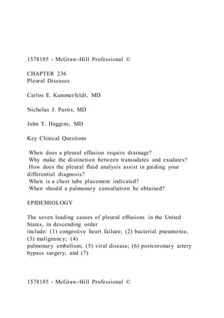
  • 3. antiplatelet or anticoagulation medications should be weighed against the risks in the individual patient and should always be discussed with the patient. Ultrasound should be performed in all patients undergoing thoracentesis to less the risk of complications associated with a blind tap. Ultrasound can identify the pleural fluid and other underlying anatomical structures, estimate the size of the effusion, and determine the presence of underlying septations or complexity that may indicate the presence of loculations (Figure 236-1) (see Chapter 124 [Thoracentesis]). TABLE 236-1 Indications for Thoracentesis 1. Pleural effusion size ≥1 cm on chest radiography, ultrasound or computed tomography (CT) 2. Fever 3. Pleuritic chest pain 4. Dyspnea 5. Suspected hospital acquired infection 6. Evidence of loculation, complexity, or septations on imaging 7. Evidence of mediastinal shift, complete hemithorax opacification or large effusion on imaging file://view/books/9780071843140/epub/EPUB/xhtml/156_Chapt er124.html 1578185 - McGraw-Hill Professional ©
  • 4. Figure 236-1 Ultrasound images show a large simple pleural effusion and a complex pleural effusion with septations. (A) Large simple anechoic pleural effusion causing atelectasis of the right lower lobe (arrow). The bright white line on the right (arrowheads) represents the diaphragm; the atelectatic lung is on the left and lower parts of the image. (B) Complex pleural effusion with septations demonstrating loculations and complexity (arrow). The small lines between the diaphragm and consolidated lung represent septations. (Images courtesy of John T Huggins, MD.) PLEURAL FLUID ANALYSIS Pleural fluid analysis is essential to determine the cause of the effusion. The hospitalist should familiarize themselves with the pleural fluid tests routinely ordered and the diagnostic clues they provide. The pleural fluid appearance, color, and even its smell may provide clues as to the diagnosis. Table 236-2 shows the differential diagnosis based on the appearance of the fluid. The following tests should always be obtained: protein, lactate dehydrogenase (LDH), pH, glucose, white cell count and differential, cytology and cultures. Other fluid tests that may assist in confirming a suspected diagnosis in selected patients include fluid amylase, triglyceride, cholesterol, adenosine deaminase (ADA), rheumatoid factor, and antinuclear antibody. Tables 236-3 through 236-5 summarize the use of these tests to narrow the differential diagnosis.
  • 5. TABLE 236-2 Differential Diagnosis of Pleural Fluid Based on Appearance Fluid Appearance Differential Diagnosis Light yellow Transudate Urinothorax (smells like urine) Exudate Dark yellow or serous Exudate Turbid Parapneumonic effusion, chylothorax, cholesterol effusion Purulent Empyema (putrid smell) Milky Chylothorax Bloody Parapneumonic, malignancy, hemothorax Clear or watery Cerebrospinal fluid leak, peritoneal dialysis, extravascular 1578185 - McGraw-Hill Professional © migration of central venous catheter Satin sheen Cholesterol effusion TABLE 236-3 Routine Pleural Fluid Tests Used for Analyzing Pleural Fluid Test Comment Protein Elevated in exudates; >5.0 g/dL associated with tuberculosis; very low <0.5 g/dL seen in urinothorax, CSF leak, or extravascular migration of central venous catheter
  • 6. Lactate dehydrogenase Elevated in exudates; if increasing with serial thoracentesis, indicates worsening degree of pleural space inflammation pH* Low pH >7.2-7.3 associated with: (1) complicated parapneumonic effusion; (2) esophageal rupture; (3) rheumatoid and lupus pleuritis; (4) tuberculosis; (5) malignancy; (6) hemothorax; (7) urinothorax Glucose Low glucose typically <60 mg/dL associated with: (1) parapneumonic effusion; (2) malignancy; (3) tuberculosis; (4) rheumatoid pleuritis (lupus has normal glucose); (5) hemothorax. When <40 mg/dL and presence of infection, chest tube insertion is indicated Cytology Positive for malignancy in up to 60%; yield increases with repeat thoracentesis Culture Yield increases when using blood culture bottles (aerobic and anaerobic); mycobacterial and fungal cultures useful when undiagnosed exudate present *The pH should ideally be measured in a heparinized syringe, placed on ice if not immediately processed and analyzed in a blood gas machine. LIGHT’S CRITERIA Distinction between whether the effusion is an exudate or transudate these two categories may assist in determining the etiology of the effusion (Figure 236-2). Light’s criteria is a set of three characteristics that compare the following:
  • 7. 1578185 - McGraw-Hill Professional © Figure 236-2 Approach to the hospitalized patient with a pleural effusion. CABG, coronary artery bypass graft; CHF, congestive heart failure; CSF, cerebrospinal fluid; SF-A, serum to pleural fluid albumin gradient; SF-P, serum to pleural fluid protein gradient; smx, syndrome; TB, tuberculosis. 1. pleural fluid to serum protein ratio >0.5; 2. pleural fluid to serum lactate dehydrogenase >0.6; or 3. pleural fluid LDH > two-thirds of the upper normal limit for serum using an “or” rule. The pleural fluid is classified as an exudate if one of the three criteria is met. The pleural fluid is a transudate if none of the three criteria are met. Light’s criteria may misclassify some transudates as exudates. This commonly occurs in patients with pleural effusions due to congestive heart failure that have received diuretic therapy. In this setting, correct classification may be possible by applying the serum to pleural fluid protein and albumin gradients. If the difference between the serum to pleural fluid protein is >3.1 g/dL or the serum to pleural fluid albumin is >1.2 g/dL, then the effusion is reclassified as a transudate (see Figure 236-2). Transudates occur as a consequence of changes in the hydrostatic or oncotic forces within the pleural space. The resulting pleural fluid is low in
  • 8. protein and LDH content. The two most commonly encountered transudates in the hospital are congestive heart failure and liver cirrhosis with ascites resulting in hepatic hydrothorax. HEART FAILURE Systolic and diastolic heart failure represents the most common cause of pleural effusion encountered in the hospital. Fluid accumulates in the pleural space by moving from the lung interstitium across leaky mesothelial cells. The triad of clinical signs and symptoms (dyspnea, orthopnea, lower-extremity edema), bilateral pleural effusions, and 1578185 - McGraw-Hill Professional © cardiomegaly on chest radiography establish the diagnosis. The majority of these effusions resolve with diuretic therapy and do not require thoracentesis for diagnosis (see Chapter 129 [Heart Failure]). A thoracentesis is indicated if no cardiomegaly is appreciated on chest radiography, if the effusion is unilateral, or if the patient meets one of the criteria listed in Table 236-1. Thoracentesis may be indicated if dyspnea does not resolve with diuretic therapy after a few days or the effusion is large or does not appear to resolve. Thoracentesis should be performed if another concomitant cause for an effusion (dual diagnosis) is suspected or if
  • 9. one of the effusions is significantly greater than the other. The hospitalist should keep in mind that about 80% of patients have bilateral effusions, 15% to 20% a unilateral right side effusion, and only 5% to 10% have a unilateral left side effusion. Pulmonary consultation should be considered in the above settings or if a patient presents with refractory symptomatic pleural effusions despite optimal diuretic therapy. In such extreme cases, an indwelling pleural catheter or talc pleurodesis may be considered for palliative measures. HEPATIC HYDROTHORAX Hepatic hydrothorax is the second most commonly encountered cause of a transudate in the hospitalized patient. It is estimated that about 6% of patients with cirrhosis develop this complication. About 80% of the effusions develop on the right side, 17% on the left, and 3% occur bilaterally. Ascitic fluid moves via diaphragmatic pores and defects into the pleural space resulting in fluid accumulation. In addition, the negative pressure gradient between the pleural and peritoneal cavities favors movement of fluid into the pleural space. Hepatic hydrothorax may occur in patients without ascites if all the ascites has moved into the pleural cavity. Thoracentesis should always be performed to exclude spontaneous bacterial pleuritis, defined as the presence of a positive bacterial culture, a pleural fluid neutrophil count >250
  • 10. cells/μL, and absence of empyema or pneumonia with parapneumonic effusion. Culture negative spontaneous bacterial pleuritis occurs if pleural fluid cultures do not grow any microorganisms and the fluid neutrophil count is >500 cells/μL. A diagnostic thoracentesis should be performed in all cases of ascites and hepatic hydrothorax in patients presenting with fever, even when spontaneous bacterial peritonitis is excluded, due to the presence of hematogenous spread. Antibiotic therapy is the treatment of choice. Chest tube insertion or indwelling pleural catheters should be avoided as they result in persistent fluid drainage and protein loss that l eads to malnourishment and higher rates of infection. Definitive treatment of hepatic hydrothorax should target control of the ascites in consultation with both pulmonary and liver specialists (see Chapter 160 [Cirrhosis and Its Complications]). OTHER TRANSUDATES Less common transudates include nephrotic syndrome, urinothorax, peritoneal dialysis, trapped lung, myxedema, pericarditis, and cerebrospinal fluid leak (Table 236-6). Pulmonary consultation should be sought whenever the cause of a transudate remains unclear. EXUDATES
  • 11. file://view/books/9780071843140/epub/EPUB/xhtml/163_Chapt er129.html file://view/books/9780071843140/epub/EPUB/xhtml/198_Chapt er160.html 1578185 - McGraw-Hill Professional © Exudates occur as a consequence of pleural membrane inflammation and disruption. The resulting pleural fluid is high in protein and LDH content. Exudates result from disruption of the pleural membranes due to inflammation (parapneumonic effusions), direct injury, or invasion as with malignancy (Table 236-7). Initial diagnosis will not establish a diagnosis in about 20% of exudates. When the exudate does not resolve spontaneously or if malignancy is being considered, pulmonary consultation should be obtained to assist with appropriate workup that may include pleural biopsy by either medical or surgical thoracoscopy. PARAPNEUMONIC EFFUSIONS About 40% of bacterial pneumonias are complicated by the development of a parapneumonic effusion. Three stages develop: (1) exudative; (2) fibrinopurulent; and (3) organized. During the first exudative stage, increased permeability in the visceral pleura results in pleural fluid formation characterized by high protein content but normal glucose, pH, and LDH. Bacterial invasion during the second fibrinopurulent stage results in
  • 12. leukocyte, bacteria and cell debris accumulation. Pleural fluid continues to accumulate, and fibrin deposits in the visceral and parietal pleura with resulting loculations. Anaerobic utilization of glucose results in a lower glucose and pH levels, and cell lysis results in increased LDH levels. During the third and final organized stage, pus formation occurs from cellular debris resulting in empyema formation and pleural thickening. Unless the effusion is small in size (<1 cm when measured from the inner border of the chest wall), the majority of parapneumonic or suspected parapneumonic effusions require thoracentesis. Pleural fluid analysis will determine if chest tube drainage is required (see Figure 236-3). Complicated parapneumonic effusions and all empyemas require chest tube drainage. Pulmonary consultation is recommended when chest tube drainage is indicated for evaluation of intrapleural tissue plasminogen activator (t-PA) combined with DNase administration. Intrapleural t-PA with DNase has been shown to reduce hospital stay as well as surgical referrals. Surgical drainage via video- assisted thoracoscopic surgery (VATS) or open thoracotomy should be considered when there is ongoing sepsis, fever and infection despite appropriate antibiotics or chest tube drainage. 1578185 - McGraw-Hill Professional ©
  • 13. Figure 236-3 Management of parapneumonic effusions. MALIGNANT EFFUSIONS Malignant effusions represent the second most common cause of exudates after parapneumonic effusions, affecting about 200,000 persons per year in the United States. Most patients present with dyspnea, cough and less often chest pain. In order of frequency, the most common causes of tumors leading to development of malignant pleural effusion include: (1) lung, most commonly adenocarcinoma (38%); (2) breast (17%); (3) lymphoma (12%); (4) genitourinary (9%); (5) gastrointestinal (7%); other (7%) and unknown cause (10%). Malignant effusions develop as a consequence of both an increased amount of fluid entry and a decreased amount of fluid exit from the pleural cavity. Factors that lead to an increased amount of fluid entry include: (1) direct pleural and pulmonary vessel invasion with increased permeability; (2) increased hydrostatic pressures due to venous obstruction; (3) increased vascular endothelial growth factor (VEGF) formation by some tumors; and (4) in some occasions, disruption of lymphatic vessels leading to chyle accumulation. Factors that lead to a decreased amount of fluid exit include: (1) lymphatic obstruction in the parietal pleura or mediastinal lymph nodes; (2) decreased intrapleural pressure from atelectasis formation; and (3) increased central venous pressure if
  • 14. underlying thrombosis is present. Pleural fluid may demonstrate a serous appearing or bloody effusion. Fluid analysis varies but typically shows an elevated LDH due to a high cell turnover with lysis, a differential showing lymphocyte predominance, and glucose and pH may be low. Fluid cytology may be positive in up to 60% of cases. If measured, amylase may be elevated in about 10% of the cases. A chylothorax may be present. If pleural fluid cytology is negative 1578185 - McGraw-Hill Professional © or indeterminate on initial thoracentesis and a malignant diagnosis is highly suspected, a repeat thoracentesis with cytology is recommended to increase the diagnostic yield. Pulmonary consultation should be sought at this time in order to assist with diagnosis. Malignant effusions are the most common cause of near complete hemithorax opacification on chest imaging (see Figure 236-4). Contralateral shift of the mediastinum usually indicates a large effusion rather than a large mass. If a large effusion does not result in contralateral mediastinal shift, then the lung may be unable to expand. Unexpandable lung or the inability of the lung to fully expand to the chest wall results from the following: (1) trapped lung; (2) visceral pleural inflammation or invasion causing
  • 15. lung entrapment; (3) endobronchial obstruction; and/or (4) chronic atelectasis. Tumor causing endobronchial obstruction and atelectasis may require bronchoscopy; visceral pleural thickening from direct tumor invasion can be better visualized via a contrast chest CT (see Figure 236-5). Figure 236-4 Postero-anterior chest imaging shows a large left- sided pleural effusion with mediastinal shift to the contralateral side. Note the right tracheal deviation, near-complete left hemithorax opacification. The patient had a malignant bloody effusion due to lung adenocarcinoma. (Image courtesy of Sharon Jessie, Radiology Department, TJ Samson Community Hospital.) 1578185 - McGraw-Hill Professional © Figure 236-5 Chest ultrasound shows a pleural opacity consistent with tumor invasion (arrow). (Image courtesy of Sharon Jessie, Radiology Department, TJ Samson Community Hospital.) The goal of therapy of malignant pleural effusions is directed toward palliation of symptoms. Due to rapid reaccumulation and symptom recurrence, repeated thoracentesis is not recommended in the majority of cases. A chest physi cian should be consulted to recommend the most appropriate treatment, based on the individual circumstances:
  • 16. 1) Breast, small cell lung cancer and lymphoma are chemosensitive and respond well to chemotherapy. 2) Talc pleurodesis via chest tube or thoracoscopy may be considered if no evidence of unexpandable lung. 3) Indwelling pleural catheter insertion (such as PleurX® catheters) if unexpandable lung present; about 50% to 60% of effusions resolve after indwelling catheter has been inserted, with subsequent catheter removal and no evidence of recurrence. 4) Thoracic duct ligation or a pleuroperitoneal shunt with pump system is recommended in the presence of chylothorax; indwelling pleural catheters may result in protein and lymphocyte depletion with subsequent malnourishment and infections. PULMONARY EMBOLISM It is estimated that about 30% of patients with pulmonary embolism have an associated pleural effusion. The effusion may be unilateral or bilateral. Computed tomography imaging with contrast can identify segmental or subsegmental filling defects consistent with embolic disease. Treatment of the pulmonary embolism results in resolution of the associated effusion (see Chapter 115 [Advanced Cardiothoracic Imaging]).
  • 17. POSTCORONARY ARTERY BYPASS SURGERY About 10% of patients who undergo CABG develop a large pleural effusion within 1 month after the surgery most commonly in the left hemithorax, although it may be bilateral with file://view/books/9780071843140/epub/EPUB/xhtml/146_Chapt er115.html 1578185 - McGraw-Hill Professional © the left effusion usually larger than the right. The effusion is typically bloody as the result of bleeding from the internal mammary harvest site. The cell count has a lymphocyte predominance. One or two therapeutic thoracentesis are required as treatment. Persistence of pleural effusion for greater than 6 months post-CABG is usually due to the presence of a trapped lung. Most often the effusions are transudative and are not associated with respiratory symptoms. However, surgical decortication should be considered if the trapped lung causes a large effusion. POSTCARDIAC INJURY SYNDROME Postcardiac injury syndrome (previously known as Dressler syndrome) occurs after myocardial infarction, cardiac surgery, pacemaker implantation or blunt chest trauma. It is characterized by the presence of fever, chest pain, a new pericardial friction rub and
  • 18. effusion, and in about 70% of cases small bilateral pleural effusions. Postcardiac injury syndrome may develop between 3 weeks and up to a year after cardiac injury. Postcardiac injury syndrome is usually treated with aspirin, colchicine, or indomethacin and in severe cases corticosteroids. TUBERCULOSIS Although uncommon in the United States, tuberculous pleuritis may result in serious health consequences both to the patient and from a public health perspective if not recognized. There is a 50% probability of developing active tuberculosis within 5 years if the patient does not receive antituberculous therapy. Tuberculous pleuritis may be a consequence of primary infection that occurred 3 to 6 months prior, or due to reactivation. Pleural fluid cultures are negative nearly 80% of the time, and a tuberculin skin test may be negative in up to one-third of patients. About 67% of patients present with an acute clinical presentation that includes cough, dyspnea and chest pain; these symptoms may be confused with pneumonia and a parapneumonic effusion. Less commonly, patients may present with a chronic illness and a unilateral effusion. Pleural fluid analysis shows lymphocyte predominance. If thoracentesis is done in early stages, pleural fluid may show neutrophil predominance. A very high protein level of greater than 5.0 g/dL is highly suggestive of the diagnosis (Table
  • 19. 236-4). Pulmonary consultation may assist in recommending specific diagnostic pleural tests such as adenosine deaminase (see Table 236-6), polymerase chain reaction for mycobacterial DNA and pleural fluid interferon-γ. Induced sputum smear and culture will be positive in half of the patients. Pleural biopsy should be obtained if suspicion is high and exudative effusion has not resolved. Tuberculous pleuritis typically resolves in several months regardless if tuberculosis treatment is given; however, if tuberculosis treatment is not provided, these patients have a high risk for relapse (see Chapter 200 [Tuberculosis]). TABLE 236-4 Differential Diagnosis Based on the Pleural Fluid Cell Count Differential Neutrophil Predominance Lymphocyte Predominance Eosinophil Predominance Parapneumonic Tuberculosis Pulmonary embolism Empyema Malignancy Asbestos exposure Pulmonary embolism Lymphoma Hemothorax file://view/books/9780071843140/epub/EPUB/xhtml/242_Chapt er200.html 1578185 - McGraw-Hill Professional © Acute pancreatitis Sarcoidosis Drug induced Intra-abdominal abscess Rheumatoid pleuritis Fungal infections
  • 20. Bilio-pleural fistula Postcoronary artery bypass surgery Eosinophilic granulomatosis with polyangiitis (formerly Churg- Strauss) Uremia Parasite infections Chylothorax TABLE 236-5 Special Pleural Fluid Tests Used for Analyzing Pleural Fluid Test Comment Albumin Useful when suspected transudate is misclassified as exudate (see Figure 236-2); if SF-A gradient >1.2 g/dL, re-classify effusion as transudate Amylase Elevated in: (1) esophageal perforation; (2) pancreatitis (3) pancreatico-pleural fistulas; (4) malignancy Triglyceride Elevated to >110 mg/dL in chylothorax Cholesterol Elevated to >250 mg/dL in cholesterol effusion due to tuberculosis, rheumatoid pleuritis, trauma, or parasitic infection Hematocrit Hemothorax if fluid to peripheral blood hematocrit ratio >50% Adenosine deaminase Elevated in patients with tuberculosis; tuberculosis excluded if
  • 21. <40 U/L Rheumatoid factor May be elevated to ≥1:320 in rheumatoid pleuritis Antinuclear antibody Elevated to >1:40 in lupus pleuritis Creatinine Elevated to higher level than serum in urinothorax SF-A, serum to fluid albumin gradient. TABLE 236-6 Other Less Common Causes of Transudates Cause Characteristics Imaging Nephrotic syndrome Due to decreased oncotic pressure from urine protein loss and increased intravascular hydrostatic pressure from salt retention Usually bilateral effusions Urinothorax Due to renal obstruction resulting in retroperitoneal urine collection and drainage across pressure gradient into pleural cavity; creatinine level in pleural fluid higher than serum Effusion on same side as obstruction 1578185 - McGraw-Hill Professional © Peritoneal dialysis Leakage of dialysate rich in glucose from peritoneal cavity through diaphragmatic defects into pleural space; high glucose and
  • 22. low protein fluid Usually right sided; may be bilateral Trapped lung Old inflammation resulting in fibrous membrane with visceral pleural thickening that causes inability of lung to fully re- expand, increasing negative pressure within pleural space; pleural manometry recommended to establish diagnosis Unilateral effusion; chest CT with air contrast shows visceral pleural thickening Myxedema Forms from decreased lymphatic drainage Bilateral; concomitant pericardial effusion many times Constrictive pericarditis Increased pulmonary and systemic capillary pressures result in fluid formation Bilateral; may be unilateral Cerebrospinal fluid leak Fistula formation between CSF and pleural
  • 23. cavity from surgery, trauma, or shunts; low protein and LDH in fluid; measurement of β2-transferrin virtually diagnostic Unilateral CT, computed tomography; LDH, lactate dehydrogenase. TABLE 236-7 Causes of Pleural Effusions Exudates Transudates Common Parapneumonic Malignancy Common Congestive heart failure Liver cirrhosis Less common Tuberculosis Pulmonary embolism Postcoronary artery bypass surgery Chylothorax Pseudo-chylothorax Hemothorax Uremia Rheumatoid pleuritis Lupus (drug induced or systemic) Less common Nephrotic syndrome Urinothorax Peritoneal dialysis Trapped lung Atelectasis
  • 24. Uncommon Asbestos exposure Drug induced Uncommon Cerebrospinal fluid leak Constrictive pericarditis 1578185 - McGraw-Hill Professional © Yellow-nail syndrome Esophageal perforation Pancreatitis Postabdominal surgery Bilio-pleural fistula Sarcoidosis Myxedema Pulmonary veno-occlusive disease Central venous occlusion Extravascular migration of central venous catheter Glycinothorax HEMOTHORAX Hemothorax or the presence of blood in the pleural cavity is defined as a pleural fluid hematocrit that is at least 50% that of blood. Table 236-8 summarizes the causes of hemothorax encountered in the hospital. Bleeding may be significant and lead to hemodynamic compromise and cardiovascular collapse if not
  • 25. recognized quickly. Clinicians should always consider hemothorax in the situations listed in Table 236-8. Management requires chest tube insertion in all cases in order to quantify the rate of bleeding and prevent any of the following complications: (1) retention of clot; (2) infection; and (3) fibrothorax. Thoracic surgical consultation is recommended if chest tube output is greater than 200 mL/h and there are no signs of slowing. Persistence of blood in the pleural space increases the risk for fibrothorax or trapped lung. TABLE 236-8 Causes of Hemothorax Traumatic Iatrogenic Nontraumatic Penetrating injury Thoracic surgery (heart or lung) Malignant effusion Nonpenetrating injury Central vein perforation after central line insertion Anticoagulation therapy Thoracentesis Ruptured aortic aneurysm Chest tube insertion Arterio-venous malformation Lung biopsy Hematological disorder (ie, hemophilia, thrombocytopenia) Transbronchial biopsy Intrapleural fibrinolytics Catamenial CHYLOTHORAX AND CHOLESTEROL EFFUSIONS
  • 26. Chylothorax is the accumulation of lipid from chyle in the pleural space due to disruption or obstruction of the thoracic duct. Chylothoraces may be unilateral or bilateral, depending on the level at which the thoracic duct disruption occurs: right sided if the disruption occurs below the fourth to sixth thoracic vertebrae, left sided or bilateral if the disruption occurs at this level or above. The pleural fluid has a characteristic milky appearance but may mimic that of empyema. Centrifugation of the fluid will result in layering and deposition of cellular debris at the bottom in empyema, whereas in chylothorax the 1578185 - McGraw-Hill Professional © appearance will remain the same. Pleural fluid analysis shows lymphocyte predominance, a relatively low LDH and high protein (protein discordance) level. Triglyceride levels are greater than 110 mg/dL (see Table 236-6). Chylomicrons should be measured if the triglyceride level falls between 50 and 110 mg/dL. Table 236-9 lists the causes of chylothorax encountered in the hospital. TABLE 236-9 Causes of Chylothorax Traumatic Nontraumatic Iatrogenic Surgery Radiotherapy
  • 27. Endoscopy Tumors Lymphoma Metastatic pleural tumors Noniatrogenic Chest wall trauma Childbirth Lymphatic involvement Lymphangioleiomyomatosis Tuberous sclerosis Amyloidosis Yellow-nail syndrome Sarcoidosis Filariasis Dasatinib and tyrosine kinase inhibitors Gorham syndrome Venous pressure Mediastinal fibrosis Superior vena cava thrombosis Chylous ascites A pulmonary consultation is recommended in all cases of chylothoraces to tailor the most appropriate therapy according to the etiology. A diet rich in medium-chain triglycerides may reduce the flow of chyle; absorbed directly into the blood, medium-chain triglycerides bypass the thoracic duct. Octreotide may reduce the rate of chyle formation as well. Thoracentesis may reduce dyspnea; a chest tube should be avoided because drainage of significant amount of protein and lymphocyte s may cause malnutrition and
  • 28. immunodeficiency. A pleuroperitoneal shunt may be considered in cases of malignant obstruction unresponsive to chemo- or radiation therapy. Thoracic duct embolization, ligation, or talc pleurodesis are all aimed at controlling chyle leak. Cholesterol effusion is the accumulation of lipid from cholesterol or lecithin-globulin due to a long-standing pleural effusion in the presence of a pleural cavity surrounded by fibrin. Cholesterol effusion develops in the presence of chronic pleural space inflammation, such as tuberculous pleuritis, rheumatoid pleuritis, parasitic infection, or trauma. Unlike chylothorax, triglyceride levels in pleural fluid are low. Cholesterol crystals may be seen on cytology and the cholesterol level is usually greater than 250 mg/dL (see Table 236-6). Treatment is aimed at the underlying cause. 1578185 - McGraw-Hill Professional © PNEUMOTHORAX Pneumothorax is defined as presence of air in the pleural cavity. Primary spontaneous pneumothorax occurs as a result of a ruptured apical pleural bleb in the majority of cases. Chest tube insertion or air evacuation is indicated if the patient is symptomatic or if the pneumothorax is greater than 20% in size. Nearly all patients with secondary spontaneous pneumothoraces require chest tube insertion given their low underlying lung reserve and
  • 29. symptomatic presentation. The most common cause of pneumothorax in the hospitalized patient is iatrogenic. Table 236-10 lists the general classification of pneumothoraces. Diagnosis is established by chest radiography or ultrasound imaging (see Figure 236-6). Ultrasound findings that indicate the presence of a pneumothorax include: (1) absence of lung-sliding; (2) absence of B-lines; and (3) presence of a lung-point. The identification of a lung-point confirms the presence of pneumothorax; while the absence of lung-sliding on M-mode or 2-D ultrasound suggests a pneumothorax. The nondependent parts of the thorax should be initially scanned with the ultrasound. The size of the pneumothorax is estimated by measuring the distance between the visceral pleural line and the chest wall at the level of the hilum or apex. About 1 cm is equivalent to 10%. The estimated size of the pneumothorax is approximately 20% if the distance is 2 cm. TABLE 236-10 Classification of Pneumothorax Spontaneous Primary (no underlying lung disease) Secondary (underlying lung disease) Traumatic Iatrogenic Catamenial Tension
  • 30. 1578185 - McGraw-Hill Professional © Figure 236-6 Chest radiography shows a large right side pneumothorax with a visceral pleural line noted and absence of blood vessels and lung markings towards the chest wall beyond the pleural line. Note that the pleural line is sharp and well demarcated. Chest tube insertion is recommended in iatrogenic pneumothoraces that result in symptoms and are greater than 20% in size. If the patient is asymptomatic and the pneumothorax is less than or equal to 20%, conservative management may be pursued with high-flow 100% continuous oxygen and observation. Tension pneumothorax may easily develop in patients on mechanical ventilation; and therefore chest tube insertion is recommended in patients who develop iatrogenic pneumothorax. The chest tube should be left in place for at least 48 hours after the air l eak stops while these patients remain on the ventilator. TENSION PNEUMOTHORAX The clinical signs of tension pneumothorax include sudden cardiovascular collapse, cyanosis, and respiratory distress. In mechanically ventilated patients, peak pressure suddenly increases if assist control volume cycled mechanical ventilation is used. In patients receiving cardiopulmonary resuscitation, difficulty in ventilating the patient may
  • 31. be the only sign. Emergent size 14 to 16 gauge needle catheter insertion at the level of the midclavicular line, anterior second intercostal space is performed. The catheter is left in place until air ceases to exit. Chest tube should then be inserted. PRACTICE POINT Indications for Chest Tube Insertion Chest tube insertion should be performed by experienced operators and in the appropriate clinical setting. Smaller bore chest tubes are noninferior to larger bore chest tubes for most indications. Although they are easier to insert and reduce the amount of pain, 1578185 - McGraw-Hill Professional © smaller bore chest tubes tend to dislodge easier. Chest tube insertion is recommended for the following: Complicated parapneumonic effusion Empyema Talc pleurodesis administration Hemothorax Tension pneumothorax Secondary spontaneous pneumothorax and symptomatic Pneumothorax greater than 20% Indwelling pleural catheters are recommended in malignant pleural effusions with an unexpandable lung.
  • 32. PRACTICE POINT Pulmonary Consultation Pulmonary consultation is recommended in the following: Thoracentesis and chest tube insertion if provider is unfamiliar or uncomfortable with procedures Unilateral effusion, absence of cardiomegaly, bilateral asymmetric effusions, fever or pleurisy in a patient with congestive heart failure Recurrent hepatic hydrothorax or suspected spontaneous bacterial pleuritis Undiagnosed transudate or exudate Suspected trapped lung and need for pleural manometry Complicated parapneumonic effusions and empyemas Evaluation for intrapleural t-PA and DNase Malignant effusions with need for indwelling pleural catheter insertion and talc pleurodesis evaluation Suspected tuberculous pleuritis Hemothorax Chylothorax and pseudochylothorax Pneumothorax SUGGESTED READINGS Broaddus VC, Light RW. Pleural effusion. In: Broaddus VC, Mason RM, Ernst JD, et al, eds. Murray & Nadel’s Textbook of Respiratory Medicine, 6th ed. Philadelphia, PA: Elsevier Saunders; 2015;1396-1424. Colice GL, Curtis A, Deslauriers J, et al. Medical and surgical treatment of parapneumonic effusions: an evidence-based guideline. Chest.
  • 33. 2000;118(4):1158-1171. 1578185 - McGraw-Hill Professional © Light RW. Textbook of Pleural Diseases. Baltimore, MD: Wolters Kluwer Lippincott Williams and Wilkins; 2013. Light RW, Macgregor MI, Luchsinger PC, Ball WC Jr. Pleural effusions: the diagnostic separation of transudates and exudates. Ann Intern Med. 1972;77(4):507-513. MacDuff A, Arnold A, Harvey J. Management of spontaneous pneumothorax: British Thoracic Society Pleural Disease Guideline 2