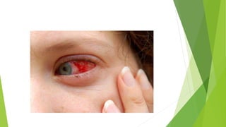
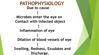
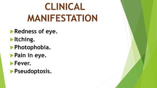
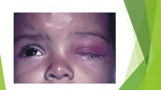
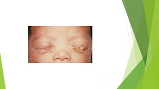
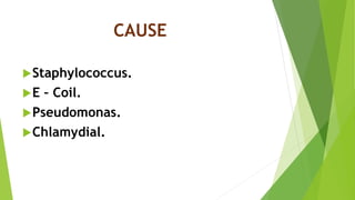
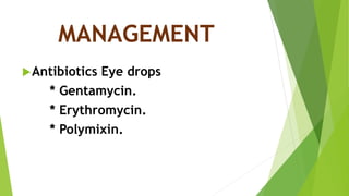
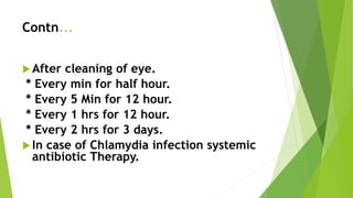
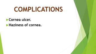
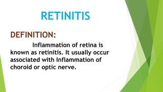
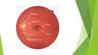
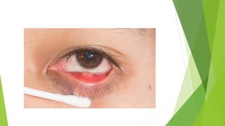
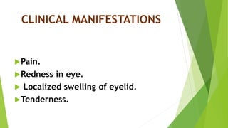
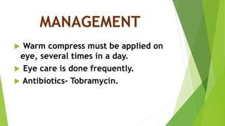
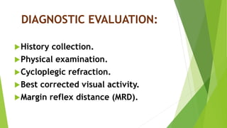
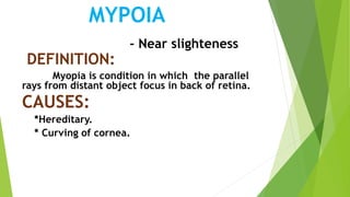
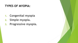
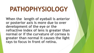
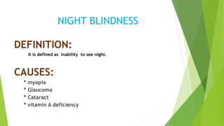
3. ANATOMY OF EYE
The eye is a specialized
Sense organ that helps us to understand our
environment.
The eye is able to refract
light and produce a focused image that can
stimulate neural response.
4. Contn....
The eye is spherical in shape.
It is about 2.5cm in diameter.
It is situated in the orbital cavity.
The eye is essentially and opaque ball
filler with a water- like fluid which is
called aqueous humour that provides
nutrition and helps to maintain the shape
of the cornea.
5. Contn......
In front of eye ball is a transparent
opening known as cornea. It is a thin
membrane that has a refraction index.
Sclera is a white outer layer of the eye ball,
tough , fibrous membrane that helps to
maintain the spherical shape of eye ball.
6. Contn....
Conjunctiva is a thin transparent mucous
membrane that covers the eye ball.
Choroid layer, is the middle layer of the
eye that supply blood vessel to the eye.
The Pupil is the black portion in the middle
of the eye ball that is rounded opening of
the iris through which light pass.
7. Contn....
Iris is the coloured part of the eye
formed by circularly and radially
arranged smooth muscle fibres. It is a
diaphragm that is capable of stretching
and reducing the size of the pupil.
Lens is a flexible, biconvex crystal like
structure that brings rays of light into
focus and procedures an image on the
retina.
8. Contn...
Retina is the innermost layer of the eye.
It contain photoreceptors like Rods and
cones (Cell responsible for converting
light into nerve impulses.)
Vitreous humour is a thick, gel-like
substance that fills the largest chamber
of the eye and helps to hold it’s
spherical shape.
10. PART OF EYE FUNCTIONS
Cornea It refract the light and protect the eye.
Iris Controls the level of light enters into the
pupil.
Lens Focuses light into the retina.
Optic nerve The bundle of sensory neurones that
carry the impulses to the brain.
Retina The rods and cones cell responsible for
converting light into the nerves system.
Aqueous humour Maintains the pressure in the eye and
nourishes cornea.
Vitreous humour It maintains the shape of the eye and
attaches to the retina.
Ciliary muscles Helps to change the shape of lens.
Sclera Helps to maintain the spherical shape of
eye ball.
Pupil Regulates the flow of light to the retina.
11. NORMAL DEVELOPMENT
OF EYE
The eye start to developing about
22 days of gestation. The development is
not Complete at birth. However, the
newborn is able to focus an object at
distance of 3 feet. Sensitivity brightness
develop rapidly in the first 2 months of
and infants display blink reflex.
12. Contn...
By the 4-6 months the infant’s have the visual
accommodation (ability of the eye to focus of an
object at all distance.) equal to an adult.
Binocularity (Fixing of two ocular Image
into one picture) is also established by 6 month of
age.
At the birth the colour of eye is blue or
light grey in fair newborns and brown in dark
newborns. Permanently eye colour may not be
appear Until one year of age.
14. INTRODUCTION
Vision , it is act of seeing, it
extremely important to human
survival. More than half the sensory
receptors in the human body are
located in the eye , and a large part of
the cerebral cortex is devoted to
processing visual information.
15. VISUAL DISODER
DEFINITION:
Visual impairment includes a range of vision
loss, from low vision to total blindness.
. * Parul Dutta
* Refractory disorders are the most
Common visual impairment in children.
17. CLASSIFICATION
There are mainly 3 type of
classification.
Infection and Inflammatory
conditions.
Non Inflammatory conditions.
Disorders of impairment of eye.
24. PATHOPHYSIOLOGY
Due to cause
|
Microbes enter the eye on
Contact with infected object
|
Inflammation of eye
|
Dilation of blood vessels of eye
|
Swelling, Redness, Exudates and
Discharge.
35. MODE OF INFECTION
* Intrauterine infection.
* Infection during the process of
delivery (most common).
* Infection after birth.
36. PATHOPHYSIOLOGY
Due to infections, the blood vessels
dilate and there is formation of new
blood vessels around the papillae of
eye. Numerous polymorphous are
present in the epithelium which leads
to purulent discharge and exudate
formation in eye.
40. Contn...
After cleaning of eye.
* Every min for half hour.
* Every 5 Min for 12 hour.
* Every 1 hrs for 12 hour.
* Every 2 hrs for 3 days.
In case of Chlamydia infection systemic
antibiotic Therapy.
44. PATHOPHYSIOLOGY
Due to causes
|
Infection or Inflammation occur
|
Exudate through the brunch’s
Membrane and reach retina
|
Exudate from retina reach the vitreous
|
Distortion of image and blurring of vision
48. MANAGEMENT
Protect the eye from light by
wearing dark goggles.
Atropine eye drops are instilled
thrice daily.
Eye care should be done using warm
sterile water.
49. STYE
DEFINITION:
Stye is an Infection of the
sebaceous glands near the eye lashes.
CAUSES :
Bacterial.
Viral.
Mostly staphylococcus infections.
58. CAUSES
Congenital or primary cause:
* Intrauterine infection in early
Months of pregnancy.
* Maternal malnutrition.
* Chromosomal abnormalities like
Down syndrome.
Acquired or secondary cause:
* Trauma.
* Glaucoma.
59. PATHOPHYSIOLOGY
Due to cause
|
The lens capsule is formed during the
fourth And 5th week of gestation
|
Clear membrane which allows light to
enter the eye and refract the rays
for a clear image on retina.
|
Interfere with lens development
|
lens become milky white and cloudy
61. GLAUCOMA
DEFINITION:
It is defined as a condition which is increased
intra ocular pressure in eye. It leads to blindness.
CAUSES:
* family history of diabetic.
* Family history of glaucoma.
* Age over 40 year.
* Elevated pressure of the eye.
TYPES:
*congenital or infantile glaucoma.
* Juvenile glaucoma.
62.
63. PATHOPHYSIOLOGY
Due to cause
|
sufficient amount of aqueous humour is not
drained out of the intraocular space
|
Accumulation of aqueous humour in the
anterior chamber of eye
|
requesting in increased intraocular
Pressure
72. PATHOPHYSIOLOGY
When the length of eyeball is anterior
or posterior axis is more due to over
development of the eye or the
refractive index of lens is greater than
normal or if the curvature of cornea is
greater than normal it causes the light
rays to focus in front of retina.
78. STRABISMUS
DEFINITION:
The deviation of visual axis from normal
alignment is known as strabismus.
INCIDENCE:
it is occur in 2-3% of children.
CAUSES:
* Family history.
* Genetic factor.
79.
80. CLINICAL MANIFESTATIONS:
Headache on reading.
Dimness of vision for distant object.
The child is seen holding books
closely to eye, while reading.
81. AMBLYOPIA
DEFINITION:
Amblyopia means poor vision in eye that
has not developed normal sight.
This condition is sometimes called lazy eye.
CAUSES:
* Brain suppress vision.
* Double vision.
82.
83. COLOUR BLINDNESS
DEFINITION:
It is a genetically determined conditions in
which colour perception is detective or absent.
Red and green colour defective usually form.
Colour blindness is not blindness, it is just a
deficiency in the way one sees colour.
CAUSE:
*Genetic factors.
* Defects in rods and cones.
* chronic illnesses.
* Accidents or stroke.
84. CLINICAL MANIFESTATIONS:
Trouble to see colours and
brightness of colour in the usual way.
Confusion.
In ability to tell the difference
between shades of the same colour.
87. DIPIOPIA
DEFINITION:
It is a squint and ptosis. It may be a warning signs of
increased intraocular pressure, brain tumor , orbital mass
CAUSES:
putting external pressure on one eye.
Having an incorrect eye glass prescription.
Extreme fatigue.
88. SIGNS AND SYMPTOMS:
Experience of double vision.
Weakness
Eye pain.
Bulging of eye