

Recommended
More Related Content
What's hot
What's hot (20)
Similar to Mss lecture
Similar to Mss lecture (20)
More from imrana tanvir
More from imrana tanvir (20)
Recently uploaded
Recently uploaded (20)
Mss lecture
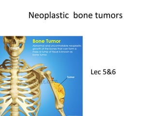
- 1. Neoplastic bone tumors Lec 5&6
- 2. CLASSIFICATION OF PRIMARY BONE TUMORS Histological Types Benign Malignant Hematopoietic (40%) Myeloma Malignant lymphoma Chondrogenic (22%) Osteochondroma Chondroma Chondroblastoma Chondromyxoid fibroma Chondrosarcoma Dedifferentiated chondrosarcoma Osteogenic (19%) Osteoid osteoma,Osteoma Osteoblastoma Osteosarcoma Unknown origin (10%) Giant cell tumour Ewing tumor Giant cell tumor Histiocytic origin Fibrogenic Notochordal Vascular, Cystic, lipogenic neurogenic Fibrous histiocytoma Fibroma MFH Fibrosarcoma
- 4. BONE TUMORS • Benign lesions are more common than malignant. • The most common benign tumour of bone is osteochondroma. • All bone forming tumors occur between the ages of 10-20yrs except osteoma which occurs in older age group (40-50ys). • Metastatic bone tumors > Primary bone T. • Common tumor sites giving bone metastasis are: prostate and breast .
- 5. BONE TUMORS cont….. • Malignant bone tumors, the most common are osteosarcoma followed by chondrosarcoma, and Ewing’s sarcoma. • Osteosarcoma occur in adolescence and affect bone around the knee in ½ of cases. • Chondrosarcoma develop in the mid to late adulthood involve the trunk, limb girdles, proximal long bones. • Chondroblastoma and giant cell tumor occur in the epiphysis of long bone. • Ewing’s sarcoma arise in diaphysis. • Diagnosis of bone tumors requires clinical history, X- ray, gross and microscopic examination.
- 8. BONE FORMING TUMORS • OSTEOMA • OSTEOID OSTEOMA • OSTEOBLASTOMA • OSTEOSARCOMA (OSTEOGENIC SARCOMA)
- 9. OSTEOMA • Osteoma are benign lesions of bone, developmental or reactive growths rather than true neoplasms. • Common in head, neck, paranasal sinuses, and facial bone. • Osteomas are localized, usually solitary, hard exophytic growths attached to the surface of the bone. • More common in males, any age, middle age.
- 10. Osteoma Gross Osteoma most commonly present as localized, usually solitary, hard, exophytic growth attached to the surface of bone.
- 11. OSTEOMA cont….. • Histologically, are composed of a mixture of woven and lamellar bone, similar to normal bone, might have bone marrow elements. • Do not undergo malignant transformation. • When multiple + intestinal polyposis + soft tissue tumors (Gardner’s syndrome) DDX: Reactive bone induced by infection, trauma, or hemangioma simulate osteoma.
- 12. OSTEOID OSTEOMA AND OSTEOBLASTOMA • Osteoid osteomas and osteoblastomas are benign neoplasms • Have very similar histologic features. • Different in size, and site of origin and symptoms.
- 13. OSTEOID OSTEOMA AND OSTEOBLASTOMA Osteoid Osteomas • Arise in proximal femur and tibia, on the cortex. • Second to third decades of life. • Males > females, 2:1 ratio. • Cause pain, severe at night, relieved by aspirin. • Measure less than 2 cm in greatest dimension. • Treatment with local excision.
- 14. OSTEOID OSTEOMA AND OSTEOBLASTOMA Osteoblastoma • Are larger. • Cause dull achy pain, not relived by aspirin. • Occur in vertebral column,& long bone. • Affect 20-30 y, M>F. • Treatment local excision , may recur.
- 15. Osteoid osteoma & Osteoblastoma • Radiographically: Are well-circumscribed lesions, involve cortex. The central area called the nidus, radiolucent surrounded by rim of sclerotic bone in both types. • Gross: round oval mass of hemorrhagic gritty tan tissue. • Microscopically: both are well circumscribed and composed of trabeculae woven bone surrounded by Osteoblast. The stoma is made up of loose, vascular connective tissue, and contains number of giant cells.
- 16. This is the central nidus of an osteoid osteoma. Radiographically, there is a small round central lucent area variably mineralized in the metatarsal bone cortex surrounded by sclerotic bone.
- 17. This is the central nidus of an osteoid osteoma composed of irregular reactive new bone. Osteoid osteomas usually occur in the axial skeleton (especially tibia and femur) in bone cortex of young males in the second decade of life. An osteoblastoma is just a big osteoid osteoma in the vertebra&rarely have well formed nidus .
- 19. OSTEOSARCOMA (OSTEOGENIC SARCOMA) • Osteosarcomas are malignant mesenchymal neoplasms in which the cancerous cells produce bone matrix. • Osteosarcoma 20% of all primary bone tumors. • It is the most common malignant primary bone tumor.
- 20. Osteosarcoma cont…. Occur: in 10-20 years of age. Affect: Male > female. Second peak in elderly who have had Pagets disease or bone infarction. Site in metaphysis (60-70% around the knee, distal femur and proximal tibia). Any bone can be involved. Flat bone (jaws and pelvis) and long bone are involved equally after age 25 years.
- 22. Osteosarcoma cont….. Morphology: Several subtypes are described, and grouped according to: 1.The anatomic location in the bone (intramedullary, intracortical or surface). 2.Degree of differentiation. 3.Multicentrecity (synchronous, metachronous). 4.Primary or secondary (arising on preexisting disease such benign tumors, Paget’s disease, infarcts…….) 5.Histological variants (osteoblastic, chrondroblastic, fibroblastic, telangiectatic, small cell, and giant cell).
- 23. Osteosarcoma cont…….. • The most common type is osteosarcoma arising in the metaphysis of long bone. • It is solitary. • Poorly differentiated and produce large bony matrix.
- 24. Osteosarcoma cont……. Grossly: • are large, fleshy ill-defined, in medullary cavity of metaphyseal end of the bone • areas of necrosis and haemorrhage. • areas of bone and cartilage formation present. • The tumor destroys the cortex and frequently the T.extends inwards in marrow cavity and outwards into soft tissues. • Invasion of epiphyseal plate uncommon.
- 26. Osteosarcoma cont….. Microscopically: • The hallmark of osteosarcoma is the formation of osteoid by malignant osteoblast. • Island of bony trabeculae hugged by a rim of malignant osteoblasts. • Cartilage also be present. • Neoplastic mesenchymal cells between osteoid are spindle or pleomorphic, with frequent mitotic figures. • Giant cells, either many or few.
- 28. Osteosarcoma cont…. X-ray: Tumor is destructive breaks the cortex, elevates the periosteum result in reactive new bone deposition. Give the so-called Codman’s triangle on radiographs.
- 30. The most common subtype is osteosarcoma that arises in the metaphysis of long bones; is primary, solitary, intramedullary, and poorly differentiated; and produces a predominantly bony matrix
- 31. Osteosarcoma cont…… • Clinical Features • Early give progressively enlarging, painful masses with limited joint movement. • Late cause weight loss, anaemia and fracture of the involved bone. Fracture can be the presenting symptom.
- 32. Osteosarcoma cont….. Spread of osteosarcomas: • Metastasize via the bloodstream to the lung. • 20% of pt. have pulmonary spread at diagnosis. • Direct spread to the soft tissue. Prognosis: • 5 years survival rate is 5-20%. • Improved prognosis, advanced surgical techniques, combined with radiation and chemotherapy.
- 34. Assessment -The most common primary malignant neoplasm of bones under the age of 20 years is: a. Osteosarcoma b. Giant cell tumor of bone c. Chondrosarcoma d. Metastasis
- 35. Lecture 6
- 36. CARTILAGE FORMING TUMORS • OSTEOCHONDROMA (EXOSTOSIS) • CHONDROMA • CHONDROBLASTOMA • CHONDROSARCOMA
- 38. Osteochondroma • Osteochondromas, exostoses are benign,& common. • Are malformation rather than neoplasm. • Most common benign bone tumors under age 21 (10-30). • M:F is 3:1. • Occur as solitary lesion, sporadic can be multiple in familial exostosis which have the risk of rare (1%) sarcomatous transformation . • Are outgrowth attached to the skeleton by bony stalk & covered by cartilage cap.
- 39. Osteochondroma cont…… • Clinically : are asymptomatic, cause deformities. • Site : arise from the metaphysis near the growth plate of long tubular bones (about the knee) rare in short tubular bone of hands and feet. • Gross : Mushroom shaped with broad-base, anchored to the cortex of the adjacent bone, the outer layer of the head is composed of benign hyaline cartilage and from 1-20 cm in size . • Microscopically : are proliferations composed of mature bone and cartilaginous cap. They stop growing once the normal growth of the skeleton is completed.
- 40. This is an osteochondroma cut into three sections. A bluish- white cartilagenous cap overlies the bony cortex. These are probably not true neoplasms, but they are a mass lesion that extends outward from the metaphyseal region of a long bone.
- 41. The microscopic appearance of an osteochondroma displays the benign cartilagenous cap at the left and the bony cortex at the right. This bone growth, though benign, can sometimes cause problems of pain and irritation that leads to removal surgically.
- 42. CHONDROMA (ENCHONDROMA) *Chrondromas are benign lesions composed of mature hyaline cartilage. Site: small bones of hands and feet. Age: 20-40y, occur at any age. Clinical: solitary or multiple, well-circumscribed. Occur in metaphysis of tubular short bone of the hand and feets. if within the medullary cavity of the bone (called enchondroma). if on surface of the bone (called subperiosteal chondromas).
- 43. CHONDROMA cont…… Ollier’s disease is multiple chondromas involving one side of the body. Maffucci’s syndrome is multiple chondromas associated with benign vascular tumors (angiomas) and brain gliomas. Pathogenesis : Develop in enchondral bone. arising from rests of growth plate cartilage. X-ray : give O ring sign, unmineralized nodule surrounded by rim of radiodense bone.
- 45. CHONDROMA cont…… Gross : are usually smaller than 3 cm , gray-blue translucent with nodular configuration. Microscopically : are composed of mature, hypocellular benign hyaline cartilage . Clinically : can be painful or cause pathological fracture. If multiple result in deformities and recur if incompletely excised. Prognosis : solitary are benign. Chondrosarcomas may develop in patients with multiple chondromas.
- 46. CHONDROMA
- 47. CHONDROBLASTOMA Rare benign neoplasm affected age is 10-20 years male > female. Site : Epiphyseal region of long bone, femur, humerous, tibia (around the knee). less common in pelvis, ribs, skull, small bone of feets and in vertebrae. Gross : Round oval soft gray pink, with calcification, hemorrhage, necrosis and cystic changes.
- 48. CHONDROBLASTOMA cont….. Micro : Uniform small round chondroblasts. calcification (chicken wire pattern), multinucleated giant cells, mitosis and necrosis present. Clinically : are painful, cause effusion in joint and motion restriction. Recurrence is common after excision or curettage. Metastasis occur if the lesions fracture or after repeated curettage. X-ray : well-defined lucency with spotty calcification DDX : giant cell tumor.
- 51. CHONDROSARCOMA **Chondrosarcomas are malignant neoplasms. **Second to osteosarcoma in frequency. Incidence : Males 2x > females. Age : older patients, with a peak incidence in the 60y Site : Central portions of skeleton, shoulder area, pelvis, proximal femur, and ribs, around the knee, these tumors may occur anywhere. *Most primary chondrosarcomas arise de novo. *Secondary occur in patients with multiple enchondromas or osteochondromas.
- 52. CHONDROSARCOMA cont… MORPHOLOGY: *Cells in chondrosarcomas do not form osteoid. *Chondrosarcoma arises centrally within the medullary cavity of the bone *Rarely on the surface of the bone (peripheral, subperiosteal). Gross: large, gray-white glistening mass, with spotty calcification; central necrosis and cyst formation. erodes the cortex and extend into soft tissue.
- 53. A chondrosarcoma arising in the pelvis. Note the extensive nodules of white to bluish-white cartilagenous tumor tissue eroding and extending outward from the bone at the lower right. Many of them are slow growing .
- 54. CONDROSARCOMA cont… Histology • Malignant cells infiltrates the marrow and surrounds bony trabeculae. • Well differentiated T. have little atypia. • Poorly differentiated T. have pleomorphic cells with many mitosis. Two or more chondroblasts in one lacuna. • 10% of low grade chondrosarcoma transform into high grade sarcoma and called dedifferentiated chondrosarcoma.
- 55. This high power microscopic appearance of a chondrosarcoma demonstrates pleomorphic chondrocytes that are piled together in a haphazard arrangement.
- 56. CHONDROSARCOMA cont… Clinical Features *Most of chondrosarcomas are indolent (grade1&2). *progressively enlarging mass. *painful masses involving the central portions of the skeleton. *poorly differentiated T. behaves in a more aggressive fashion (grade 3 ). *Large chondrosarcoma (>10 cm ) are more aggressive. *Chondrosarcomas metastasize via blood, to lungs and skeleton.
- 58. MISC. TUMORS of BONE • EWING sarcoma • GIANT CELL TUMOR • METASTASES
- 59. EWING’S SARCOMA Ewing’s sarcoma is a primitive malignant small round cell tumor of bone and soft tissues. Accounts for 6-10% of primary malignant bone tumors. Is a common malignancy of bone in children, second to osteosarcoma. It is the third most common malignant bone tumor, exceeded only by osteosarcoma and chondrosarcoma.
- 60. EWING’S SARCOMA cont… Age: Children and adolescents. Peak incidence younger than 20 years. M > F more in whites. Site of origin: Can be skeletal or extra skeletal in the soft tissues
- 61. EWING’S SARCOMA cont… Gross Soft, tan-white expansile mass with areas of necrosis and hemorrhage. Site Diaphysis region in the medullary cavity of long tubular bone as femur, tibia, and pelvis are favored sites of origin Extends to the cortex and periosteum & produce soft tissue mass. X-ray: Destructive lytic tumor. Give periosteal reaction with lamellae of reactive bone deposited in an onion-skin pattern.
- 62. EWING’S SARCOMA cont….. Microscopy *Ewing’s sarcoma is composed of sheets of primitive small round blue cells . *Have uniform nuclei and little cytoplasm. *The cytoplasm contains glycogen( seen with periodic acid-Schiff (PAS) stains or by electron microscopy ).
- 64. EWING’S SARCOMA cont…. CLINICAL FEATURES *Presents with pain as local inflammation with fever. *The diagnosis requires biopsy. Prognosis: is bad but recent advances in treatment, the 5-year survival rate close to 75%. Spread: Via blood.
- 65. Giant Cell Tumor of Bone *Giant cell tumor of bone, known as osteoclastoma. *neoplasm that contains large numbers of osteoclast-like giant cells admixed with mononuclear cells. Incidence relatively uncommon tumors and locally aggressive, 20% of all benign tumors of the bone.
- 66. Giant Cell Tumor of Bone Site: *In adult involve the epiphyses and metaphysis *in adolescent are confined to growth plate. *55% around the knee, the distal femur, proximal tibia, proximal humerus, and distal radius, sacrum, pelvis, and small bones of hand and feets. Age: 20 and 40, female > male. Histogenesis Is incompletely understood. The giant cells are reactive ( are macrophages). The accompanying mononuclear cells are neoplastic.
- 67. Giant Cell Tumor of Bone Grossly Are dark brown due to abundant vascularity. Areas of necrosis, hemorrhage and cystic change. The cortex either thinned or eroded due to tumor expansion.
- 68. Here is a giant cell tumor of bone. The proximal femur has been amputated and cut in half to reveal an irregular dark red-black hemorrhagic mass in the epiphyseal region. Giant cell tumors are lytic on
- 69. Giant Cell Tumor of Bone Histologically are composed of two major cell populations: • Large multinucleated giant cells ,resemble osteoclasts, have 100 nuclei identical to the mononuclear cells. • Neoplastic component is made up of round to spindle-shaped mononuclear cells, contains a number of mitotic figures. • Rare occasions malignancy may develop. • Necrosis, hemorrhage, hemosiderin and reactive bone formation are common.
- 70. GCT (Giant Cell Tumor), BONE
- 72. METASTASES • Metastatic bone tumors are more common than primary malignant bone tumors • Common tumor sites giving bone metastasis are: prostate and breast . • Some metastatic deposits are osteolytic , others are osteoblastic and most of them induce a mixed lytic and blastic reactions
- 73. Here is a closer view of bone metastases. Virtually all bone metastases are from carcinomas. The primary sites of carcinomas that commonly go to bone are: breast, prostate, kidney, thyroid, lung. Renal cell carcinomas tend to be osteolytic (they destroy the bone) whereas prostatic adenocarcinomas tend to be osteoblastic (they initiate new bone formation).
- 74. At high magnification, metastatic infiltrating ductal carcinoma of breast is seen within bone and filling the marrow cavity. There is reactive new bone with pale pink osteoid being laid down next to a bony spicule at the upper left.
- 75. Assessment • A 37 year old lady with a known history of breast carcinoma presents with a fracture of the neck of femur. The most likely cause is : a. Trauma b. Osteoporosis c. Osteomyelitis d. Pathological fracture due to metastasis
Editor's Notes
- Ewing's sarcoma is one of the "small round blue cell" tumors histologically.
- GCT occurs in patients, 20s-40-s, and GCT has a MACROPHAGE lineage, as most multinucleated giant cells do. Histologically, giant cell tumors of bone as seen here are composed of multinucleated giant cells in a sea of round to oval mononuclear cells