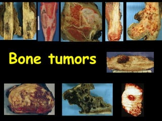
SlideShare a Scribd company logo
1 of 92
1
Bone tumors
2
Bone tumors
Benign Malignant
Primary Secondary
= metastatic
3
• Metastasis is the MC malignancy of bone.
– Breast cancer is the MC primary site.
– Others include:
• Prostate ,Lung ,Kidney and thyroid
• Nature of metastasis
– Osteolytic:
• Destructive
• Most mets (breast**, lung, kidney etc)
– Osteoblastic:
• Reactive new bone formation
• Carcinoma prostate**, breast Ca.
4
Osteolytic metastasis
5
Spine, prostate carcinoma metastatic to vertebrae
6
Radiodense lesion
Osteoblastic Metastasis
7
8
Primary Bone tumors: Clinical presentation
• Benign tumors:
– Usually asymptomatic
– Present as painless mass
• *Osteoid osteomas are painful,
• *Enchondromas may cause a stress #.
– Usually detected incidentally.
• Malignant tumors:
– Present with :
• Pain
• Swelling or a
• Pathological fracture
9
Important parameters for diagnosis of
bone tumors
1. Age of patient
2. Bone involved
3. Specific area within the bone (epiphysis,
metaphysis, diaphysis)
4. Radiographic appearance
5. Gross and microscopic features
10
Age and bone tumors
• Remember these general age ranges:
– Metastatic neuroblastoma*: infants &
toddlers
– Ewing's sarcoma: older children &
adolescents (5-20)
– Osteosarcoma: adolescents and young
adults(10—20).
– Giant cell tumors: young adults & middle age
(20-40)
– Chondrosarcoma: middle age (40-60)
– Multiple myeloma : middle to old age (>40)
– Metastatic cancer: middle and old age
11
Location in bone
12
Location in bone
• Diaphysis:
– Ewing's sarcoma**, chondrosarcoma ,
enchondroma.
• Epiphysis:
– Giant cell tumor** (osteoclastoma)
• Metaphysis:
– Osteogenic sarcoma**
13
Risk factors for bone sarcoma
• Include
– some familial syndromes (e.g Li Fraumeni)
– radiation therapy.
• However, most cases are unrelated to any of
these.
• In the previously irradiated patient, the
commonest primary bone cancer:
– osteosarcoma **
14
Treatment
• Benign tumors may be treated by
– curettage and
– packing with bone chips from elsewhere.
• Malignant tumors require
– resection,
– radiation, and/or chemotherapy.
• Osteosarcoma and Ewing's sarcoma often
respond well to chemotherapy.
15
Classification of primary bone tumors
• Primary BT classified in to ten groups
1. Osteogenic (bone forming)
– Benign: osteoma, osteoid osteoma* and
osteoblastoma
– malignant: Osteosarcoma (osteogenic
sarcoma)*
2. Chondrogenic (cartilage forming)
– Benign: osteochondroma*, chondroma*,
chondroblastoma
– malignant: chondrosarcoma*
3. Unknown origin:
– Giant cell tumor* (benign / malignant)
– Ewing’s sarcoma* (malignant)
16
4. Fibrogenic (from fibroblasts)
– Benign: Fibrous cortical defect / nonossifying
fibroma *, Fibrous dysplasia*
– Malignant: fibrosarcoma
5. Hematopoietic (from hematopoietc elements)
– multiple myeloma
6. Histiocytic origin (from histiocytes)
7. Notochordal
8. Vascular
9. Lipogenic
10.Neurogenic
17
BONE-FORMING TUMORS
Benign
18
Osteoma
• A benign bone forming tumor.
• Occurs in middle aged adults (40-50yrs)
• Sites:
– usually occurs in the skull (sinuses – most
common) ,facial bones and jaw.
• Usually harmless, they may impinge on the
brain, obstruct sinus drainage, or look ugly.
• Histologically resemble normal bone
• If multiple, suspect Gardener's syndrome
19
Osteoma
20
• A benign painful* tumor of osteoblasts.
• Occurs in individuals 5-25 yrs old.
• Sites:
– Metaphysis/diaphysis of femur and tibia
– cortex of bone (size: <2cm*)
• Clinical:
– well-localized pain (Nocturnal).
– pain is relieved by aspirin
• Micro:
– Central nidus of osteoid surronded by dense sclerotic
rim of bone
• X ray reveals:
– a small radiolucent focus (nidus) surrounded by densely
sclerotic bone.
Osteoid osteoma***
21
Osteoid osteoma
Radiolucent focus
(nidus)
surrounded by
densely sclerotic bone
22
Radiolucent focus (nidus)
surrounded by densely sclerotic bone
23
• *Osteoblastoma ("giant
osteoid osteoma"):
– Similar to osteoid
osteoma
– But is larger (>2cm)
and
– Arises in the
vertebral bodies
– Pain not worse at
night and not relived
by aspirin.
24
Malignant bone forming tumor
25
26
Osteosarcoma (= osteogenic sarcoma)
• Cancer of the osteoblasts
• The malignant tumor cells make osteoid.
• The 2nd MC primary malignant tumor of bone
(1st is multiple myeloma).
• Age: (bimodal)
– 10-20 years old
• Arises de novo.
– >40 years old
• Usually secondary to risk factors (Paget’s
disease of bone etc.)
27
• Location:
– Most in the
metaphyseal region
of long bones.
• Lower end of
femur (most
common)
• Upper end of tibia
• Upper end of
humerus
– Less commonly in flat
bones  jaw.
28
29
• Osteosarcoma: TYPES:
– Primary Osteosarcoma
• Arise in absence of associated bone
disease.
• In patients < 20 years
• From metaphyseal region of long bones.
• Genetic association:
– 2/3rd show inactivation of Rb gene**
– Common in Li-Fraumeni syndrome** (p53
gene inactivation)
– Secondary Osteosarcoma
• Occurs in older people
• Both in flat and long bones
• In a background of preexisting bone
disease
30
Risk factors for osteosarcoma
• Paget’s disease of bone
– Osteosarcoma of the pelvic bones
• Irradiation (radium watch dial workers)
• Bone infarct
• Osteomyelitis
• Clinical features:
– Local pain, tenderness , swelling and
pathological fracture
31
Destroyed cortex
Tumor massGrowth plate
32
Osteosarcoma
33
Codman triangle
34
Soft tissue mass
Sunburst
appearance
35
• Gross:
– Gray white mass with areas of
hemorrhage and necrosis
– Destroys cortex and invades adjacent
soft tissue
• Elevates the Periosteum , producing
Codman’s triangle on X ray.
• Sunburst appearance : due to
calcified osteoid extending into the
adjacent soft tissue.
36
Osteoid
37
Spindle shaped cells Osteoid
38
• Microscopy:
– Tumor cells produce osteoid.
– Tumor cells may be spindle, oval or round.
• Spread:
– Metastasize through blood stream  lung
the most common site.
• Treatment:
– Surgery (limb sparing) with
– Preoperative and postoperative Chemotherapy.
• Prognosis
– five-year survival 50-60%.
39
Benign
Cartilaginous tumors of bone
40
41
• Exostosis ("osteochondroma”):
– Most common** benign bone tumor.
• Tumor: Mushroom shaped
– Composed of outgrowth of bone (exostoses)
capped by benign proliferating cartilage.
• Age: 10-30
• Location: Usually located in the metaphysis of
long bone.
• Can be: solitary or multiple
• If Multiple then k/a osteochondromatosis:
– Hereditary multiple exostoses
– Increased risk of transformation into
chondrosarcoma (if multiple).
42
Osteochondroma
43
• Chondromas: benign tumor composed of hyaline
cartilage
• If located within shaft of bone then k/a
enchondroma.
– Site: small bones of hand and feet (proximal
phalanges).
– Can be
• Solitary
• Multiple (called enchondromatosis)
– Chondrosarcoma risk with multiple
tumors.
• Ollier’s disease : multiple enchondromas.
• Maffucci's syndrome : multiple enchondromas
plus hemangiomas of soft tissues.
44
Chondroma
45
46
Malignant cartilaginous tumor
47
48
Chondrosarcoma
• A malignant tumor of chondroblasts.
• MC primary malignant cartilaginous tumors.
• Age: 40-60 years
• Location:
– Central skeleton: ribs, shoulder and
Pelvic bones and
– Upper end of femur and humerus
• Can arise
– de novo or
– secondary to osteochondromatosis or
enchondromatosis.
49
Chondrosarcoma
50
51
• Gross:
– grayish blue, glistening and semi-
translucent.
• Micro:
– composed of atypical chondrocytes and
chondroblasts often with multiple nuclei in
a lacuna.
• Biologic behavior and Prognosis
– depends on the grade of tumor.
• Metastasis
– Hematogenously to lungs
52
Tumors of uncertain origin
53
54
Ewing's Sarcoma
• An extremely malignant tumor composed of
small round blue cells.
– Arises within the marrow cavity.
• Histogenesis uncertain: Current evidence:
– Probably of neuroectodermal origin.
– Reasons:
• Specific translocation : t(11;22)
• Same translocation also present in similar
tumor of soft tissue known as PNET
(primitive neuro-ectodermal tumor).
55
• Age:
– Most occur in teenagers 5-20 yrs
• Genetics:
– classic translocation t(11;22) which produces
the EWS-FLI1 fusion gene.
• Gross:
– Arises in medullary cavity in the diaphysis of
long bones.
– Most common sites are the femur, pelvis and
the tibia.
– Penetrates the cortex to produce :
• White tan mass with necrosis and
hemorrhage.
56
Ewings Sarcoma
Tan to red to brown tumor mass
57
"small round blue cells"
58
Glycogen, seen as reddish granular material in
cytoplasm by PAS stain.
59
Onion skin appearance
of reactive new bone
60
• Microscopy:
– Tumor composed of
• Sheets of "small round blue cells"
with little cytoplasm.
• Cells are loaded with glycogen.
• Glycogen can be seen by PAS stain.
• Homer Wright rosettes (tumor cells
arranged in circle about a central
fibrillary space)
61
• X ray:
– Concentric “onion skin” layering of new
periosteal bone.
• Clinical features:
– It presents “as an infection” with
• fever,
• Painful enlarging mass often tender
• Heat over the tumor.
– Therefore simulates osteomyelitis.
• Prognosis:
– Radiation + Chemotherapy+ surgery 75% 5-
year survival.
62
• Differential diagnosis:
– Other small round blue cell tumors
• metastatic neuroblastoma
• metastatic malignant lymphoma
• acute lymphoblastic leukemia
• Rhabdomysarcoma
• IHC will help differentiate ES from these
small round blue cell tumors (SRBCT).
63
Giant cell tumor
64
65
66
Giant cell tumor ("osteoclastoma")
• A locally aggressive potentially malignant tumor
composed of :
– multinucleated giant cells (nonneoplastic)
admixed with mononuclear cells (neoplastic).
• More common in females than males.
• Seen in patients 20-40 years of age
• Arises in the epiphysis and extends into
metaphysis
• Favored locations:
– Classical : epiphysis of long bone.
– Sites most commonly affected:
• Lower end of femur> Upper end of
tibia >Lower end of radius.
67
Giant cell tumor
68
Giant cell tumor
69
"Soap bubble"
lesion.
70
Soap bubble
appearance
71
Round to oval
Mononuclear cells
Multinucleated giant
cells
72
73
• Gross: Cut section of the tumor:
– Solid red brown mass with hemorrhage and
cystic degeneration
• Microscopy: Two components
• Osteoclast like giant cells:
– Large with 20-30 nuclei.
– Are Not neoplastic.
– Formed due to fusion of monocytes.
• Mononuclear stromal cells.
– Are the neoplastic cells and determine
the behavior.
• X-ray:A lytic expansile lesion in the epiphysis.
– May have a “soap bubble” appearance.
• Clinical behavior: Unpredictable
74
Epiphysis
Femur, tibia
radius
20-40 yrsGiant cell tumor
Diaphysis
Pelvis, ribs,
shoulder bones.
40-60Chondrosarcoma
Diaphysis
Femur , pelvis,
tibia
5-20 yrsEwing’s sarcoma
LocationAgeTumor
Metaphysis
Femur, tibia,
humerus
10-20 yrsOsteosarcoma
75
Fibrous and fibro-osseous tumor
76
Fibrous cortical defects and
Non-ossifying fibromas
• Essentially the same disease except for a
difference in size.
– Fibrous cortical defects : 1-4 cm
– Non-ossifying fibromas : 5-10 cm.
• Both are benign tumor like lesions
77
Fibrous cortical defects and
Non-ossifying fibromas
78
Fibrous cortical defects and
Non-ossifying fibromas
• Clinical:
– Extremely common: (1/3rd of normal children)
– Asymptomatic / fracture
– Occur in the long leg bones of children
(cortical aspect of metaphysis).
– Femur>tibia>fibula
– Made of fibroblasts,and lipid-laden
macrophages
• X ray:
– irregular, sharply demarcated radiolucent
defect in the metaphyseal cortex.
• Treatment:
– often resolves spontaneously
79
Fibrous dysplasia
• Benign non-neoplastic process of bones.
• Characterized by
1. replacement of marrow by fibrous tissue.
2. presence of poorly formed woven bone
arranged in Chinese letter pattern.
• Primarily targets ribs, femur, or cranial bones
of children and young adults.
80
Wavy spicules of woven bone ("chinese characters");
Fibrous tissue
81Fibrous dysplasia
82
Fibrous dysplasia:Types
– Monostotic fibrous dysplasia
• Involvement of single bone
• Usually asymptomatic
– Polystotic fibrous dysplasia
• Involvement of Multiple bones.
• Often symptomatic
• deformities and fractures of craniofacial
bones
• Shepherd Crook deformity of proximal
femur.
– McCune-Albright syndrome
• Polystotic disease with endocrine
abnormalities
83
Fibrous dysplasia
• McCune-Albright syndrome :
– features include -
• Polystotic disease
• precocious sexual development
• irregular skin pigmentation (café-au-lait
spots) (akin to “coast of Maine”)
• Fibrous dysplasia:
– Clinical course
• Secondary sarcoma may develop after
irradiation (rarely).
84
Shepherd Crook deformity
Café-au-lait spots
85
Additional Images
For Self Study
86
87
88
89
90
91
92

More Related Content

What's hot

What's hot (20)

Bone tumors
Bone tumorsBone tumors
Bone tumors
 
Chondrosarcoma
ChondrosarcomaChondrosarcoma
Chondrosarcoma
 
Cystic lesion of bones
Cystic lesion of bonesCystic lesion of bones
Cystic lesion of bones
 
Aproach to bone tumours
Aproach to bone   tumoursAproach to bone   tumours
Aproach to bone tumours
 
Primary bone tumors
Primary bone tumorsPrimary bone tumors
Primary bone tumors
 
Chronic osteomyelitis
Chronic osteomyelitisChronic osteomyelitis
Chronic osteomyelitis
 
Chondrosarcoma
ChondrosarcomaChondrosarcoma
Chondrosarcoma
 
Systemic approach to bone tumor radiology
Systemic approach to bone tumor radiologySystemic approach to bone tumor radiology
Systemic approach to bone tumor radiology
 
Malignant bone tumours
Malignant bone tumoursMalignant bone tumours
Malignant bone tumours
 
Bone tumor
Bone tumorBone tumor
Bone tumor
 
Osteochondroma
OsteochondromaOsteochondroma
Osteochondroma
 
Benign bone tumors
Benign bone tumorsBenign bone tumors
Benign bone tumors
 
Orthopaedic oncology
Orthopaedic oncologyOrthopaedic oncology
Orthopaedic oncology
 
Metastatic bone disease
Metastatic bone disease Metastatic bone disease
Metastatic bone disease
 
Bone tumors
Bone tumorsBone tumors
Bone tumors
 
Radiological and pathological correlation of bone tumours Dr.Argha Baruah
Radiological and pathological correlation of bone tumours  Dr.Argha BaruahRadiological and pathological correlation of bone tumours  Dr.Argha Baruah
Radiological and pathological correlation of bone tumours Dr.Argha Baruah
 
Classification of bone tumors
Classification of bone tumorsClassification of bone tumors
Classification of bone tumors
 
Bone tumours
Bone  tumoursBone  tumours
Bone tumours
 
Tumors of bone
Tumors of boneTumors of bone
Tumors of bone
 
Bone tumours
Bone tumoursBone tumours
Bone tumours
 

Similar to 02 bone Tumors

Tumors Musculoskeletal.. VAPMS college of physiotherapy, vskp.
Tumors Musculoskeletal.. VAPMS college of physiotherapy, vskp.Tumors Musculoskeletal.. VAPMS college of physiotherapy, vskp.
Tumors Musculoskeletal.. VAPMS college of physiotherapy, vskp.
Rajani Cartor
 

Similar to 02 bone Tumors (20)

Bone tumours -.pptx
Bone tumours -.pptxBone tumours -.pptx
Bone tumours -.pptx
 
bonetumors...............-180123083659.pptx
bonetumors...............-180123083659.pptxbonetumors...............-180123083659.pptx
bonetumors...............-180123083659.pptx
 
Bone tumors.pptx
Bone tumors.pptxBone tumors.pptx
Bone tumors.pptx
 
bonetumors-180123083659.pdf
bonetumors-180123083659.pdfbonetumors-180123083659.pdf
bonetumors-180123083659.pdf
 
Bone tumors
Bone tumorsBone tumors
Bone tumors
 
Bone Tumors
Bone TumorsBone Tumors
Bone Tumors
 
Bone.pptx
Bone.pptxBone.pptx
Bone.pptx
 
Mss lecture
Mss lectureMss lecture
Mss lecture
 
MUSCULOSKELETAL SYSTEM LEC 5&6
MUSCULOSKELETAL SYSTEM LEC 5&6MUSCULOSKELETAL SYSTEM LEC 5&6
MUSCULOSKELETAL SYSTEM LEC 5&6
 
BT.pptx
BT.pptxBT.pptx
BT.pptx
 
Osteosarcoma
OsteosarcomaOsteosarcoma
Osteosarcoma
 
Bone tumours_Dr.Manal Ismail Abd-Elghany_2020.pptx
Bone  tumours_Dr.Manal Ismail Abd-Elghany_2020.pptxBone  tumours_Dr.Manal Ismail Abd-Elghany_2020.pptx
Bone tumours_Dr.Manal Ismail Abd-Elghany_2020.pptx
 
Bone tumours by dr narmada prasad tiwari
Bone tumours by dr narmada prasad tiwariBone tumours by dr narmada prasad tiwari
Bone tumours by dr narmada prasad tiwari
 
Bone Tumor (Benign and malignant)
Bone Tumor (Benign and malignant)Bone Tumor (Benign and malignant)
Bone Tumor (Benign and malignant)
 
Bone tumors ug
Bone tumors ugBone tumors ug
Bone tumors ug
 
Common Malignant tumors in orthopedics
Common Malignant tumors in orthopedicsCommon Malignant tumors in orthopedics
Common Malignant tumors in orthopedics
 
Bone tumors
Bone tumorsBone tumors
Bone tumors
 
BONE SARCOMAS.pptx
BONE SARCOMAS.pptxBONE SARCOMAS.pptx
BONE SARCOMAS.pptx
 
Bone 2
Bone 2Bone 2
Bone 2
 
Tumors Musculoskeletal.. VAPMS college of physiotherapy, vskp.
Tumors Musculoskeletal.. VAPMS college of physiotherapy, vskp.Tumors Musculoskeletal.. VAPMS college of physiotherapy, vskp.
Tumors Musculoskeletal.. VAPMS college of physiotherapy, vskp.
 

More from med_students0

08 respiratory restrictive
08 respiratory   restrictive08 respiratory   restrictive
08 respiratory restrictive
med_students0
 

More from med_students0 (20)

Anti diabetic medications
Anti diabetic medicationsAnti diabetic medications
Anti diabetic medications
 
Renal pathology iii
Renal pathology iiiRenal pathology iii
Renal pathology iii
 
Renal pathology iv
Renal pathology ivRenal pathology iv
Renal pathology iv
 
Renal pathology ii
Renal pathology iiRenal pathology ii
Renal pathology ii
 
Renal pathology i
Renal pathology iRenal pathology i
Renal pathology i
 
Cervical ca screening ..
Cervical ca screening ..Cervical ca screening ..
Cervical ca screening ..
 
01 cardiac pathology
01 cardiac pathology01 cardiac pathology
01 cardiac pathology
 
05 cardiac pathology
05 cardiac pathology05 cardiac pathology
05 cardiac pathology
 
04 cardiac pathology
04 cardiac pathology04 cardiac pathology
04 cardiac pathology
 
03 cardiac pathology
03 cardiac pathology03 cardiac pathology
03 cardiac pathology
 
02 cardiac pathology
02 cardiac pathology02 cardiac pathology
02 cardiac pathology
 
09 respiratory pleura
09 respiratory   pleura09 respiratory   pleura
09 respiratory pleura
 
08 respiratory restrictive
08 respiratory   restrictive08 respiratory   restrictive
08 respiratory restrictive
 
07 respiratory obstructive2
07 respiratory   obstructive207 respiratory   obstructive2
07 respiratory obstructive2
 
06 respiratory obstructive1
06 respiratory   obstructive106 respiratory   obstructive1
06 respiratory obstructive1
 
05 respiratory tumors
05 respiratory   tumors05 respiratory   tumors
05 respiratory tumors
 
04 respiratory infection2
04 respiratory   infection204 respiratory   infection2
04 respiratory infection2
 
03 Respiratory infection1
03 Respiratory   infection103 Respiratory   infection1
03 Respiratory infection1
 
02 respiratory vascular
02 respiratory   vascular02 respiratory   vascular
02 respiratory vascular
 
01 respiratory RDS
01 respiratory   RDS01 respiratory   RDS
01 respiratory RDS
 

Recently uploaded

Call Girls in Gagan Vihar (delhi) call me [🔝 9953056974 🔝] escort service 24X7
Call Girls in Gagan Vihar (delhi) call me [🔝  9953056974 🔝] escort service 24X7Call Girls in Gagan Vihar (delhi) call me [🔝  9953056974 🔝] escort service 24X7
Call Girls in Gagan Vihar (delhi) call me [🔝 9953056974 🔝] escort service 24X7
9953056974 Low Rate Call Girls In Saket, Delhi NCR
 
College Call Girls in Haridwar 9667172968 Short 4000 Night 10000 Best call gi...
College Call Girls in Haridwar 9667172968 Short 4000 Night 10000 Best call gi...College Call Girls in Haridwar 9667172968 Short 4000 Night 10000 Best call gi...
College Call Girls in Haridwar 9667172968 Short 4000 Night 10000 Best call gi...
perfect solution
 

Recently uploaded (20)

Russian Call Girls Service Jaipur {8445551418} ❤️PALLAVI VIP Jaipur Call Gir...
Russian Call Girls Service  Jaipur {8445551418} ❤️PALLAVI VIP Jaipur Call Gir...Russian Call Girls Service  Jaipur {8445551418} ❤️PALLAVI VIP Jaipur Call Gir...
Russian Call Girls Service Jaipur {8445551418} ❤️PALLAVI VIP Jaipur Call Gir...
 
Premium Call Girls Cottonpet Whatsapp 7001035870 Independent Escort Service
Premium Call Girls Cottonpet Whatsapp 7001035870 Independent Escort ServicePremium Call Girls Cottonpet Whatsapp 7001035870 Independent Escort Service
Premium Call Girls Cottonpet Whatsapp 7001035870 Independent Escort Service
 
Call Girls Siliguri Just Call 8250077686 Top Class Call Girl Service Available
Call Girls Siliguri Just Call 8250077686 Top Class Call Girl Service AvailableCall Girls Siliguri Just Call 8250077686 Top Class Call Girl Service Available
Call Girls Siliguri Just Call 8250077686 Top Class Call Girl Service Available
 
♛VVIP Hyderabad Call Girls Chintalkunta🖕7001035870🖕Riya Kappor Top Call Girl ...
♛VVIP Hyderabad Call Girls Chintalkunta🖕7001035870🖕Riya Kappor Top Call Girl ...♛VVIP Hyderabad Call Girls Chintalkunta🖕7001035870🖕Riya Kappor Top Call Girl ...
♛VVIP Hyderabad Call Girls Chintalkunta🖕7001035870🖕Riya Kappor Top Call Girl ...
 
Call Girls Nagpur Just Call 9907093804 Top Class Call Girl Service Available
Call Girls Nagpur Just Call 9907093804 Top Class Call Girl Service AvailableCall Girls Nagpur Just Call 9907093804 Top Class Call Girl Service Available
Call Girls Nagpur Just Call 9907093804 Top Class Call Girl Service Available
 
Call Girls Jabalpur Just Call 8250077686 Top Class Call Girl Service Available
Call Girls Jabalpur Just Call 8250077686 Top Class Call Girl Service AvailableCall Girls Jabalpur Just Call 8250077686 Top Class Call Girl Service Available
Call Girls Jabalpur Just Call 8250077686 Top Class Call Girl Service Available
 
Night 7k to 12k Chennai City Center Call Girls 👉👉 7427069034⭐⭐ 100% Genuine E...
Night 7k to 12k Chennai City Center Call Girls 👉👉 7427069034⭐⭐ 100% Genuine E...Night 7k to 12k Chennai City Center Call Girls 👉👉 7427069034⭐⭐ 100% Genuine E...
Night 7k to 12k Chennai City Center Call Girls 👉👉 7427069034⭐⭐ 100% Genuine E...
 
Call Girls Bareilly Just Call 8250077686 Top Class Call Girl Service Available
Call Girls Bareilly Just Call 8250077686 Top Class Call Girl Service AvailableCall Girls Bareilly Just Call 8250077686 Top Class Call Girl Service Available
Call Girls Bareilly Just Call 8250077686 Top Class Call Girl Service Available
 
Call Girls Dehradun Just Call 9907093804 Top Class Call Girl Service Available
Call Girls Dehradun Just Call 9907093804 Top Class Call Girl Service AvailableCall Girls Dehradun Just Call 9907093804 Top Class Call Girl Service Available
Call Girls Dehradun Just Call 9907093804 Top Class Call Girl Service Available
 
Call Girls in Gagan Vihar (delhi) call me [🔝 9953056974 🔝] escort service 24X7
Call Girls in Gagan Vihar (delhi) call me [🔝  9953056974 🔝] escort service 24X7Call Girls in Gagan Vihar (delhi) call me [🔝  9953056974 🔝] escort service 24X7
Call Girls in Gagan Vihar (delhi) call me [🔝 9953056974 🔝] escort service 24X7
 
VIP Service Call Girls Sindhi Colony 📳 7877925207 For 18+ VIP Call Girl At Th...
VIP Service Call Girls Sindhi Colony 📳 7877925207 For 18+ VIP Call Girl At Th...VIP Service Call Girls Sindhi Colony 📳 7877925207 For 18+ VIP Call Girl At Th...
VIP Service Call Girls Sindhi Colony 📳 7877925207 For 18+ VIP Call Girl At Th...
 
Top Rated Hyderabad Call Girls Erragadda ⟟ 9332606886 ⟟ Call Me For Genuine ...
Top Rated  Hyderabad Call Girls Erragadda ⟟ 9332606886 ⟟ Call Me For Genuine ...Top Rated  Hyderabad Call Girls Erragadda ⟟ 9332606886 ⟟ Call Me For Genuine ...
Top Rated Hyderabad Call Girls Erragadda ⟟ 9332606886 ⟟ Call Me For Genuine ...
 
Top Quality Call Girl Service Kalyanpur 6378878445 Available Call Girls Any Time
Top Quality Call Girl Service Kalyanpur 6378878445 Available Call Girls Any TimeTop Quality Call Girl Service Kalyanpur 6378878445 Available Call Girls Any Time
Top Quality Call Girl Service Kalyanpur 6378878445 Available Call Girls Any Time
 
Call Girls Coimbatore Just Call 9907093804 Top Class Call Girl Service Available
Call Girls Coimbatore Just Call 9907093804 Top Class Call Girl Service AvailableCall Girls Coimbatore Just Call 9907093804 Top Class Call Girl Service Available
Call Girls Coimbatore Just Call 9907093804 Top Class Call Girl Service Available
 
Premium Call Girls In Jaipur {8445551418} ❤️VVIP SEEMA Call Girl in Jaipur Ra...
Premium Call Girls In Jaipur {8445551418} ❤️VVIP SEEMA Call Girl in Jaipur Ra...Premium Call Girls In Jaipur {8445551418} ❤️VVIP SEEMA Call Girl in Jaipur Ra...
Premium Call Girls In Jaipur {8445551418} ❤️VVIP SEEMA Call Girl in Jaipur Ra...
 
College Call Girls in Haridwar 9667172968 Short 4000 Night 10000 Best call gi...
College Call Girls in Haridwar 9667172968 Short 4000 Night 10000 Best call gi...College Call Girls in Haridwar 9667172968 Short 4000 Night 10000 Best call gi...
College Call Girls in Haridwar 9667172968 Short 4000 Night 10000 Best call gi...
 
Call Girls Tirupati Just Call 8250077686 Top Class Call Girl Service Available
Call Girls Tirupati Just Call 8250077686 Top Class Call Girl Service AvailableCall Girls Tirupati Just Call 8250077686 Top Class Call Girl Service Available
Call Girls Tirupati Just Call 8250077686 Top Class Call Girl Service Available
 
VIP Hyderabad Call Girls Bahadurpally 7877925207 ₹5000 To 25K With AC Room 💚😋
VIP Hyderabad Call Girls Bahadurpally 7877925207 ₹5000 To 25K With AC Room 💚😋VIP Hyderabad Call Girls Bahadurpally 7877925207 ₹5000 To 25K With AC Room 💚😋
VIP Hyderabad Call Girls Bahadurpally 7877925207 ₹5000 To 25K With AC Room 💚😋
 
Premium Bangalore Call Girls Jigani Dail 6378878445 Escort Service For Hot Ma...
Premium Bangalore Call Girls Jigani Dail 6378878445 Escort Service For Hot Ma...Premium Bangalore Call Girls Jigani Dail 6378878445 Escort Service For Hot Ma...
Premium Bangalore Call Girls Jigani Dail 6378878445 Escort Service For Hot Ma...
 
Call Girls Agra Just Call 8250077686 Top Class Call Girl Service Available
Call Girls Agra Just Call 8250077686 Top Class Call Girl Service AvailableCall Girls Agra Just Call 8250077686 Top Class Call Girl Service Available
Call Girls Agra Just Call 8250077686 Top Class Call Girl Service Available
 

02 bone Tumors

  • 2. 2 Bone tumors Benign Malignant Primary Secondary = metastatic
  • 3. 3 • Metastasis is the MC malignancy of bone. – Breast cancer is the MC primary site. – Others include: • Prostate ,Lung ,Kidney and thyroid • Nature of metastasis – Osteolytic: • Destructive • Most mets (breast**, lung, kidney etc) – Osteoblastic: • Reactive new bone formation • Carcinoma prostate**, breast Ca.
  • 5. 5 Spine, prostate carcinoma metastatic to vertebrae
  • 7. 7
  • 8. 8 Primary Bone tumors: Clinical presentation • Benign tumors: – Usually asymptomatic – Present as painless mass • *Osteoid osteomas are painful, • *Enchondromas may cause a stress #. – Usually detected incidentally. • Malignant tumors: – Present with : • Pain • Swelling or a • Pathological fracture
  • 9. 9 Important parameters for diagnosis of bone tumors 1. Age of patient 2. Bone involved 3. Specific area within the bone (epiphysis, metaphysis, diaphysis) 4. Radiographic appearance 5. Gross and microscopic features
  • 10. 10 Age and bone tumors • Remember these general age ranges: – Metastatic neuroblastoma*: infants & toddlers – Ewing's sarcoma: older children & adolescents (5-20) – Osteosarcoma: adolescents and young adults(10—20). – Giant cell tumors: young adults & middle age (20-40) – Chondrosarcoma: middle age (40-60) – Multiple myeloma : middle to old age (>40) – Metastatic cancer: middle and old age
  • 12. 12 Location in bone • Diaphysis: – Ewing's sarcoma**, chondrosarcoma , enchondroma. • Epiphysis: – Giant cell tumor** (osteoclastoma) • Metaphysis: – Osteogenic sarcoma**
  • 13. 13 Risk factors for bone sarcoma • Include – some familial syndromes (e.g Li Fraumeni) – radiation therapy. • However, most cases are unrelated to any of these. • In the previously irradiated patient, the commonest primary bone cancer: – osteosarcoma **
  • 14. 14 Treatment • Benign tumors may be treated by – curettage and – packing with bone chips from elsewhere. • Malignant tumors require – resection, – radiation, and/or chemotherapy. • Osteosarcoma and Ewing's sarcoma often respond well to chemotherapy.
  • 15. 15 Classification of primary bone tumors • Primary BT classified in to ten groups 1. Osteogenic (bone forming) – Benign: osteoma, osteoid osteoma* and osteoblastoma – malignant: Osteosarcoma (osteogenic sarcoma)* 2. Chondrogenic (cartilage forming) – Benign: osteochondroma*, chondroma*, chondroblastoma – malignant: chondrosarcoma* 3. Unknown origin: – Giant cell tumor* (benign / malignant) – Ewing’s sarcoma* (malignant)
  • 16. 16 4. Fibrogenic (from fibroblasts) – Benign: Fibrous cortical defect / nonossifying fibroma *, Fibrous dysplasia* – Malignant: fibrosarcoma 5. Hematopoietic (from hematopoietc elements) – multiple myeloma 6. Histiocytic origin (from histiocytes) 7. Notochordal 8. Vascular 9. Lipogenic 10.Neurogenic
  • 18. 18 Osteoma • A benign bone forming tumor. • Occurs in middle aged adults (40-50yrs) • Sites: – usually occurs in the skull (sinuses – most common) ,facial bones and jaw. • Usually harmless, they may impinge on the brain, obstruct sinus drainage, or look ugly. • Histologically resemble normal bone • If multiple, suspect Gardener's syndrome
  • 20. 20 • A benign painful* tumor of osteoblasts. • Occurs in individuals 5-25 yrs old. • Sites: – Metaphysis/diaphysis of femur and tibia – cortex of bone (size: <2cm*) • Clinical: – well-localized pain (Nocturnal). – pain is relieved by aspirin • Micro: – Central nidus of osteoid surronded by dense sclerotic rim of bone • X ray reveals: – a small radiolucent focus (nidus) surrounded by densely sclerotic bone. Osteoid osteoma***
  • 22. 22 Radiolucent focus (nidus) surrounded by densely sclerotic bone
  • 23. 23 • *Osteoblastoma ("giant osteoid osteoma"): – Similar to osteoid osteoma – But is larger (>2cm) and – Arises in the vertebral bodies – Pain not worse at night and not relived by aspirin.
  • 25. 25
  • 26. 26 Osteosarcoma (= osteogenic sarcoma) • Cancer of the osteoblasts • The malignant tumor cells make osteoid. • The 2nd MC primary malignant tumor of bone (1st is multiple myeloma). • Age: (bimodal) – 10-20 years old • Arises de novo. – >40 years old • Usually secondary to risk factors (Paget’s disease of bone etc.)
  • 27. 27 • Location: – Most in the metaphyseal region of long bones. • Lower end of femur (most common) • Upper end of tibia • Upper end of humerus – Less commonly in flat bones  jaw.
  • 28. 28
  • 29. 29 • Osteosarcoma: TYPES: – Primary Osteosarcoma • Arise in absence of associated bone disease. • In patients < 20 years • From metaphyseal region of long bones. • Genetic association: – 2/3rd show inactivation of Rb gene** – Common in Li-Fraumeni syndrome** (p53 gene inactivation) – Secondary Osteosarcoma • Occurs in older people • Both in flat and long bones • In a background of preexisting bone disease
  • 30. 30 Risk factors for osteosarcoma • Paget’s disease of bone – Osteosarcoma of the pelvic bones • Irradiation (radium watch dial workers) • Bone infarct • Osteomyelitis • Clinical features: – Local pain, tenderness , swelling and pathological fracture
  • 35. 35 • Gross: – Gray white mass with areas of hemorrhage and necrosis – Destroys cortex and invades adjacent soft tissue • Elevates the Periosteum , producing Codman’s triangle on X ray. • Sunburst appearance : due to calcified osteoid extending into the adjacent soft tissue.
  • 38. 38 • Microscopy: – Tumor cells produce osteoid. – Tumor cells may be spindle, oval or round. • Spread: – Metastasize through blood stream  lung the most common site. • Treatment: – Surgery (limb sparing) with – Preoperative and postoperative Chemotherapy. • Prognosis – five-year survival 50-60%.
  • 40. 40
  • 41. 41 • Exostosis ("osteochondroma”): – Most common** benign bone tumor. • Tumor: Mushroom shaped – Composed of outgrowth of bone (exostoses) capped by benign proliferating cartilage. • Age: 10-30 • Location: Usually located in the metaphysis of long bone. • Can be: solitary or multiple • If Multiple then k/a osteochondromatosis: – Hereditary multiple exostoses – Increased risk of transformation into chondrosarcoma (if multiple).
  • 43. 43 • Chondromas: benign tumor composed of hyaline cartilage • If located within shaft of bone then k/a enchondroma. – Site: small bones of hand and feet (proximal phalanges). – Can be • Solitary • Multiple (called enchondromatosis) – Chondrosarcoma risk with multiple tumors. • Ollier’s disease : multiple enchondromas. • Maffucci's syndrome : multiple enchondromas plus hemangiomas of soft tissues.
  • 45. 45
  • 47. 47
  • 48. 48 Chondrosarcoma • A malignant tumor of chondroblasts. • MC primary malignant cartilaginous tumors. • Age: 40-60 years • Location: – Central skeleton: ribs, shoulder and Pelvic bones and – Upper end of femur and humerus • Can arise – de novo or – secondary to osteochondromatosis or enchondromatosis.
  • 50. 50
  • 51. 51 • Gross: – grayish blue, glistening and semi- translucent. • Micro: – composed of atypical chondrocytes and chondroblasts often with multiple nuclei in a lacuna. • Biologic behavior and Prognosis – depends on the grade of tumor. • Metastasis – Hematogenously to lungs
  • 53. 53
  • 54. 54 Ewing's Sarcoma • An extremely malignant tumor composed of small round blue cells. – Arises within the marrow cavity. • Histogenesis uncertain: Current evidence: – Probably of neuroectodermal origin. – Reasons: • Specific translocation : t(11;22) • Same translocation also present in similar tumor of soft tissue known as PNET (primitive neuro-ectodermal tumor).
  • 55. 55 • Age: – Most occur in teenagers 5-20 yrs • Genetics: – classic translocation t(11;22) which produces the EWS-FLI1 fusion gene. • Gross: – Arises in medullary cavity in the diaphysis of long bones. – Most common sites are the femur, pelvis and the tibia. – Penetrates the cortex to produce : • White tan mass with necrosis and hemorrhage.
  • 56. 56 Ewings Sarcoma Tan to red to brown tumor mass
  • 58. 58 Glycogen, seen as reddish granular material in cytoplasm by PAS stain.
  • 59. 59 Onion skin appearance of reactive new bone
  • 60. 60 • Microscopy: – Tumor composed of • Sheets of "small round blue cells" with little cytoplasm. • Cells are loaded with glycogen. • Glycogen can be seen by PAS stain. • Homer Wright rosettes (tumor cells arranged in circle about a central fibrillary space)
  • 61. 61 • X ray: – Concentric “onion skin” layering of new periosteal bone. • Clinical features: – It presents “as an infection” with • fever, • Painful enlarging mass often tender • Heat over the tumor. – Therefore simulates osteomyelitis. • Prognosis: – Radiation + Chemotherapy+ surgery 75% 5- year survival.
  • 62. 62 • Differential diagnosis: – Other small round blue cell tumors • metastatic neuroblastoma • metastatic malignant lymphoma • acute lymphoblastic leukemia • Rhabdomysarcoma • IHC will help differentiate ES from these small round blue cell tumors (SRBCT).
  • 64. 64
  • 65. 65
  • 66. 66 Giant cell tumor ("osteoclastoma") • A locally aggressive potentially malignant tumor composed of : – multinucleated giant cells (nonneoplastic) admixed with mononuclear cells (neoplastic). • More common in females than males. • Seen in patients 20-40 years of age • Arises in the epiphysis and extends into metaphysis • Favored locations: – Classical : epiphysis of long bone. – Sites most commonly affected: • Lower end of femur> Upper end of tibia >Lower end of radius.
  • 71. 71 Round to oval Mononuclear cells Multinucleated giant cells
  • 72. 72
  • 73. 73 • Gross: Cut section of the tumor: – Solid red brown mass with hemorrhage and cystic degeneration • Microscopy: Two components • Osteoclast like giant cells: – Large with 20-30 nuclei. – Are Not neoplastic. – Formed due to fusion of monocytes. • Mononuclear stromal cells. – Are the neoplastic cells and determine the behavior. • X-ray:A lytic expansile lesion in the epiphysis. – May have a “soap bubble” appearance. • Clinical behavior: Unpredictable
  • 74. 74 Epiphysis Femur, tibia radius 20-40 yrsGiant cell tumor Diaphysis Pelvis, ribs, shoulder bones. 40-60Chondrosarcoma Diaphysis Femur , pelvis, tibia 5-20 yrsEwing’s sarcoma LocationAgeTumor Metaphysis Femur, tibia, humerus 10-20 yrsOsteosarcoma
  • 76. 76 Fibrous cortical defects and Non-ossifying fibromas • Essentially the same disease except for a difference in size. – Fibrous cortical defects : 1-4 cm – Non-ossifying fibromas : 5-10 cm. • Both are benign tumor like lesions
  • 77. 77 Fibrous cortical defects and Non-ossifying fibromas
  • 78. 78 Fibrous cortical defects and Non-ossifying fibromas • Clinical: – Extremely common: (1/3rd of normal children) – Asymptomatic / fracture – Occur in the long leg bones of children (cortical aspect of metaphysis). – Femur>tibia>fibula – Made of fibroblasts,and lipid-laden macrophages • X ray: – irregular, sharply demarcated radiolucent defect in the metaphyseal cortex. • Treatment: – often resolves spontaneously
  • 79. 79 Fibrous dysplasia • Benign non-neoplastic process of bones. • Characterized by 1. replacement of marrow by fibrous tissue. 2. presence of poorly formed woven bone arranged in Chinese letter pattern. • Primarily targets ribs, femur, or cranial bones of children and young adults.
  • 80. 80 Wavy spicules of woven bone ("chinese characters"); Fibrous tissue
  • 82. 82 Fibrous dysplasia:Types – Monostotic fibrous dysplasia • Involvement of single bone • Usually asymptomatic – Polystotic fibrous dysplasia • Involvement of Multiple bones. • Often symptomatic • deformities and fractures of craniofacial bones • Shepherd Crook deformity of proximal femur. – McCune-Albright syndrome • Polystotic disease with endocrine abnormalities
  • 83. 83 Fibrous dysplasia • McCune-Albright syndrome : – features include - • Polystotic disease • precocious sexual development • irregular skin pigmentation (café-au-lait spots) (akin to “coast of Maine”) • Fibrous dysplasia: – Clinical course • Secondary sarcoma may develop after irradiation (rarely).
  • 86. 86
  • 87. 87
  • 88. 88
  • 89. 89
  • 90. 90
  • 91. 91
  • 92. 92