
Recommended
More Related Content
What's hot
What's hot (20)
Similar to Necrosis class
Similar to Necrosis class (20)
Recently uploaded
Recently uploaded (20)
Necrosis class
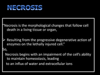
- 1. “Necrosis is the morphological changes that follow cell death in a living tissue or organ, Resulting from the progressive degenerative action of enzymes on the lethally injured cell.” So, Necrosis begins with an impairment of the cell’s ability to maintain homeostasis, leading to an influx of water and extracellular ions
- 2. Anoxia Ischemia Physical agents Chemical agents Biological agents Hypersenstivity
- 3. Necrotic changes in tissues are caused By Digestion of cell by enzymes Denaturation of proteins Digestion of cell by enzymes This digestion is of two types Autolysis: Digestion of cell by enzymes derived from their own lyosomes Heterolysis: Digestion of cell by enzymes derived from lysosmes of leukocytes.
- 4. Denaturation of proteins caused by intracellular acidosis and due to this result is that: Injury to the cell membrane Severe impairment of phosphorylation of cell Increase permeability of the cell Influx of Na+ and Ca+ in the cell Decreased intracellular activity of the cell
- 5. Changes inside the cell Changes in mitochondria Changes in Nucleus Changes in cytoplasm
- 6. Endoplasmic reticulum is disorganized There is rupture of membrane Ribosomes are shed off Disorganization of polysomes & their structures Mitochondria become swallon Loss of interamitochondrial granules Loss of cristae & change their shape Rupture of outer membrane of Mitochondria
- 7. Nucleus becomes smaller Chromatin loses & become clumped Nucleus shows following changes Pyknosis Karyorrhexis Karyolysis
- 8. PYKNOSIS “When the dna is broken down by endonucleases fragments are formed & the nucleus becomes acid and stains basophillic” KARYORRHEXIS “The pyknotic nucleus may break up into fragments and disappear. This process is called karyorrhexis” KARYOLYSIS “The pyknotic nucleus may undergo lysis by the enzyme DNAse”
- 9. Cytoplasm becomes more eosinophilic: Due to loss of Rna & denaturation of cytoplasmic proteins Cytoplasm becomes opaque.
- 10. Basic types Coagulative necrosis Liquefactive necrosis Caseous necrosis In special sites Fat necrosis Fibrinoid necrosis Gangrenous necrosis
- 11. “In this type of necrosis, the necrotic cell retains its cellular outline for several days” Coagulative necrosis typically occurs in solid organs such as kidney, heart and adrenal gland usually as a result of deficient blood supply and anoxia. Examples
- 12. Denaturation of protein is the basic mechanism of coagulative necrosis The injury and the subsequent increasing acidosis denatures not only the structural proteins but also the enzymic proteins, thus blocking the cellular proteolysis. Morphology Preservation of basic structural outline of the coagulated cells Appears as a mass of coagulated, pink staining homogenous cytoplasm
- 15. It is the type of necrosis that occurs due to autolytic and heterolytic actions of enzymes that convert the proteins of cells into liquid. It is characterized by softening and liquifaction of tissue. Examples Ischemic necrosis of brain Suppurative inflammation.
- 16. Enzymatic degradation of proteins is the basic mechanism of liquefactive necrosis Morphology o Complete loss of cellular detail o Cellular outline is also destroyed
- 19. Combination of coagulative and liquefactive necrosis Characterized by the presence of soft, dry, cheesy homogenous necrotic material. It is not liquified Examples Principaly in the center of tuberculous granuloma Morphology Microscopically the necrotic focus is composed of structureless amorphous granular debris enclosed within a ring of granulomatous inflammation.
- 21. It occurs in two forms: Enzymatic fat necrosis Traumatic fat necrosis
- 22. Most commonly seen in acute pancreatitis. “Refers to the necrosis in adipose tissue, induced by the action of pancreatic enzymes which are lead due to trauma to the pancreas” Morphology Chalky white opaque spots surrounded by inflammatory margins are seen Necrotic area shows acute inflammatory changes with dissolved fat cells
- 23. It occur following severe injury to the tissues with high fat content such as the breast, subcutaneous tissue and abdomen. Morphology Foam cells and giant cells are seen. necrotic foci contain a lot of phagocytes containing fat known as foam cells
- 25. Type of connective tissue necrosis especially affecting arterial walls. Mostly seen in two conditions Auto immune diseases e.g Rheumaic fever Malignant hypertension
- 27. Gangrene is the necrosis of tissue with superadded putrefaction (enzymatic decomposition). It is the clinical condition in which extensive tissue necrosis is complicated to a variable degree by secondary bacterial infection. Gangrene= Necrosis + infection + putrefaction
- 29. Arterial obstructon due to: Thrombosis of atherosclerotic artery Embolus Diabetes:- atherosclerotic artery , loss of sensation results repeated trauma & increase chances of infection Infection Gas gangrene Gangrene of scrotum Trauma Crush injuries Physical agents Burns Chemicals
- 30. Dry gangrene Wet gangrene Gas gangrene
- 31. It is usually secondary to slow occlusive vascular disease Etiology Gradual loss of arterial supply to an organ or tissue as happens in Arteriosclerosis Trauma Ergot poisoning Common sites limbs; especially foot
- 32. It is a traditional term used to describe the infarction of the limbs. It is not true gangrene because the infection in necrotic tissue is insignificant and putrefaction is absent or minimal. The necrotic area becomes black due to breakdown of hemoglobin and formation of iron sulfide
- 34. It is a type of gangrene in which tissue appears moist. It results from severe bacterial infection superimposed on necrosis Pathogenesis It is a true gangrene because it shows the severe infection and putrefaction of tissue with edema and foul smell. Arterial obstruction present. blackening of the tissue is due to formation of iron sulphide It is not clearly demarcated from adjacent healthy tissues. Common sites Intestine Appendix Limbs
- 35. Wet gangrene of intestine Wet gangrene of appendix
- 36. “In this type of gangrene bacterial infection causes necrosis and then gangrene with abundant gas formation in the tissue” Gas gangrene=wet gangrene + gas formation Predisposing factors Foreign bodies in wound cause tissue ischemia Foreign bodies favour infection Contamination of wound by soil is dangerous because its ionisable calcium salts and silicic acid may lead to tissue necrosis. Infection by aerobic organisms at the same time serve to produce anaerobic environment that is favorable for anaerobic clostridia.
- 37. Two groups of clostridia cause gas gangrene Saccharolytic: Clostridium perfringens Proteolytic: Clostridium isolyticum Pathogenesis Deep wound----anerobic condition---caused by spores of clostridia Necrosis of muscle fiber occur Fermentation of muscle carbohydrate occur with formation of lactic acid and gas. Arterial supply of the area is cut down Muscles become greenish- black due to iron sulphide & foul smell
- 38. Muscles Liver Complicatons Rapidly spreading gangrene Shock and hemolytic anemia Treatment of gangrene Treatment of predisposing factor: Amputation: Surgical removal of gangrene tissue to prevent spreading of the infection to the healthy tissue.
- 39. Gas gangrene of muscles Gas gangrene of Liver
- 40. “It is wet type of gangrene in which necrosis is superadded by infection and putrefaction” Predisposing factors: Sensory neuropathy Ischemia Lower resistance to infection Management: Control diabetes Keep the tissue dry and clean Antibiotics Surgical drainage of necrotic tissue