
Recommended
More Related Content
What's hot
What's hot (20)
Similar to Pediatric urology:Prune Belly Syndrome(PBS)
Similar to Pediatric urology:Prune Belly Syndrome(PBS) (20)
More from GovtRoyapettahHospit
More from GovtRoyapettahHospit (20)
Recently uploaded
Recently uploaded (20)
Pediatric urology:Prune Belly Syndrome(PBS)
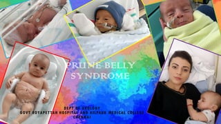
- 1. PRUNE BELLY SYNDROME DEPT OF UROLOGY GOVT ROYAPETTAH HOSPITAL AND KILPAUK MEDICAL COLLEGE CHENNAI 1
- 2. MODERATORS: Professors: Prof. Dr. G. Sivasankar, M.S., M.Ch., Prof. Dr. A. Senthilvel, M.S., M.Ch., Asst Professors: Dr. J. Sivabalan, M.S., M.Ch., Dr. R. Bhargavi, M.S., M.Ch., Dr. S. Raju, M.S., M.Ch., Dr. K. Muthurathinam, M.S., M.Ch., Dr. D. Tamilselvan, M.S., M.Ch., Dr. K. Senthilkumar, M.S., M.Ch. DEPT OF UROLOGY,GRH ANDKMC,CHENNAI. 2
- 3. PRUNE 3 DEPT OF UROLOGY,GRH ANDKMC,CHENNAI.
- 4. HISTORY 4 DEPT OF UROLOGY,GRH ANDKMC,CHENNAI.
- 5. ALSO KNOWN AS… 1. Triad syndrome 2. Eagle-Barrett Syndrome 3. Abdominal musculation syndrome 5 DEPT OF UROLOGY,GRH ANDKMC,CHENNAI.
- 6. EPIDEMIOLOGY Incidence 1in 29000 to 1 in 40000 live births. 95% in Males Higher incidence in twins, blacks and children born to younger mothers. Incidence in twins is discordant. 6 DEPT OF UROLOGY,GRH ANDKMC,CHENNAI.
- 7. GENETICS Normal karyotype is the rule. Association Noted in: Turner syndrome Monosomy 16 Trisomy 13 Trisomy 18 Beckwith-Wiedemann Syndrome 7 DEPT OF UROLOGY,GRH ANDKMC,CHENNAI.
- 8. 1. Abdominal musculature deficiency 2. Bilateral intraabdominal testes 3. Anomalous urinary tract 3 MAJOR FINDINGS 8 DEPT OF UROLOGY,GRH ANDKMC,CHENNAI.
- 9. PSEUDOPRUNE Females with prune belly syndrome and Males who do not have the complete triad of prune belly syndrome. Hence, males with a lax abdominal wall but without undescended testes or without a dilated urinary tract and all females are considered pseudoprunes. 9 DEPT OF UROLOGY,GRH ANDKMC,CHENNAI.
- 10. DIFFERENTIAL DIAGNOSES Nonimmune ascites, Posterior urethral valves, Amniotic band syndrome, Visceromeglia, Polycystic kidneys, Impaired lymphatic drainage in Turner’s syndrome, Giant liver cyst and Cystic adenomatoid malformation of the lung. 10 DEPT OF UROLOGY,GRH ANDKMC,CHENNAI.
- 11. PATHOGENESIS - THEORIES 1. Early in utero posterior urethral obstruction resulting in severe dilatation of urinary tract and possible fetal ascites and oligohydramnios. 2. Primary defect in the lateral plate mesoderm 3. An intrinsic defect of the urinary tract leading to ureteral dilatation 4. Yolk sac defect 11 DEPT OF UROLOGY,GRH ANDKMC,CHENNAI.
- 12. URETHRAL OBSTRUCTION THEORY 12 DEPT OF UROLOGY,GRH ANDKMC,CHENNAI.
- 13. OBSTRUCTION CAUSES True areas of stenosis, atresia, valves, or pinpoint diaphragms at the junction of the posterior and membranous urethra. “Functional obstruction” present in utero, secondary to prostatic hypoplasia, as the specific etiology of the syndrome. This “obstruction” is thought to be the result of conformational changes in the prostatic urethra during voiding, creating a valve-like mechanism. 13 DEPT OF UROLOGY,GRH ANDKMC,CHENNAI.
- 14. MESENCHYMAL DYSPLASIA 14 DEPT OF UROLOGY,GRH ANDKMC,CHENNAI.
- 15. RENAL DYSPLASIA – MECHANISMS (STEPHEN) (1) Defects of the ureteric bud or its branches, (2) Qualitative or quantitative deficiencies of the nephrogenic mesenchyme, and (3) Vascular ischemic insults with resultant ureteric obstruction and renal cystic dysplasia. (4) Due to a combination of a ureteric bud and metanephric defect. 15 DEPT OF UROLOGY,GRH ANDKMC,CHENNAI.
- 16. Mesenchymal Abnormality Urethral Obstruction Renal Dysplasia PCS and ureter dilatation Decreased Urine Output Oligohydramnios Urinary Ascites Abdominal Wall Defect Pulmonary Hypoplasia Orthopedic Abnormalities Patent Urachus 16 DEPT OF UROLOGY,GRH ANDKMC,CHENNAI.
- 17. 1. Renal anomalies 2. Ureteric anomalies 3. Bladder anomalies 4. Prostate and accessory sex organs 5. Urethral anomalies 6. Testicular anomalies UROGENITAL ANOMALIES 17 DEPT OF UROLOGY,GRH ANDKMC,CHENNAI.
- 18. RENAL ANOMALIES Renal dysplasia present in 50% of cases. Spectrum varies from normal renal parenchyma to dysplasia. Potter Type II and Type IV varieties are present. Renal collecting system is dilated. Non obstructive hydronephrosis is the rule. Ureteropelvic junction obstruction can occur in primary or secondary basis. 18 DEPT OF UROLOGY,GRH ANDKMC,CHENNAI.
- 19. URETERAL ANOMALIES Dilated, tortuous and redundant ureters. Proximal ureters are less abnormal than distal ureters. Massive dilation and stenosis can occur at all levels. VUR present in 75% of children. Ratio of collagen to smooth muscle is elevated in refluxing ureters. 19 DEPT OF UROLOGY,GRH ANDKMC,CHENNAI.
- 20. Infection Stasis Ineffective Peristalsis Dilated Ureters 20 DEPT OF UROLOGY,GRH ANDKMC,CHENNAI.
- 21. BLADDER ANOMALIES Enlarged with pseudodiverticulum at the urachus. Patent urachus in 25% to 30%. Ratio of collagen to smooth muscle is increased without obstruction. Smooth muscle hypertrophy seen in obstructed prune bladder. 21 DEPT OF UROLOGY,GRH ANDKMC,CHENNAI.
- 22. Dilated ureter and PCS Dilated bladder Urethral obstruction 22 DEPT OF UROLOGY,GRH ANDKMC,CHENNAI.
- 23. Ureteric Reflux Displaced Ureteric Orifices Splaying of Trigone Dilated Bladder 23 DEPT OF UROLOGY,GRH ANDKMC,CHENNAI.
- 24. UNBALANCED VOIDING Dilated Bladder Delayed First Sensation Ineffective Contraction Increased PVR 24 DEPT OF UROLOGY,GRH ANDKMC,CHENNAI.
- 25. PROSTATIC ANOMALIES Dilated posterior urethra Obstructive lesions – Urethral atresia, urethral stenosis, urethral membrane and Urethral diverticulum (20%) Type IV valve – Angulation of the urethra during voiding 25 DEPT OF UROLOGY,GRH ANDKMC,CHENNAI.
- 26. Dilated Posterior urethra Prostatic Hypoplasia Abnormal Mesenchymal Development 26 DEPT OF UROLOGY,GRH ANDKMC,CHENNAI.
- 27. Retrograde Ejaculation Incompetent Bladder Neck Dilated Prostatic Urethra 27 DEPT OF UROLOGY,GRH ANDKMC,CHENNAI.
- 28. ACCESSORY SEXUAL ORGANS ANOMALIES Atretic vas and seminal vesicles Poor attachment of epididymis to the testis Lack of continuity between efferent ductules and rete testis. Infertility is a normal in Prune Belly syndrome. 28 DEPT OF UROLOGY,GRH ANDKMC,CHENNAI.
- 29. URETHRAL ANOMALIES Urethral atresia/Hypoplasia (Associated with Patent urachus) Megalourethra – Transient in utero obstruction of junction between glanular and penile urethra Two types: 1. Scaphoid variety – Deficiency of spongiosum only 2. Fusiform variety - Deficiency of spongiosum and cavernosum. 29 DEPT OF UROLOGY,GRH ANDKMC,CHENNAI.
- 30. MEGALOURETHRA A newborn with prune belly syndrome with evidence of megalourethra and penoscrotal transposition. 30 DEPT OF UROLOGY,GRH ANDKMC,CHENNAI.
- 31. MEGALOURETHRA (a) A newborn with prune belly syndrome and megalourethra. (b) Voiding cystourethrogram in this patient demonstrates a large dilated bladder, open bladder neck, dilated posterior urethra, and wide, patent megalourethra. 31 DEPT OF UROLOGY,GRH ANDKMC,CHENNAI.
- 32. ABDOMINAL WALL DEFECT Medial and inferior muscular segments are typically involved. The order of severity of involvement from most to least involved is: Transversus abdominis, Rectus abdominis below the umbilicus, Internal oblique, External oblique, and Rectus abdominis above the umbilicus. 32 DEPT OF UROLOGY,GRH ANDKMC,CHENNAI.
- 33. ABDOMINAL WALL DEFECT Most severely affected areas may have skin, subcutaneous fat and a single fibrous layer on the peritoneum. Some patients show improvement in muscle tonus as they grow. Poor support of lower chest wall. Vulnerable to respiratory infections. 33 DEPT OF UROLOGY,GRH ANDKMC,CHENNAI.
- 34. EXTRA GENITOURINARY ABNORMALITIES Cardiopulmonary (49%) Orthopedic (65%) Gastrointestinal (65%) 34 DEPT OF UROLOGY,GRH ANDKMC,CHENNAI.
- 35. EXTRA GENITOURINARY ABNORMALITIES Cardiopulmonary (49%) Orthopedic (65%) Gastrointestinal (65%) Pulmonary hypoplasia Pneumothorax Pneumomediastinum Lobar atelectasis Pneumonia Chronic bronchitis Patent ductus arteriosus Atrial septal defect Ventricular septal defect Tetralogy of Fallot 35 DEPT OF UROLOGY,GRH ANDKMC,CHENNAI.
- 36. EXTRA GENITOURINARY ABNORMALITIES Cardiopulmonary (49%) Orthopedic (65%) Gastrointestinal (65%) Congenital dislocation of hips Chest wall deformity: Pectus excavatum/ carinatum Scoliosis Genu valgum Talipes equinovarus Severe leg maldevelopment: Arthrogryposis Knee Dimple 36 DEPT OF UROLOGY,GRH ANDKMC,CHENNAI.
- 37. ORTHOPEDIC ANOMALIES Pectus excavatum, scoliosis noted in a older patient with Prune belly syndrome 37 DEPT OF UROLOGY,GRH ANDKMC,CHENNAI.
- 38. KNEE DIMPLE Lateral dimple of the knee is a mild manifestation of the compression effects of intrauterine oligohydramnios. 38 DEPT OF UROLOGY,GRH ANDKMC,CHENNAI.
- 39. EXTRA GENITOURINARY ABNORMALITIES Cardiopulmonary (49%) Orthopedic (65%) Gastrointestinal (65%) Malrotation Intestinal atresia Intestinal stenosis Volvulus Anorectal agenesis Imperforate anus Omphalocele Gastroschisis Hepatobiliary anomalies Acquired megacolon VACTERL anomalies 39 DEPT OF UROLOGY,GRH ANDKMC,CHENNAI.
- 40. PBS WITH IMPERFORATE ANUS Newborn with prune-belly syndrome and imperforate anus 40 DEPT OF UROLOGY,GRH ANDKMC,CHENNAI.
- 41. PRENATAL DIAGNOSIS USG is similar to other causes of congenital bladder outlet obstruction. Diagnosis can be made as early as 11 to 14 weeks. Classical finding: HUN, Distended bladder and irregular abdominal wall circumference. But not consistently present even at 30 weeks. 41 DEPT OF UROLOGY,GRH ANDKMC,CHENNAI.
- 42. PRENATAL USG (A) Massively dilated bladder that fills most of the abdominal cavity. Note the lack of amniotic fluid. (B) The cephalic portion of the bladder reaching the level of both kidneys with hydronephrosis and renal parenchyma noted. (C) A dilated bladderwith a urachal diverticulum (arrow) and an elongated and dilated posterior urethra. 42 DEPT OF UROLOGY,GRH ANDKMC,CHENNAI.
- 43. PRENATAL MANAGEMENT Some have recommended in utero intervention for relief of urinary tract dilatation and oligohydramnios. Prenatal intervention has no benefit in terms of postnatal renal function. Urethral atresia with progressive oligohydramnios- Only circumstance in which prenatal intervention is justified. 43 DEPT OF UROLOGY,GRH ANDKMC,CHENNAI.
- 44. NEONATAL PRESENTATION Appearance of the abdominal wall immediately suggests the diagnosis of PBS. Cardiac and pulmonary abnormalities take precedence over urogenital abnormalities in the absence of true bladder outlet obstruction. 44 DEPT OF UROLOGY,GRH ANDKMC,CHENNAI.
- 45. WOODARD’S CLASSIFICATION 45 DEPT OF UROLOGY,GRH ANDKMC,CHENNAI.
- 46. MULTIDISCIPLINARY EVALUATION Neonatologist Nephrologist Urologist Cardiologist Orthopedician 46 DEPT OF UROLOGY,GRH ANDKMC,CHENNAI.
- 47. INITIAL EVALUATION Sr. Creatinine and electrolytes – rule out systemic acidosis and electrolyte imbalances. Baseline creatinine of < 0.7 mg/dl – Adequate renal function. In the presence of renal insufficiency, VCUG is warrented. 47 DEPT OF UROLOGY,GRH ANDKMC,CHENNAI.
- 48. VCUG Voiding cystourethrogram of a 1-year- old child with prune belly syndrome exhibiting the classic elongates, tortuous, and dilated ureter representing one of the radiographic hallmarks of the syndrome. The large, distended bladder is noted. 48 DEPT OF UROLOGY,GRH ANDKMC,CHENNAI.
- 49. VCUG Voiding cystourethrogram details the grossly enlarged, but non-trabeculated bladder. The bladder neck is widely patent, with hypoplastic urethra. 49 DEPT OF UROLOGY,GRH ANDKMC,CHENNAI.
- 50. VCUG Voiding cystourethrogram of a 1-year- old child demonstrating the classic radiographic appearance of the posterior urethra in prune belly syndrome. The open bladder neck and dilated prostatic/posterior urethra is evident. No urethral obstruction is present. 50 DEPT OF UROLOGY,GRH ANDKMC,CHENNAI.
- 51. CATEGORY I Prognosis is bad. Most of succumb to the disease in the newborn period. Supportive care Simple bladder drainage 51 DEPT OF UROLOGY,GRH ANDKMC,CHENNAI.
- 52. CATEGORY II Individualised evaluation and treatment Evaluation of renal function and drainage is required IVU provide dramatic images; no sufficient information on comparative function. 52 DEPT OF UROLOGY,GRH ANDKMC,CHENNAI.
- 53. EVALUATION – CATEGORY II Parenchymal function – DMSA at 4 to 6 weeks of age Renal outflow obstruction – MAG3 In case of poor renal function – Selective use of Whitaker test 53 DEPT OF UROLOGY,GRH ANDKMC,CHENNAI.
- 54. TREATMENT – CATEGORY II Aggressive interventions recommended. Infection and progressive renal insufficiency – Greatest threat to quality of life. Early reconstruction of the urinary system to reduce stasis and infection. Reconstruction delayed upto 3 months to allow for pulmonary maturation. Can be combined with circumcision, orchiopexy and abdominoplasty. 54 DEPT OF UROLOGY,GRH ANDKMC,CHENNAI.
- 55. CATEGORY II- ALTERNATIVE APPROACH Limited surgical intervention Close surveillance with medical management of bacteriuria. Surgical intervention only in patients with proven obstruction or intractable infection. 55 DEPT OF UROLOGY,GRH ANDKMC,CHENNAI.
- 56. CATEGORY II Progressive renal function detoriatiation is common in prune belly syndrome. Many patients ultimately require renal transplantation. 56 DEPT OF UROLOGY,GRH ANDKMC,CHENNAI.
- 57. CATEGORY III Good renal function Rarely require early urological intervention Regular monitoring of urinary tract dilatation using USG and renal function using serum creatinine. Regular monitoring for UTI. Correct cryptorchidism in 1st year of life. Abdominoplasty may be needed. Reflux correction may be needed in mid to long term follow up. 57 DEPT OF UROLOGY,GRH ANDKMC,CHENNAI.
- 58. SURGICAL MANAGEMENT Urinary tract procedures Orchiopexy Abdominal wall reconstruction 58 DEPT OF UROLOGY,GRH ANDKMC,CHENNAI.
- 59. URINARY TRACT PROCEDURES Reserved for children with: Progressive or severe HUN, High grade VUR, Recurrent infection, True obstructive uropathy, Progressive renal failure. 59 DEPT OF UROLOGY,GRH ANDKMC,CHENNAI.
- 60. EXTENSIVE RECONSTRUCTION The operation is performed transperitoneally. No attempt is made to straighten the lower ureter. The ureter may require tapering. The dome of the bladder is excised 60 DEPT OF UROLOGY,GRH ANDKMC,CHENNAI.
- 61. SUPRAVESICAL URINARY DIVERSION Repeated upper tract infections or deterioration of renal function Cutaneous vesicostomy is usually enough. Cutaneous pyelostomy/ distal ureterostomy in the presence of PUJ or UVJ obstruction respectively. Urachal diverticulum if present can be excised during cutaneous vesicostomy. 61 DEPT OF UROLOGY,GRH ANDKMC,CHENNAI.
- 62. MEGALOURETHRA RECONSTRUCTION A, The prepuce is reduced, and a circumcising incision is formed, preserving a mucosal collar. B, With a catheter in place to assist with identification of the urethra, the penis is degloved along the subdartos plane. C, The involved segment of the urethra is opened longitudinally, and the redundant urethral wall is excised to allow tapering of the urethra over a catheter of approximate size. 62 DEPT OF UROLOGY,GRH ANDKMC,CHENNAI.
- 63. MEGALOURETHRA RECONSTRUCTION D, The urethra is closed with absorbable running sutures and is bolstered with a second layer of sutures placed in an interrupted fashion if possible. E, The penile skin is brought forward, and the excess foreskin is removed with a second circumferential incision. F, The penile shaft skin is approximated to the mucocutaneous border. 63 DEPT OF UROLOGY,GRH ANDKMC,CHENNAI.
- 64. UNDESCENDED TESTES - ORCHIDOPEXY Transabdominal bilateral orchidopexy at 6 months of age is the approach of choice. Done in conjuction with other abdominal surgeries. In the absence of need for other abdominal surgeries, it can be done laparoscopically. 64 DEPT OF UROLOGY,GRH ANDKMC,CHENNAI.
- 65. UNDESCENDED TESTES Operative photograph showing the ease with which the testes reach the scrotum after neonatal transabdominal mobilization of the spermatic cords 65 DEPT OF UROLOGY,GRH ANDKMC,CHENNAI.
- 66. ABDOMINAL WALL RECONSTRUCTION Indicated in moderate to severe degree of abdominal wall laxity. Can be done as early as 6 months of age. Can be combined with orchidopexy. Improve cosmesis. Improvement in bowel, bladder and pulmonary function is controversial. 66 DEPT OF UROLOGY,GRH ANDKMC,CHENNAI.
- 67. TECHNIQUES 1. Randolph technique 2. Ehrlich technique 3. Monfort technique 4. Denes technique 5. Furness technique 67 DEPT OF UROLOGY,GRH ANDKMC,CHENNAI.
- 68. RANDOLPH ABDOMINOPLASTY A curvilinear incision extends from the tip of the 12th rib, along the anterior superior iliac spine to the pubic tubercle and onto the opposite 12th rib. 68 DEPT OF UROLOGY,GRH ANDKMC,CHENNAI.
- 69. RANDOLPH ABDOMINOPLASTY A parallel incision is made, avoiding the removal of too much abdominal wall. 69 DEPT OF UROLOGY,GRH ANDKMC,CHENNAI.
- 70. RANDOLPH ABDOMINOPLASTY Rhomboid-like pieces of abdominal wall are removed. 70 DEPT OF UROLOGY,GRH ANDKMC,CHENNAI.
- 71. RANDOLPH ABDOMINOPLASTY Critical non-absorbable sutures are placed at the anterior iliac spines and pubic tubercle. These are placed through full thickness of the abdominal wall including the periosteum. 71 DEPT OF UROLOGY,GRH ANDKMC,CHENNAI.
- 72. EHRLICH ABDOMINOPLASTY (a) Sharp dissection separates the skin the subcutaneous tissue from the musculofascial layer. (b–d) A vest–over-pants closure of the musculofascial layers. The umbilicus can be saved on a separate pedicle. (e) The excess abdominal skin is removed. 72 DEPT OF UROLOGY,GRH ANDKMC,CHENNAI.
- 73. MONFORT ABDOMINOPLASTY (a) An almond-shaped incision is made on the abdomen from the xyphoid to the pubic symphysis. (b) The full thickness of skin is removed, sparing the umbilicus. 73 DEPT OF UROLOGY,GRH ANDKMC,CHENNAI.
- 74. MONFORT ABDOMINOPLASTY (c) The peritoneum is opened lateral to the musculofascial later. Intra-abdominal surgery can be easily performed through these incisions. (d) The parietal peritoneum is incised at a level to achieve a normal-appearing waistline. 74 DEPT OF UROLOGY,GRH ANDKMC,CHENNAI.
- 75. MONFORT ABDOMINOPLASTY (e) The edges of the musculofascial plate are sewn to the peritoneal incisions. (f) The lateral skin flaps are trimmed and sutured together in the midline. 75 DEPT OF UROLOGY,GRH ANDKMC,CHENNAI.
- 76. DENES TECHNIQUE 76 DEPT OF UROLOGY,GRH ANDKMC,CHENNAI.
- 77. DENES TECHNIQUE 77 DEPT OF UROLOGY,GRH ANDKMC,CHENNAI.
- 78. 78 DEPT OF UROLOGY,GRH ANDKMC,CHENNAI.