
Recommended
Recommended
More Related Content
What's hot
What's hot (20)
Similar to Pediatric urology :Posterior Urethral Valve (PUV)- diagnosis & management
Similar to Pediatric urology :Posterior Urethral Valve (PUV)- diagnosis & management (20)
More from GovtRoyapettahHospit
More from GovtRoyapettahHospit (20)
Recently uploaded
Recently uploaded (20)
Pediatric urology :Posterior Urethral Valve (PUV)- diagnosis & management
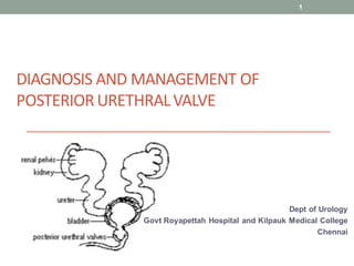
- 1. DIAGNOSIS AND MANAGEMENT OF POSTERIOR URETHRALVALVE Dept of Urology Govt Royapettah Hospital and Kilpauk Medical College Chennai 1
- 2. Moderators: Professors: • Prof. Dr. G. Sivasankar, M.S., M.Ch., • Prof. Dr. A. Senthilvel, M.S., M.Ch., Asst Professors: • Dr. J. Sivabalan, M.S., M.Ch., • Dr. R. Bhargavi, M.S., M.Ch., • Dr. S. Raju, M.S., M.Ch., • Dr. K. Muthurathinam, M.S., M.Ch., • Dr. D. Tamilselvan, M.S., M.Ch., • Dr. K. Senthilkumar, M.S., M.Ch. Dept of Urology, GRH and KMC, Chennai. 2
- 3. POSTERIOR URETHRAL VALVES • Congenital obstructing membranous folds within the lumen of posterior urethra causing LUTO starts in foetus in utero. • Primary tissues mature in an abnormal environment of high intraluminal pressures and organ distension Universal injury in the urinary tract 3 Dept of Urology, GRH and KMC, Chennai.
- 4. POSTERIOR URETHRAL VALVES • MC structural cause of urinary outflow obstruction in paediatric practice • MC type of obstructive uropathy leading to childhood renal failure. • Incidence - 1.4-2.1 per 10,000 live births • 10 % of prenatally diagnosed hydronephrosis. • Higher incidence in Blacks 4 Dept of Urology, GRH and KMC, Chennai.
- 5. HISTORY • First recognized by Morgagni in 1769. • Confirmed by Langenbeck in 1802 • First endoscopic description and the term posterior urethral valve coined by Hugh Hampton Young in 1919 • Young classified valves based on endoscopic appearance into 3 types • First endoscopic valve ablation by Randall in 1920 5 Dept of Urology, GRH and KMC, Chennai.
- 6. PUV – CLASSIFICATION Young classified valves based on endoscopic appearance into 3 types: Type – 1 • 95% cases • Valves arise from veru and insert in midline proximal to EUS • Distal end of WD into UGS Type – 2 • Valve extends from veru proximally, posterosuperiorly to BN Type – 3 • Annular ring • Persistence of urogenital membrane after uro-rectal septum divides cloacal membrane 6 Dept of Urology, GRH and KMC, Chennai.
- 7. 7 Dept of Urology, GRH and KMC, Chennai.
- 8. 8 Dept of Urology, GRH and KMC, Chennai.
- 9. 9 Dept of Urology, GRH and KMC, Chennai.
- 10. 10 Dept of Urology, GRH and KMC, Chennai.
- 11. 11 Dept of Urology, GRH and KMC, Chennai.
- 12. 12 Dept of Urology, GRH and KMC, Chennai.
- 13. EMBRYOLOGY Theories • Hypertrophy of urethral mucosal folds • Cloacal remnants after division of urogenital membrane • Abnormal development of WD/MD • Persistent oblique urogenital membrane 13 Dept of Urology, GRH and KMC, Chennai.
- 14. PATHOPHYSIOLOGY OF PUV PUV cause BOO in early life Bladder hypertrophy High voiding pressures to maintain complete voiding of bladder ( compensated phase) Gradual remodelling of bladder wall ( deposition of ECM ) Further increase in voiding pressures 14 Dept of Urology, GRH and KMC, Chennai.
- 15. Incomplete emptying of bladder as bladder begins to fail High PVR Overflow incontinence Urine reflux and upper tract dilatation Further renal damage Inc urine output ( normal growth and development) Polyuria 1. Existing dysplasia 2. Inc glomerular pressure with tubular injury 15 Dept of Urology, GRH and KMC, Chennai.
- 16. Bladder in a state of partial or complete stretch dilated bladder with poor contractility ( decompensated phase) 16 Dept of Urology, GRH and KMC, Chennai.
- 17. PATHOPHYSIOLOGY OF PUV Lower Urinary Tract Root cause Dysfunctional bladder BOO Bladder overdistention with bladder wall remodelling UUT dilation Renal dysplasia Features of BOO on cystoscopy •Posterior urethral dilation •BN Hypertrophy •Flattening of veru •Dilated ejaculatory ducts 17 Dept of Urology, GRH and KMC, Chennai.
- 18. PATHOPHYSIOLOGY OF PUV Upper urinary tract Ureteral dillation •Direct transmission of pressure •VUR 70% Chronic ureteral dilation •Wall thickening •Loss of co-aptation, peristalsis • Urine stasis/infection • Intra-renal pressure Renal dysfucntion 18 Dept of Urology, GRH and KMC, Chennai.
- 19. PATHOPHYSIOLOGY OF PUV Renal dysfunction in PUV Obstructive uropathy •Damage to luminal cells in renal tubule •Concentration defects •Loss of medullary gradient •Polyuria Renal Dysplasia •In-utero obstruction during renal development •Irreversible 19 Dept of Urology, GRH and KMC, Chennai.
- 20. PATHOPHYSIOLOGY OF PUV 20 Dept of Urology, GRH and KMC, Chennai.
- 21. PATHOPHYSIOLOGY OF PUV VURD Syndrome Vesicoureteral Reflux and Dysplasia 13% patients with PUV Theory •Dysplastic kidney with VUR protects C/L kidney •Pop-off mechanism Disproved now C/L kidney also at risk of renal dysfunction and ESRD 21 Dept of Urology, GRH and KMC, Chennai.
- 22. POP-OFF MECHANISMS • Reflux – VURD syndrome-high bladder pressures on the refluxing kidney, sparing and protecting the nonrefluxing kidney. • Bladder diverticula • Urinary ascites • Patent urachus -RARE 22 Dept of Urology, GRH and KMC, Chennai.
- 23. POP- OFF MECHANISMS 23 Dept of Urology, GRH and KMC, Chennai.
- 24. 24 Dept of Urology, GRH and KMC, Chennai.
- 25. 25 Dept of Urology, GRH and KMC, Chennai.
- 26. PRE-NATAL DIAGNOSIS • 1 in 1250 on USG • 10% of all antenatal GU anomalies • 1/3rd of B/L renal disease • Pathognomonic Findings B/L HUN Oligohydramnios Dilated posterior urethra (KEY-HOLE SIGN) renal echogenecity 26 Dept of Urology, GRH and KMC, Chennai.
- 27. OLIGOHYDRAMINOS-DEFINATION • Amniotic fluid volume < 500ml at 32-36wks gestation • AFV < 5th percentile for gestational age • AFI < 5 ( normal= 7-25 ) • Single vertical pocket < 2cms 27 Dept of Urology, GRH and KMC, Chennai.
- 28. AMNIOTIC FLUID INDEX • Four quadrant technique • AFI is measured by dividing the uterus into four quadrants • Linea nigra- divides into right and left halves • Umblicus serves as the dividing point for the upper and lower halves • AFI= A+B+C+D Each individual pocket of fluid should be= 2-8cm 28 Dept of Urology, GRH and KMC, Chennai.
- 29. FETAL MRI – To distinguish the degree of obstruction based on urethral dilation – Distended bladder size with thickening – Reduced amniotic fluid levels. • Lung hypoplasia and cystic changes in renal parenchyma are also apparent on MRI • Do not provide added analysis of causes of obstruction, in diagnosing the actual cause of LUTO 29 Dept of Urology, GRH and KMC, Chennai.
- 30. MANAGEMENT- APPROACH AT BIRTH • Management starts immediately after birth • Establish urinary drainage( 5-6fr feeding tube/coude tip ) • Sample for urine routine and urine c/s collected • IV line secured and antibiotics started/ IV fluids • Blood sample withdrawn after 48-72hrs for CBC, S. creatinine, and electrolytes • Look for dehydration and acidosis with other electrolyte imbalance 30 Dept of Urology, GRH and KMC, Chennai.
- 31. MANAGEMENT-APPROACHAT BIRTH Sonography KUB: • Kidneys for cortical echogenicity, CMD, degree of HN, • Dilatation of ureters • Bladder for distension, thickness, presence of diverticula, high bladder neck with hyperplasia • Posterior uretheral dilatation, • Urinoma, urinary ascities and other anomalies 31 Dept of Urology, GRH and KMC, Chennai.
- 32. Urinoma 3-10% •Forniceal rupture •Trans-peritoneal transudation •Bladder rupture Neonatal ascites 32 Dept of Urology, GRH and KMC, Chennai.
- 33. MANAGEMENT-APPROACHAT BIRTH • VCUG- is a definitiveinvestigation • Timing: hemodynamicallystable • Antibiotic prophylaxis: not needed if urine c/s report is sterile and within 72hrs of this sterile report • How to make baby to urinate: suprapubic massage, pinching glans, sprinkling cold water on glans o Condition of bladder, diverticula o Reflux o Bladder neck hypertrophy o Dilatation and length of posterior urethra o Abrupt funneling and configurationof obstruction and its level 33 Dept of Urology, GRH and KMC, Chennai.
- 34. MANAGEMENT- APPROACH AT BIRTH • Definitive study VCUG • Findings on VCUG Thickened trabeculated bladder with multiple diverticulae High grade VUR – 50% Hypertrophied elevated bladder neck Grossly dilated posterior urethra Abrupt funneling of urethra at level of valve 34 Dept of Urology, GRH and KMC, Chennai.
- 35. MCU COMPLICATION • Rupture of bladder due to over distension EBC- NEONATES(< 1 mth); infants(1mth-1yr)= 7x wt in kgs Rest capacity= (age+2)x30, capacity in mls 5-6fr catheter, passed aseptically. Water soluble contrast 20%(w/v), slow filling with gravity under fluoroscopic control • Other complications: dysuria, infection, hematuria, retention of urine 35 Dept of Urology, GRH and KMC, Chennai.
- 36. RADIONUCLIDE RENAL SCAN • The radionuclide renal scan offers • quantification of differential renal function • cortical deficits seen on the study may imply renal dysplasia. • MAG3 or DMSA • Placement of a urinary catheter is essential in a patient with VUR to minimize error in the calculation of renal function. 36 Dept of Urology, GRH and KMC, Chennai.
- 37. • Definitive management Cystoscopy + Endoscopic valve ablation First line option To be done in hemodynamically stable pts 37 Dept of Urology, GRH and KMC, Chennai.
- 38. ADVANTAGESOF EARLY VALVEABLATION • Bladder cycling, obstruction free drainage • Thus improves - Complaince - Bladder stability in infancy with a possibility of less bladder dysfunction in older children 38 Dept of Urology, GRH and KMC, Chennai.
- 39. PUVFULGRATION-PRECAUTIONS • Use appropriate size equipment • Pure current • Engage leaflets adequately for optimum cut • Never an over doing i.e avoid aggressive resection 39 Dept of Urology, GRH and KMC, Chennai.
- 40. PUVFULGRATION-INSTRUMENTATION • Urethrocystoscopes- mostly 7-7.5fr used • Resectoscopes- 9.5fr 40 Dept of Urology, GRH and KMC, Chennai.
- 41. BUGBEE- ELECTRODE 41 Dept of Urology, GRH and KMC, Chennai.
- 42. PUV FULGRATION- WHERE TO ABLATE • 5 and 7o’clock- most time • 5,7 and 12o’clock- if promonent 12o’clock leaflet 42 Dept of Urology, GRH and KMC, Chennai.
- 43. Fogarty or Foley's balloon catheter • A size 4 Fr Fogarty balloon catheter or an appropriate size Foley's catheter is placed into the bladder • balloon inflated with 0.75 ml of saline. • With gentle withdrawal, the operator places the balloon at the level of the valves. • Sharp withdrawal of the catheter ruptures the membrane without injury to the urethra. • This procedure should be preferably done under fluoroscopic or ultrasonographic guide to ensure that the balloon is only inflated proximal to the valve to avoid urethral injury. 43 Dept of Urology, GRH and KMC, Chennai.
- 44. 44 Dept of Urology, GRH and KMC, Chennai.
- 45. PUV FULGRATION- FOLLOW UP • Post fulgration= in 2-3days, usg/s.creatinine/s.electrolytes • Monthly urine analysis, urine c/s, ht and wt • 3 monthly USG KUB to look for - dilatation of PCS and ureters - residual urine within the bladder • Repeat MCUG at 3 months for any residual valve • Cystoscopy only if MCUG shows persistent significant obstructive changes • DMSA at 3 months for cortical functiopn and quantification of scars 45 Dept of Urology, GRH and KMC, Chennai.
- 46. • Vesicostomy •VLBW infant not accomodating endoscope •Severe urosepsis •Failed ablation Continued impaired renal function High bladder urine volumes Upper tract deterioration •BLOCKSOM TECHNIQUE Dome of bladder fixed to fascia Most important Ensure taut posterior wall 46 Dept of Urology, GRH and KMC, Chennai.
- 47. 47 Dept of Urology, GRH and KMC, Chennai.
- 48. 48 Dept of Urology, GRH and KMC, Chennai.
- 49. 49 Dept of Urology, GRH and KMC, Chennai.
- 50. 50 Dept of Urology, GRH and KMC, Chennai.
- 51. • Supravesical Diversion •Direct low pressure renal drainage to optimise renal function •Indication Complete decompression of the lower urinary tract with Worsening renal function Increasing upper tract dilation Clinical picture of sepsis •Possible cause Compression of intramural ureter by thick walled bladder •Disadvantages Complicatedundiversion procedure needed Defunctionalised bladder Loss of contractilityand compliance 51 Dept of Urology, GRH and KMC, Chennai.
- 52. vesicostomy End stomal ureterostomy loop ureterostomy 52 Dept of Urology, GRH and KMC, Chennai.
- 53. Sober Y ureterostomy Ring ureterostomy Cutaneous pyelostomy 53 Dept of Urology, GRH and KMC, Chennai.
- 54. 54 Dept of Urology, GRH and KMC, Chennai.
- 55. 55 Dept of Urology, GRH and KMC, Chennai.
- 56. • Nephroureterectomy •Historically for VURD •Nowadays only in intractable UTI •Preserve ureter for later use in augmentation 56 Dept of Urology, GRH and KMC, Chennai.
- 57. Management of VUR •Seen in 50-80% cases •Resolves in 25-40% post valve ablation •Focus Maintaining low storage pressures •High complication rates of VUR surgery Persistence of VUR: • Residual valve ( 15-33%) • Altered bladder dynamics mainly low complaint bladder • Paraureteric diverticulum 57 Dept of Urology, GRH and KMC, Chennai.
- 58. Bladder neck incision •No evidence of benefit •Not recommended routinely Circumcision Recommended -Risk of UTI 50-60% -Incidence reduced by 90% with circumcision -should certainly be completed before giving any consideration to a ureteral reimplant in a scenario of frequent febrile urinary tract infections despite conservative measures. 58 Dept of Urology, GRH and KMC, Chennai.
- 59. BLADDER DYSFUNCTION IN PUV • Cascading pathological changes •3 distinct evolutionary patterns Infancy and Early Childhood •Detrusor hyperreflexia Late Childhood •Decreasing intravesical pressures and improving compliance Adolescence •Increased capacity bladder with hypocontractility and atony 59 Dept of Urology, GRH and KMC, Chennai.
- 60. BLADDER DYSFUNCTION IN PUV • Long term follow-up mandatory Clinical examination Periodic assessment of voiding Observation, UFR, PVR,UDS Upper tract assessment USG •Bladder Management Voiding training •Timed voiding and Double voiding •CIC if necessary Anti-cholinergics •Oxybutynin 0.1mg/kg preferred •Stop if no effect or high PVR α – blockade •To relax bladder neck •Tamsulosin 0.2mg 60 Dept of Urology, GRH and KMC, Chennai.
- 61. BLADDER DYSFUNCTION IN PUV • Valve Bladder Syndrome Coined by Mitchell in 1982 Worsening renal function and HUN No evidence of BOO Three important determinants •Polyuria •Poor bladder compliance with high-pressure voiding and elevated wall tension •Residual urine volume 61 Dept of Urology, GRH and KMC, Chennai.
- 62. BLADDER DYSFUNCTION IN PUV 62 Dept of Urology, GRH and KMC, Chennai.
- 63. VALVE BLADDER- DEFINITION • Low capacity • High pressure • Low complaince bladder with • Upper tract deterioration • In the absence of outlet obstruction 63 Dept of Urology, GRH and KMC, Chennai.
- 64. BLADDER DYSFUNCTION IN PUV • Management Routine bladder management CIC mandatory Nocturnal bladder drainage •Breaks the cycle •Extender period of bladder decompression Inability to catheterise Appendicovesicostomy (Mitrofanoff Procedure) Refractory to therapy with small contracted bladder •Augmentation cystoplasty •Ureteral augmentation preferred if possible 64 Dept of Urology, GRH and KMC, Chennai.
- 65. INDICATORS OF RENAL FUNCTION •Incidence of ESRD 20-50% •Nadir creatinine at 1 year of age < 0.8 Minimal risk > 1.2 High risk •Other factors Age at diagnosis Renal dysplasia Recc UTI Bladder dysfunction 65 Dept of Urology, GRH and KMC, Chennai.
- 66. FAVOURABLE PROGNOSTIC FACTORS 66 Dept of Urology, GRH and KMC, Chennai.
- 67. 67 Dept of Urology, GRH and KMC, Chennai.
- 68. 68 Dept of Urology, GRH and KMC, Chennai.
- 69. TRANSPLANTATION IN PUV PATIENTS • The 2006 annual report of the North American Pediatric Renal Trials and Collaborative Studies listed obstructive uropathy as the second most common cause for transplantation, accounting for 1424 of 8990 transplantation cases (15.8%) since 1987 (Smith et al, 2007). 69 Dept of Urology, GRH and KMC, Chennai.
- 70. TRANSPLANTATION IN PUV PATIENTS 70 Dept of Urology, GRH and KMC, Chennai.
- 71. TRANSPLANTATION IN PUV PATIENTS Several comorbidities: • High-grade vesicoureteral reflux into native, nonfunctioning kidneys and valve bladder syndrome with a thick-walled, poorly contractile or hypercontractile bladder. • The thickened bladder wall of PUV patients may contribute to the significantly increased incidence of ureteral obstruction 71 Dept of Urology, GRH and KMC, Chennai.
- 72. RENAL Tx IN PATIENT TREATEDFOR PUV 72 Dept of Urology, GRH and KMC, Chennai.
- 73. • Video-urodynamics should be obtained for transplant candidates to determine – the safe storage pressures – contractile function of the future reservoir. • Overnight bladder drainage or CIC - prior to transplantation to optimize the reservoir and establish proper bladder management. • Pretransplantation nephrectomy- rarely required and only considered in cases in which proteinuria or severe polyuria is creating hemodynamic challenges. TRANSPLANTATION IN PUV PATIENTS 73 Dept of Urology, GRH and KMC, Chennai.
- 74. • Augmentation is necessary if unsafe storage pressures in the bladder. • Pretransplantation augmentation is preferable – to prevent the undertaking in a immunocompromised child – one too young to take responsibility for catheterization and pouch management, • Recent experience argues that transplantation into even a vesicostomy is a safe alternative until the child grows to an appropriate age for cystoplasty and the attendant reliance on CIC. TRANSPLANTATION IN PUV PATIENTS 74 Dept of Urology, GRH and KMC, Chennai.
- 75. AUGMENTATION OF BLADDER PRE TRANSPLANT • Augment before immunosupression • To young to take responsibility of CIC or manage stoma • Increase risk of UTI POST TRANSPLANT • Risking graft loss by doing Tx in unsafe bladder • Tx in vesicostomy is safe • Later cystoplasty and CIC can be practiced effectively and safely as child is more prepared and complaint • Infection rates are less or same 75 Dept of Urology, GRH and KMC, Chennai.
- 76. ANTENATAL INTERVENTION CONTROVERSIAL • Accurate diagnosis with current technology is difficult • Natural history of each disease process causing AH is variable and has not been fully elucidated • Lack of data regarding the success and complications of intervention in cases of PUV 76 Dept of Urology, GRH and KMC, Chennai.
- 77. WHY?? • An infant affected by posterior urethral valves may be affected by serious comorbidities – pulmonary hypoplasia – physical stigmata of oligohydramnios, including • Potter facies • clubfeet and deformed hands • poor abdominal muscle tone • require intensive initial management. 77 Dept of Urology, GRH and KMC, Chennai.
- 78. ANTENATAL INTERVENTION •Indications Oligohydramnios Dilated bladder with severe HUN No renal cortical cystic lesions Normal Karyotype Fetal urine analysis after 20 weeks ( 2-3 samples at the interval of 24-48hrs) •Na < 100meq/L •Cl < 90 meq/L •Osmolarity < 200meq/L •β2 microglobulin < 6mg/L •43% fetal mortality rate 78 Dept of Urology, GRH and KMC, Chennai.
- 79. VESICOAMNIOTIC SHUNTING • Improves survival in worst cases by improving pulmonary function and renal function • But high complication rate: - 40% migration, obstruction, and displacement 79 Dept of Urology, GRH and KMC, Chennai.
- 80. FETAL CYSTOSCOPY • 8.8% fistula formation rates • 5.9% recurrence of LUTO • Preterm delivery and death after birth 80 Dept of Urology, GRH and KMC, Chennai.
- 81. SFU- STAGES OF FETAL LUTO 81 Dept of Urology, GRH and KMC, Chennai.
- 82. PLUTO TRIAL • Percutaneous vesicoamniotic shunting vs conservative management for LUTO • It was RCT • Paucity of data: poor recruitment and pregnancy termination • Only 12 live births in each group • Improved survival in shunted group for at 28 days, but overall survival was same • High mortality bec of pulmonary hypoplasia • Greater risk of pregnancy loss and maternal complications 82 Dept of Urology, GRH and KMC, Chennai.
- 83. PLUTO TRIAL 83 Dept of Urology, GRH and KMC, Chennai.
- 84. 84 Dept of Urology, GRH and KMC, Chennai.
- 85. 85 Dept of Urology, GRH and KMC, Chennai.
- 86. FINAL TAKE • Primary valve ablation is the preferred T/t • Those unstable bladder- bladder level diversion • Supervesical div.- pts who fail to respond to bladder level diversion • Diversion yields similar results- disadv of needing more surgical procedure • At puberty - 2/3rd progress to CKD - 1/3RD progress to ESRD • PUV- is it never ending story?? • Life long follow up “Primary lesion is simple to treat but total care of boy with PU valve is complicated undertaking”- SIR DAVID INNES WILLIAM 86 Dept of Urology, GRH and KMC, Chennai.
- 87. THANK U 87 Dept of Urology, GRH and KMC, Chennai.