Top profile Call Girls In Purnia [ 7014168258 ] Call Me For Genuine Models We...
DR. RABIN-Anatomy Lecture 2.pdf
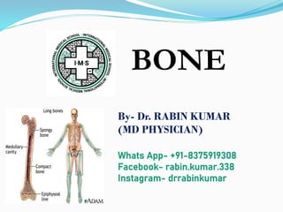
1. BONE
By- Dr. RABIN KUMAR
(MD PHYSICIAN)
Whats App- +91-8375919308
Facebook- rabin.kumar.338
Instagram- drrabinkumar
2. Parts of the skeletal system
• Bones (skeleton)- 206
• Joints
• Cartilages
• Ligaments
Divided into 2 divisions
• Axial skeleton-80
• Appendicular skeleton-126
3. 7-3
Skeletal System
The axial skeleton is composed of the bones along the
central axis of the body,
the skull
the vertebral column
the thoracic cage
The appendicular skeleton consists of the bones of the
appendages
upper and lower limbs
the bones that hold the limbs to the trunk of the body.
13. (1) Long Bones
• Longer than they are wide
• Has a shaft and 2 ends
• Weight bearing bones (like steel beams)
• Provide the greatest structure and support
• Examples:
– All limb bones
– Except…. Kneecap, Wrist and Ankle bones
14. 6-14
Structure of a Long Bone
Diaphysis
Epiphysis
proximal
distal
Metaphysis
Epiphyseal line
Articular cartilage
Medullary cavity
15. Parts of Bone
Epiphysis
The ends of long bones
Wider than diaphysis
Joint surface of each epiphysis is covered with hyaline
cartilage
Metaphysis
The area of bone between the epiphysis and
diaphysis
Diaphysis
– This is the main body of the bone – long region
– Center, main shaft
– Long part of bone
16.
17. • Growth plate – in
between epiphysis
and metaphysis
• Epiphyseal Line:
– Remnant of
Epiphyseal Plate
– Found in adult
bones
Epiphyseal Plate -
18.
19. (2) Short Bones
• Cube Shaped
• Allow for wider range of movement
• Examples:
– Wrist
– Ankle
23. (4) Irregular Bones
• Complicated, unusual shapes
• Muscles, tendons, ligaments usually
attach to these
• Examples:
– Vertebrae
– Hip bones
24.
25. • Membranes:
– Periosteum = Around the outside
• Richly supplied with nerve fibers, lymphatic vessels and
blood vessels
• Provides anchoring points for tendons and ligaments
– Endosteum = Around the inside
• Surrounds the spongy bone
27. Medullary Cavity
• In the diaphysis of
the long bone deep to
the compact bone is
the medullary cavity.
in an adult it is full of
yellow bone marrow.
• The medullary cavity
is lined with endosteum.
45. Naming Skeletal Muscles
1 – Location of the muscle
2 – Shape of the muscle
3 – Size of the muscle
4 – Direction/Orientation of the muscle
fibers/cells
5 – Number of Origins
6 – Location of the Attachments
7 – Action of the muscle
46. Muscles Named by Location
Location:
● frontalis – frontal bone
● lateralis – lateral or on the side
● tibialis anterior – front of tibia
● fibularis longus – near fibula
● supra – above
● infra – below
● sub – underneath
● Pectoral- Chest
● Brachial- Arm
● Gluteus- Buttock
48. Muscles Named by Size
Size:
● maximus – largest
● minimis – smallest
● vastus - huge
● longus – longest
● brevis – short
● major – large
● minor – small
Example: Pectoralis Major
49. Muscles Named by Direction of Fibers
Direction/Orientation:
● rectus (straight) - parallel to the
muscle’s long axis
ex: rectus abdominis
● transversus (transverse) – at
right angles to the muscle’s long
axis
● oblique – diagonal
50. Muscles Named for Number of Origins
Number of Origins:
● biceps – two origins
ex: biceps brachii
● triceps – three origins
ex: triceps brachii
● quadriceps – four origins
51. Muscles Named for
Origin and Insertion Points
Origin and Insertion:
sterno = sternum
cleiodo = clavicle
mastoid = location on
the temporal bone
sternocleiodomastoid muscle
52. •Origin
Muscle attachment that remains
fixed
•Insertion
Muscle attachment that moves
•Action
What joint movement a muscle
produces
i.e. flexion, extension, abduction,
etc.
ASSOCIATED TERMS
• Tendon
cord of fibrous tissue
• Belly
Fleshy part of muscle
53. Muscles Named for Action
Action:
● flexor carpi radialis –
flexes wrist
● abductor magnus –
abducts the thigh
● extensor digitorum –
extends the fingers
● levator – lifts a structure