Call Girls in Delhi Triveni Complex Escort Service(🔝))/WhatsApp 97111⇛47426
lower limb musclesjajsjsjsjsjsjsjsjsjsjjsjsj
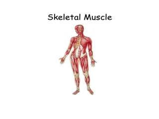
1.
2. Muscles of the Hand
• Muscles of the hand can be divided into two
groups: extrinsic and intrinsic muscles.
The extrinsic muscles are located in
the anterior and posterior compartments of
the forearm. They control crude movements
and produce a forceful grip.
The intrinsic muscles of the hand are located
within the hand itself. They are responsible for
the functional movement of the hand.
3. Extrinsic muscles of hand
muscle origin insertion action innervations
Abductor digiti
minimi
pasiform Medial side of
proximal
phalanx
Abduct little
finger
Ulnar nerve
Flexor digiti
minimi bravis
Humate Same as above Abducts little
finger
Ulnar nerve
Opponens digiti
minimi
Humate Medial side of
metacarpal 5
Opposition Ulnar nerve
4. Lumbricals
• These are four lumbricals in the hand, each associated
with a finger. They are very crucial to finger movement,
linking the extensor tendons to the flexor tendons.
• (The flexor tendons are strong smooth cords that
connect the muscles of the forearm to the bones
in the fingers and thumb).
5. Interossei
• The interossei muscles are located between
the metacarpals. They can be divided into two
groups: the dorsal and palmar interossei.
7. Introductions
• The muscles of the back are divided into
three:
• Superficial – associated with movements
of the shoulder.
• Intermediate – associated with
movements of the thoracic cage.
• Deep – associated with movements of
the vertebral column.
8. The Superficial Back Muscles
• The superficial back muscles are situated
underneath the skin and superficial fascia.
• They originate from the vertebral column
and attach to the bones of the shoulder –
the clavicle, scapula and humerus.
• All these muscles are therefore associated
with movements of the upper limb.
9. Cont.
• The muscles in this group are the
trapezius, latissimus dorsi, levator
scapulae and the rhomboids.
• The trapezius and the latissimus dorsi lie
the most superficially, with the trapezius
covering the rhomboids and levator
scapulae.
10. The Intermediate Back
Muscles
• The intermediate group contains two muscles –
the serratus posterior superior and serratus
posterior inferior.
• These muscles run from the vertebral column to the
ribcage, and assist with elevating and depressing the
ribs.
• They are thought to have a slight respiratory function.
11. Serratus Posterior Superior
The serratus posterior superior is a
thin, rectangular shaped muscle. It lies
deep to the rhomboid muscles on the
upper back.
• Origin: cervical and thoracic spines
(usually C7 – T3).
• Insrtion:posterior surface os 2nd
through 5th rib
• Innervation: Intercostal nerves.
• Actions: Elevates ribs during
inhalation
12. Serratus Posterior Inferior
• The serratus posterior inferior is
broad and strong. It lies underneath
the latissimus dorsi.
• Origin: spinous process and
supraspinous ligament t11 -12.
• Insertion: posterior aspect of ribs 9-
12.
• Innervation: Intercostal nerves.
• Actions: Depresses ribs and assists
forced expiration.
13. Deep muscles
• The deep muscles of the back are well-developed,
and collectively extend from the sacrum to the base
of the skull.
• They are associated with the movements of the
vertebral column, and the control of posture.
14. Deep muscles
• The deep back muscles lie
immediately adjacent to the vertebral
column and ribs.
Superficial:
Splenius capitis
Splenius cervicis
15. • Splenius capitis:
. It is a long, broad, strap-like muscle
found deep to the trapezius muscle.
Origin: spinous process C7-T3.
Insertion: posterior mastoid process
and inferior nuchal line of the
occipital bone.
Action: rotation of
the head. extension of
the head and cervical spine.
16. Splenius cervicis:
Origin: from the spinous
processes of T3-T6 vertebra
inserts: onto
the transverse processes of C1-C3.
Blood supply:
the occipital or transverse cervical
arteries.
Action: rotate the cervical spine.
Bilaterally, it causes extension of
the cervical spine.
18. gluteus maximus
• Origin: outer surface of ilium,
sacrum, coccyx, sacrotuberous
ligament
• Insertion: lesser trochanter
and gluteal tuberosity of femur
• Nerve supply: inferior gluteal
nerve
• Action: extends & laterally
rotates thigh; it extends knee
joint
19. gluteus medius
• Origin: outer surface of
ilium
• Insertion: greater
trochanter of femur
• Nerve supply : superior
gluteal nerve
• Action : abducts thigh. Tilts
pelvis when walking
20. gluteus minimus
• Origin: outer surface of
ilium
• Insertion: greater
trochanter of femur
• Nerve supply: superior
gluteal nerve
• Action : abduct thigh;
medially rotate thigh
21. cont.
• Other muscles
• Piriformis, superior gemellus, obturator
internus, inferior gemellus, obturator
externus all these muscles are same
insertion(greater trochanteric fossa)and
same as action(lateral rotator of thigh)